NASM Ch5 Muscular System
1/91
There's no tags or description
Looks like no tags are added yet.
Name | Mastery | Learn | Test | Matching | Spaced | Call with Kai | Chat |
|---|
No analytics yet
Send a link to your students to track their progress
92 Terms
muscular system
links the nervous and skeletal systems and is responsible for generating the forces that move the human body
muscle
contract to create internal tension that, under the control of the nervous system, manipulates the bones to produce movements around the joints
cardiac muscle
what makes up the heart
smooth muscle
primarily makes up the tissues of internal organs
skeletal muscle
holds the most importance for the fitness professional’s base of knowledge
skeletal muscle
The type of muscle tissue that connects to bones and generates the forces that create movement.
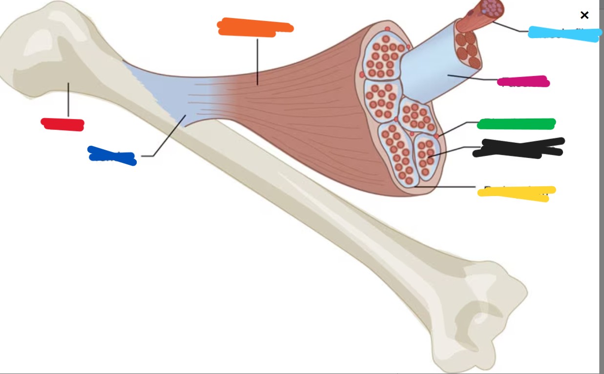
fascia
Connective tissue that surrounds muscles and bones.
epimysium
Inner layer of fascia that directly surrounds an entire muscle, commonly referred to as the “deep fascia.”
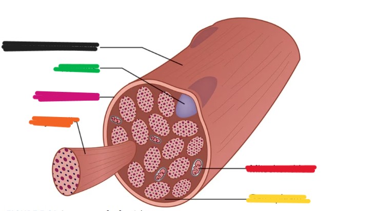
fascicles
Largest bundles of fibers within a muscle and surrounded by perimysium.
perimysium
Connective tissue surrounding a muscle fascicle.
endomysium
Connective tissue that wraps around individual muscle fibers within a fascicle
connective tissue
They allow the forces generated by the muscle to be transmitted from the contractile components of the muscle to the bones, creating motion
bone
red

tendon
dark blue

epimysium
orange

muscle fiber
light blue

fascicle
pink

blood vessels
green

endomysium
black

perimysium
yellow
sarcolemma
Within the endomysium of the fascicles, individual muscle fibers are themselves encased by a plasma membrane known as
glycogen
Glucose that is deposited and stored in bodily tissues, such as the liver and muscle cells; the storage form of carbohydrate.
myoglobin
Protein-based molecule that carries oxygen molecules into the muscles.
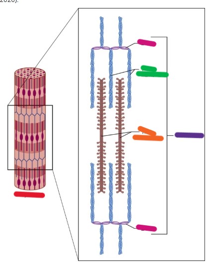
myofibrils
The contractile components of a muscle cell; the myofilaments (actin and myosin) are contained within
myofilaments
The filaments of a myofibril; include actin and myosin.
actin
The thin, stringlike, myofilament that acts along with myosin to produce muscular contraction.
myosin
The thick myofilament that acts along with actin to produce muscular contraction.
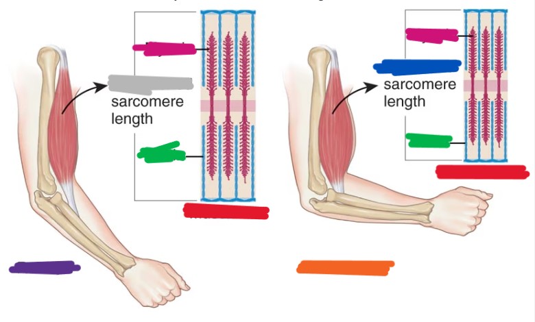
sarcomere
The structural unit of a myofibril composed of actin and myosin filaments between two Z-lines.
z-line
The meeting point of each sarcomere.
skeletal muscle fiber
black

nucleus
green

sarcolemma
pink

myofibril
orange

mitochondrion
red

sarcoplasm
yellow
z-line
pink
thin filaments
green

sarcomere
purple

thick filaments
orange

myofibril
red

tropomyosin
located on the actin filament and blocks myosin-binding sites located on the actin filament, keeping myosin from attaching to actin when the muscle is in a relaxed state
troponin
located on the actin filament, plays a role in muscle contraction by providing binding sites for both calcium and tropomyosin when a muscle needs to contract
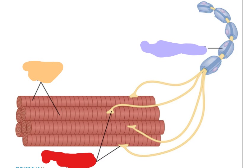
neural activation
The nervous system’s signal that tells a muscle to contract.
neuromuscular junction
The specialized site where the nervous system communicates directly with muscle fibers.
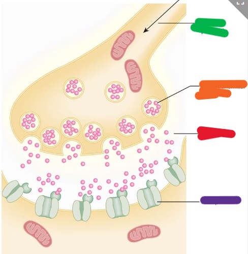
synapse
A junction or small gap between the motor neuron and muscle cells.
motor unit
A motor neuron and all of the muscle fibers that it innervates.
neuromuscular junction
red

axon of motor neurons
purple

skeletal muscle fibers
orange
action potential
Nerve impulse that is relayed from the central nervous system, through the peripheral nervous system, and into the muscle across the neuromuscular junction.
neurotransmitters
Chemical messengers that cross the synapse between neuron and muscle and assist with nerve transmission.
acetylcholine
A neurotransmitter that helps the action potential cross the synapse into the muscle, which initiates the steps in a muscle contraction.
sliding filament theory
The series of steps in muscle contraction involving how myosin (thick) and actin (thin) filaments slide past one another to produce a muscle contraction, shortening the entire length of the sarcomere.
excitation-contraction coupling
The physiological process of converting an electrical stimulus to a muscle contraction.
muscle contraction
shortening of the sarcomeres, which contain actin and myosin myofilament
Once the action potential from the CNS stops
the muscle becomes relaxed and resets itself in preparation for the next impulse from the CNS
calcium
helps stimulate actin and myosin activity inside the muscle
sodium and potassium
help transmit the motor signal down the nerve axon
water
electrically conductive, can be considered the main electrolyte of the body because it is involved in most scenarios of bodily function
electrolyte or water imbalance
may lead to exercise-associated muscle cramps
power stroke
The myosin heads bind to actin and pull them toward the sarcomere center, which slides the filaments past each other, shortening the muscle.
adenosine triphosphate (ATP)
A high-energy molecule that serves as the main form of energy in the human body; known as the energy currency of the body.
resting length
The length of a muscle when it is not actively contracting or being stretched.
type 1 muscle fibers
Muscle fibers that are small in size, generate lower amounts of force, and are more resistant to fatigue.
type 2 muscle fibers
Muscle fibers that are larger in size, generate higher amounts of force, and are faster to fatigue.
all-or-nothing principle
Motor units cannot vary the amount of force they generate; they either contract maximally or not at all.
capillaries
The smallest blood vessels and the site of exchange of elements between the blood and the tissues.
Type IIx
have a lower oxidative capacity and fatigue very quickly
type IIa fibers
commonly known as “intermediate fast-twitch fibers.” They can use both aerobic and anaerobic metabolism almost equally to create energy
action potential
green

neurotransmitter molecule
orange

synaptic gap
red

receptor site
purple
myosin
pink

actin
green
shortened
blue

lengthened
gray

muscle cell
red

relaxed
purple

contracted
orange

axon terminal
black

synaptic cleft
orange

ACh
gray

calcium
yellow

T tubule
light green

plasma membrane
pink

sarcoplasmic reticulum
light blue

stage 1 of ECC
ACh released

stage 2 of ECC
action potential releases t tubules

stage 3 of ECC
sarcoplasmic reticulum reaches calcium ions
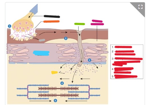
stage 4 of ECC
myosin head binds to actin

stage 5 of ECC
contraction begins
