Anatomy- The Integumentary system
1/81
There's no tags or description
Looks like no tags are added yet.
Name | Mastery | Learn | Test | Matching | Spaced |
|---|
No study sessions yet.
82 Terms
What are the components of the integumentary system?
Skin
Hair
Nails
Glands
(hair, nails, and glands are appendages)
What are the main functions of the integumentary system?
protection
thermoregulation
sensation
metabolism
What are the three layers of the skin?
Epidermis
Dermis
Hypodermis (subcutaneous)
What is the structure of the epidermis?
stratified squamous epithelium
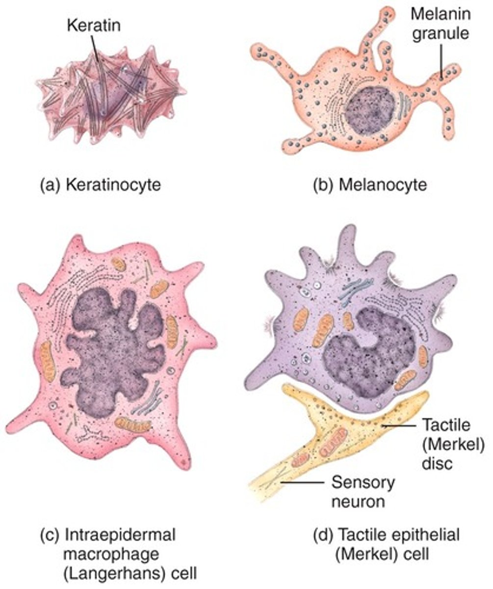
Cells types in the epidermis
keratinocytes, melanocytes, langerhans cells, merkel cells
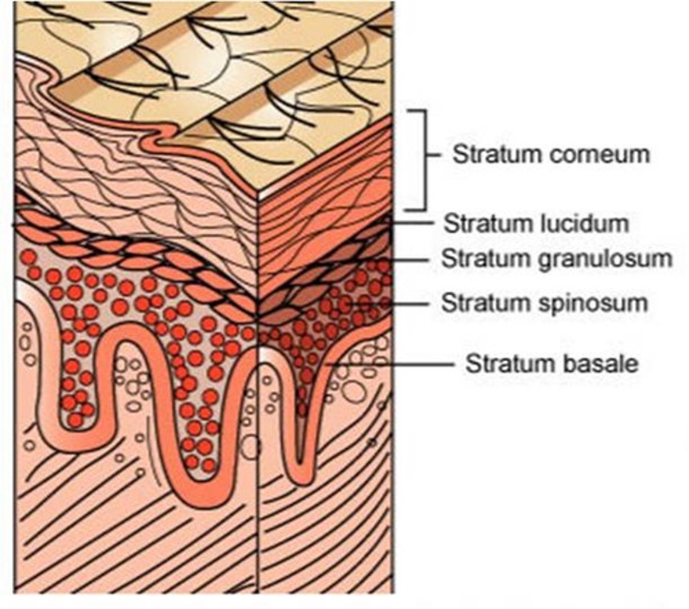
What layers make up the epidermis?
Basale
Spinosum
Granulosum
Lucidum (palms/soles)
Corneum

Is the epidermis or the hypodermis the youngest in terms of cell types
Hypodermis - contains the newest formed cells
What layer of the epidermis is constantly producing epidermal cells (keratinocytes)?
stratum basale
What layer of the epidermis is responsible for protein synthesis?
stratum spinosum
What layer of the epidermis has intercellular spaces that are filled with lipid-rich material ?
Stratum granulosum
What layer of the epidermis provides thickness and strength to withstand friction?
stratum lucidum
What layer of the epidermis is composed of dead keratinized cells that are continuously being shed?
Stratum corneum
What two layers make up the dermis?
reticular layer and papillary layer

Describe the reticular layer of the dermis. The top or bottom? The thickness? What does it have in it? Surrounded by? Protects?
- Deep/bottom layer
- Thick
- Has blood vessels, glands, hair follicles, lymphatics, nerves, fat cells
- Surrounded by a net-like structure of elastin and collagen fibers
- Protects skin structure and lets it move and stretch
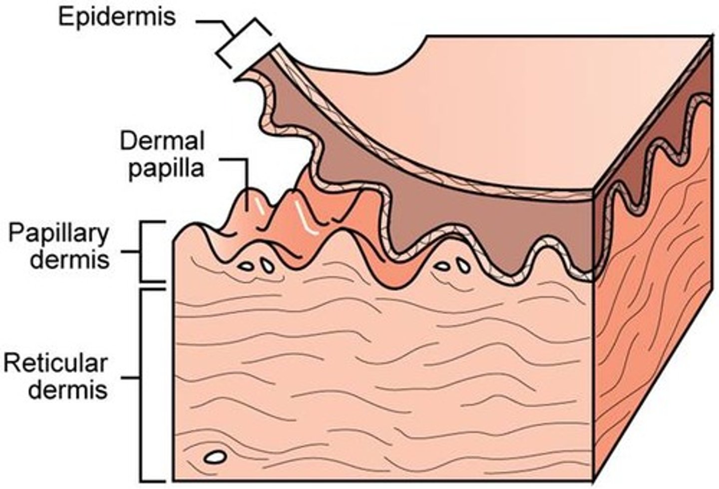
Describe the papillary layer of the dermis. The top or bottom? The thickness? What does it have in it? Surrounded by? Protects?
- Superficial/top layer
- thin
- Contains touch receptors (Meissner Corpuscles), phagocytes, blood vessels (capillary loops)
- Extends to basement layer of epidermis and forms a strong bond with it
What is the function of the hypodermis?
Insulation, energy storage
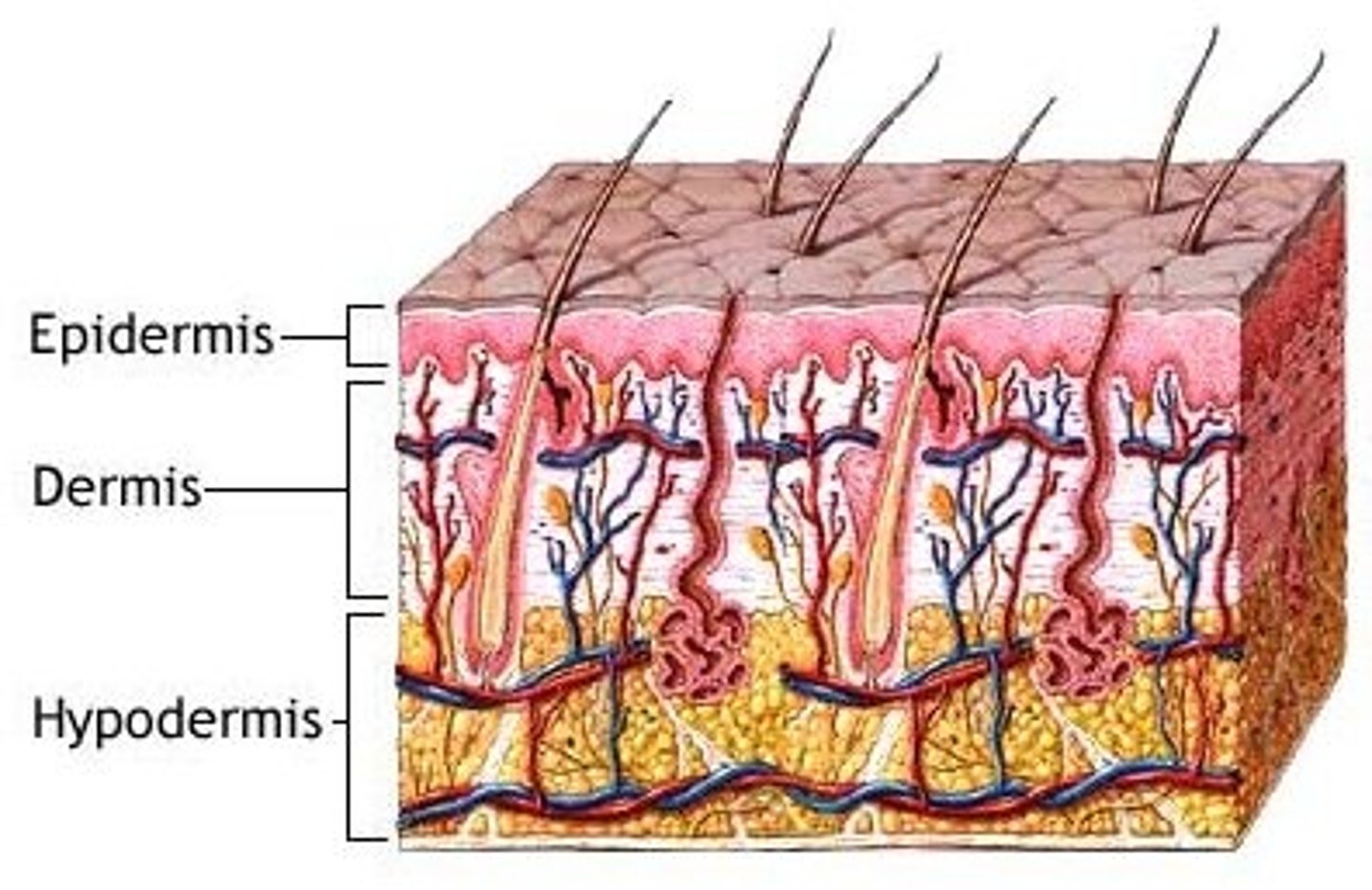
What structures can be found in the hypodermis?
adipose tissue and blood vessels
How does skin function as a barrier?
Shields body from pathogens, UV radiation, physical injury, water loss
How does skin function in thermoregulation?
Sweat glands and blood vessels help maintain stable body temperature
How does the skin function with sensory input?
Nerve endings in the skin allow us to feel touch, temperature, pain, etc.
How does the skin function as an immune defense?
in the stratum corneum, keratinocytes and lipids form a physical barrier against pathogens
Langerhans cells and dendritic cells are crucial for detecting and eliminating pathogens
How does the skin function in vitamin D synthesis?
Exposure to sunlight leads the skin to produce vitamin D which is essential for bone health and immune function
What is the function of hair as a skin appendage?
Provide insulation, sensory input, protection from UV radiation and physical trauma
What do hair follicles contain?
hair shafts, sebaceous glands, and arrector pili muscles
What are the function of nails as skin appendages?
Protects the end of fingers and toes - important for grasping and manipulation
What is the function of sebaceous glands?
Produce sebum which contributes to moisture and acts as a barrier against pathogens. Lubericates the skin and hair
What is the function of eccrine sweat glands?
Secrete sweat, which helps to regulate body temperature and eliminate waste product
What is the function of apocrine sweat glands?
Secrete a thicker, sometimes scented fluid, which can contribute to body odor and has a role in thermoregulation
What are some examples of clinically significant pathologies of hair follicles (what diseases can we see within hair follicles)?
Alopecia -
Folliculitis -
Pediculosis -
Hirsutism -
Hypertrichosis -
alopecia
hair loss
folliculitis
infections of the hair follicles
pediculosis
lice
Hirsutism
excessive hair growth from endocrine glands (PCOS)
Hypertrichosis
excessive growth of hair (wolf man)
What are some examples of clinically significant pathologies of the nails (what diseases can we see within the nails?)
Onychomycosis - fungal infection
Psoriasis - irregular pitting, splitting, or "oil drop"
Lichen planus - longitudinal ridging and splitting of nails
Alopecia areata - geometric pitting of the nails
Melanonychia - pigmentation of nails
onchomycosis
fungal infection of the nail
psoriasis
irregular pitting
lichen planus
longitudinal ridging
alopecia areata
geometric pitting of the nails
melanonychia
pigmentation of the nails
What are some examples of clinically significant pathologies of the sebaceous glands (what diseases can we see within the sebaceous glands?)
Acne
Seborrheic dermatitis
Sebaceous cyst
What are some examples of clinically significant pathologies of the sweat glands (what diseases can we see within the sweat glands?)
Hyperhidrosis - excessive sweat production
Bromhidrosis - excessive body odor
Anhidrosis - absence of sweating
Hidradenitis suppurativa - chronic skin condition causing bumps, abscess, tunneling in intertriginous areas
What skin layer are abrasions often limited to?
epidermis
What skin layers are involved in lacerations?
dermis or deeper
What skin layer is affected in a superficial (1st degree) burn?
Epidermis only
What skin layers are affected in a partial-thickness (2nd degree) burn?
Epidermis and dermis
Blistering due to a burn is indicative of what degree burn?
Second
What skin layers are affected in a full-thickness (3rd degree) burn?
Epidermis, dermis, subcutaneous tissue
What skin layers are involved in a 4th degree burn?
Epidermis, dermis, hypodermis (subcutaneous) and muscle or bone
Loss of sensation (nerve damage) due to a sunburn is indicative that the sunburn has damaged what layer of skin?
deeper dermis into hypodermis (full thickness burn)
Blistering due to a sunburn is caused by the separation of what two skin layers?
Epidermis and dermis
What is an eschar formation?
full-thickness burn with dead tissue
How are pressure ulcers staged?
Based on how deeply the wound penetrates the anatomical layers
What stage of a pressure ulcer is characterized by having intact skin with non-blanchable erythema?
Stage 1
What stage of a pressure ulcer is characterized by having a partial-thickness loss of the dermis, which may appear as a blister or shallow ulcer
Stage 2
What stage of a pressure ulcer is characterized by a full-thickness loss extending into subcutaneous fat?
Stage 3
What stage of a pressure ulcer is characterized by having a full thickness with exposed bone, tendon, or muscle?
Stage 4
Why would a pressure wound be unstageable?
They are obscured by slough or eschar
What is the role of the epidermis in wounds/burns?
It is the first line of defense
Regenerates in partial-thickness injuries
What is the role of the dermis in wounds/burns?
Determines burn severity and healing potential
Contains vasculature and nerves
What is the role of the hypodermis in wounds/burns?
Involvement signals more severe injuries, poor healing potential
What is the role of hair follicles/sweat glands in wounds/burns?
Serves as sources of epithelial cells in wound healing
What is the role of nerve endings in wounds/burns?
Helps to localize and quantify pain - damage alters sensation
What are examples different infectious dermatological conditions? Bacterial, viral, and fungal.
Bacterial - impetigo, cellulitis
Viral - HSV, HPV
Fungal - Tinea
Ulcerated nodules, common with basal cell carcinoma (BCC) often occurs on sun-exposed areas. Why?
This is the area where basal cells are more active.
Scaly, red plaques or nodules, indicative to what, relate to disrupted function of what type of cells?
Keratinocytes, SCC
What does the ABCDE rule for melanoma correspond to?
Disordered growth of melanocytes in the epidermis and beyond
- asymmetry, boarder, color, diameter, and evolution
Name the structure and the associated cancer that can be found in the epidermis.
Keratinocytes
Basal cell carcinoma
Squamous cell carcinoma
Name the structure and the associated cancer that can be found in the basal layer.
Stem cells
Basal cell carcinoma
Name the structure and the associated cancer that can be found in the spinous layer .
- Differentiating keratinocytes
-
Squamous cell carcinoma
Name the structure and the associated cancer that can be found in the stratum basale (epidermis)
melanocytes
melanoma
Name the structure and the associated cancer that can be found in the Dermis
- blood vessels, nerves
BCC, SCC, and melanoma (melanoma is most common)
Name the structure and the associated cancer that can be found in the appendages.
- hair follicles, sebaceous glands
rare skin cancers called
- adnexal tumors
Describe the predicted behavior and spread of basal cell carcinoma
Usually slow growing
Rarely metastasizes - stays within the epidermis or superficial dermis
Describe the predicted spread of squamous cell carcinoma
Has a greater risk of metastasis, especially if it invades dermis or deeper
Describe the spread of melanoma
Spreads both lymphatically and hematogenously
Breslow depth is critical for prognosis as it is a directly anatomical measurement
What is the Breslow depth?
how deep the melanoma invades into the dermis/subcutaneous layer
What would you expect to see from a biopsy of a basal cell carcinoma
basaloid cells from basal layer
What would you expect to see from a biopsy of squamous cell carcinoma
keratin pearls and intercellular bridges, indicating keratinocyte origin
What would you expect to see from a biopsy of melanoma
atypical melanocytes in the basal layer or invading the dermis
How are surgical margins determined in a skin cancer biopsy?
by understanding how far cancer might extend anatomically
What do treatments of skin cancer aim to do?
Clear the cancer while preserving key anatomical structures (particularly on the face and hands)