5) Anomalies Color vision
 Call Kai
 Learn
Learn Practice Test
 Spaced Repetition
 Match
Match Flashcards
 Knowt Play
1/97

There's no tags or description
Looks like no tags are added yet.
Name | Mastery | Learn | Test | Matching | Spaced | Call with Kai | Chat |
|---|
No analytics yet
Send a link to your students to track their progress
98 Terms
Provide examples of two inherited colour defect.
Red-green anomalies (X-linked recessive)
Men 0.8%; woman 0.4%
Tritan (autosmal dominant)
What are the prevalence of the different color vision defects?
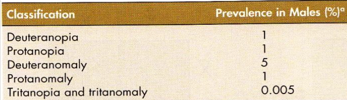
What are the Societal Implications of Anomalous Colour vision?
not physically debilitating
some may not be aware
learn they have it after FAILING a color vision test
List occupations related to public safety that have color vision standards.
Airline Pilot, Firefighter
US custom & border protection
USDA meat inspector (Ishihara)
List occupations where normal colour discriminations can be an advantage.
Chemical or electrical engineering
Pharmacy
Optometry
Ophthalmology
Dentistry
Surgery
Videographer
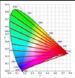
How do red–green color anomalies affect color perception?
→ Difficulty distinguishing dark or desaturated (pastel) colors that lie along red–green confusion lines
💡 Brighter light improves color discrimination and color naming
Do colored SUNGLASSES interfere with the ability of patients with inherited anomalies to quickly and correctly identify colored traffic signals?
Yes
certain non-neurtal tins (except Gray) make it harder to see traffic light
ppl with anomalous defects (eso protons seeing red) shouldn’t wear it
What does Photopic luminosity curve receive it’s input from?
2-3 LWS : 1 MWS (no input from SWS cones)
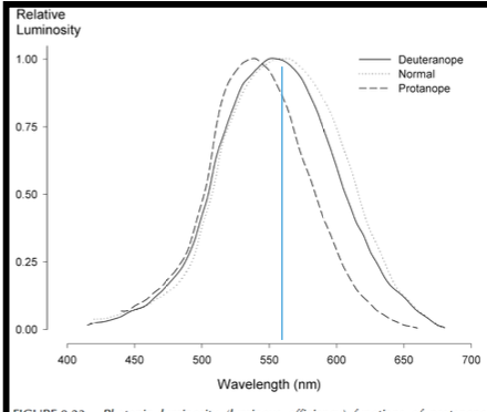
How do protanopes and protanomalous trichromats perceive red lights?
→ severe loss of luminosity to RED LIGHTS sensitivity, perceiving red as dim or dark
.
Such as:
brake lights → delayed rxn times
red traffic lights
red surface → looks black
emitting Diode
laser pointers
How do deuteranopes & deuteranomalous trichromats perceive red lights?
No loss of luminosity
Why should color vision testing be done at a young age?
→ Provides baseline color vision data early in life (helpful for careers)
parents and teachers should be informed about child’s color vision status
Parents counselled about career choices
Write the Male and Female phenotypes.
XY - Male
XY -Anomalous Male
XX - Female
XX - Carrier Female
XX - Anomalous Female
What genes does the X-chromosome have?
1 L-cone opsin gene
1+ M-cone opsin genes
What are the genetic causes of color vision deficiencies?
Intergenic (between genes):
Normal color vision
Dichromacy
.
Intragenic (within genes):
Normal color vision
Dichromacy
Anomalous trichromacy → caused by a hybrid gene (basis of abnormal cone photopigment)
What are Acquired color vision anomalies?
→ 2er to disease or toxicity
either Red–Green or Blue–Yellow
unilateral or asymmetric
Must be assumed that any in the color vision of the two eyes as demonstrated on a color vision test…
due to an Acquired Anomaly
What is Achromatopsia?
→ where have manifests monochromatic vision
fully expressed at birth
What is Autosomal Recessive (AR) Achromatopsias? Mention the 2 types, signs + sx, and treatment.
Complete (AR) Achromatopsia → only Rods are present
Incomplete (AR) Achromatopsia → residual L/M cone function
common
.
Signs + sx
No or very poor Color discrimination
Nystagmus
Photophobia
VA~20/200
.
Treatment
Dark red lenses (minimizes rhodopsin bleaching) for bright light conditions
What is X-linked (XL) Achromatopsias? Mention the 2 types, signs + sx, and treatment.
→ aka Blue or S cone monochromacy (have only Rods & S cones)
.
Signs + sx
similar to AR Achromatopsia
.
Treatment
Magenta colored lenses
What is Cone Monochromacy?
monochromatic color matching
VA = normal
defect in postreceptoral processing of color info
Where does retinal achromatopsia occur?
Retina
Where does cerebral achromatopsia occur?
Extrastriate Cortex
What is Chromatopsia?
→ NOT TRUE color vision anomalies b/c they don’t cause a ↓ ability to discriminate colors
represent “distortion” of color vision
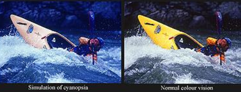
List 2 common causes of Chromatopsia.
1) Cataract extraction → Blueness perception (Cyanopsia)
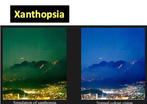
2) 2er to drugs/medications → Yellowness perception (Xanthopsia)
Digitalis (yellow vision)
Fluorescein in Fluorescein Angiography
What is Synesthesia?
→ neurological phenomenon where stimulating ONE sensory/cognitive pathway → automatic, involuntary experiences in a 2nd sensory/cognitive pathway
Cause: abnormal link between diff cortical modules
e.g., GREEN whenever someone’s presented with the number “5,”
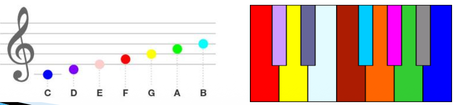
What is Chromesthesia?
→ associating SOUNDS with colors
common form of synesthesia
ppl tend to have perfect pitch
What is Köllner’s rule?
Outer retinal disease, Media changes → Blue–Yellow anomalies
Inner retina, Optic nerve, Visual pathways, Visual cortex → Red–Green anomalies
Post receptoral lesions → Blue–Yellow & Red–Green anomalies
How can acquired color defects change as eye disease progresses?
Early disease may cause a blue–yellow defect → progress to red–green defect
Some diseases can show the reverse pattern
Severe disease may cause nonselective color loss (loss of multiple color pathways)
What is color vision testing is based on?
knowledge of confusion lines
List the Goals of CV Testing.
Screening – i.e. Congenital versus Acquired
Diagnosis – i.e. type and severity
Vocation or Occupational testing
Who will have greater occupational consequences? Protans, Deutan or Tritans?
Protan
What do red–green vs blue–yellow acquired color defects indicate?
Red–green defects = cone and optic nerve disease
Blue–yellow defects = retinal and choroidal disease
Give examples of Pseudoisochromatic (PIC) plate tests.
Ishihara
HRR
CVTME
Give examples of Arrangement tests.
→ color cap tests
Farnsworth-Munsell 100
Panel D-15
Lanthony Desaturated D15
Give examples of Matching tests.
Anomaloscopes
C100
CUT
Give examples of Naming tests.
Vocation/occupations (Lantern tests)
Give examples of other occupational Color tests.
Rabin Cone Contrast Test
What are PIC plates?
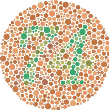
→ Patterns of objects, letters or numbers placed on isoluminant bg
Figures usually fall along the dichromatic confusion line
Normals = identify chromatic differences
CVD = will have difficulties
DON’T distinguish between Dichromats & anomalous trichromats
CAN distinguish between Protan and Deutan
List the 7 different PIC plate types.
Demonstration
Disappearing (vanishing)
Diagnostic
Ambiguous or alteration (transformation)
Combination
Quantitative
Hidden-digit
What is Demonstration plate?
→ plate luminous reflectance cues and/or color differences that don’t lie on confusion line
you don’t need color vision for a correct response
helps demonstrate how the test symbols look
helps pick up malingerers (pretends or exaggerates incapacity)
e.g., Ishihara & HRR Demo Plates
What are Disappearing plates?
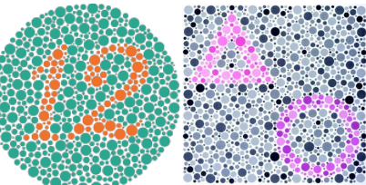
→ target/stimulus is defined by a color difference from the bg that straddle confusion lines
Object is invisible to those with certain color deficiencies
helps detect color deficiencies but not distinguish them
(-) Slower to administer for CVD
(-) Frustrating for color defectives to repeatedly see ‘blank’ plate
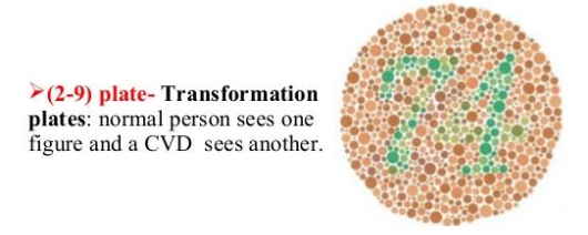
What are Ambiguous or Alteration (Transformation) plates?
→ helps provide an alternate response for the color vision defectives
has colours that can be confused with the bg and others that are not
Normal color vision: see one thing
Color deficiency: see another thing that’s just slightly altered
helps confirms that the test is understood
(+) prevents the frustration found with blank plates
What are Combination plates?
→ incorporates disappearing and demonstration figures
Color normal = might see 2 figures
CVD = sees 1 figure
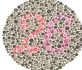
What are Diagnostic plates?
→ Multiple disappearing plates for different CVD
have 2 figures
Normals: should see 2 & 6
Deutan = see 2
Protan = see 6
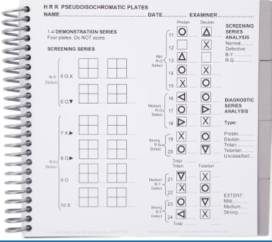
What are Quantitative plates?
→ set of plates with increasing color difference
Confusion with small color difference = Mild
Confusion with large color difference = Moderate/Severe
▪ e.g., Hardy-Rand-Rittler (HRR)
What are Hidden-digit plates?
→ Opposite of a disappearance test
Visible only to those with CVD, normals should see nothing
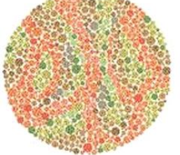
d see 5
What defects can Ishihara vs HRR test detect?
Ishihara (Sensitive) = Protan & Deutan defects
HRR (Sensitive) = Protan, Deutan, Tritan & severity
What is the Ishihara PIC test? Mention the plates used.
→ figures on each plate have a random pattern of variably-sized dots → figures & bg lie close to confusion lines
DOESN’T distinguish between Dichromats and Anomalous trichromats, it only differentiates b/w protan vs deutan
(+) ↑ sensitivity & specificity (close to 1.0)
(+) useful in children >5 years
(+) can perform under diff illumination
Plates = demonstration, disappearing, diagnostic plates, alteration & hidden
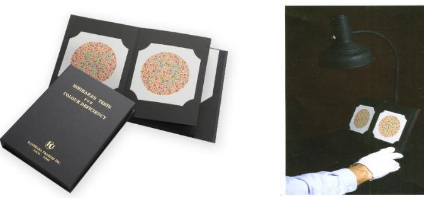
Why is an Ishihara “disappearing plate” not seen by both protans and deutans?
figure and bg colors are chosen to fall along red–green confusion lines
little color difference b/w protans and deutans in this region of color space
protans and deutans confuse the figure with the background, causing the figure to “disappear”
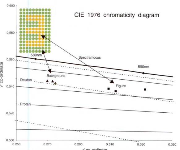
What is the “cut-off” for normal vs deficient colour vision in Ishihara PIC test
13+ = Normal
<9 = deficient
Although the Ishihara test as a whole performs "very close to 1" in both Sensitivity and Specificity, what does it have the tendency to do?
PASS: Very Mild Deutans
FAIL: Some Normals
List the 6 shortcomings of Ishihara (compared to HRR).
No TRITAN plates
Doesn’t provide a severity diagnosis
Relatively mildly affected CVDs (Protanamalous, deuteranomalous) may make as many errors like dichromats (Protanopes, deuteranope)
Can be memorized
Hard to administer in children younger than 5 years.
Doesn’t distinguish b/w Dichromats and Anomalous trichromats (i.e., protanope from protanamalous, deutaranope from deuteranomalous)
List the Common Mistakes in the Interpretation of the Ishihara.
Using the wrong failure criterion → misdiagnosis
Assuming that patients who make many errors have a “severe” CVD
# of errors on the Ishihara test is NOT a measure of severity, except that a very small number of errors may indicate a mild CVD
Diagnostic plates to differentiate Protan and Deutan CVD, don’t ALWAYS yield a diagnosis.
30 to 40 % of cases diagnosis is wrong
How many Hardy-Rand Rittler (HRR) Color test plates are there?
Plates = Demonstration, Diagnostic & Screening plates
Demonstration plates = 4 plates
Diagnostic Plates = 14 plates (10 = Red-Green, 4= Blue-Yellow)
Screening Plates = 6 plates (2=Blue-Yellow, 4= Red-Green CVD)
List the advantages of HRR test.
Has Tritan screening & diagnostic plates
have target that lie on the “Tetartanopia” confusion locus but this doesn’t exist
Doesn’t discriminate Dichromacy from Anomalous Trichromacy
Classifies Protan, Deutan, Tritan & severity
Used on very young childre b/c it uses symbols (circle, triangle, cross)
How do you diagnose normal vs defective colour vision using the HRR color test?
Normal
All 6 screening plates
1+ errors in the screening plates but 0 in the subsequent diagnostic plates
.
Red-Green Deficiency
Protan = Protan column > Deutan column
Deutan = Deutan column > Protan column
Unclassified = Protan column = Deutan column OR errors in screening plates
.
Blue-Yellow Deficiency
Tritan = Tritan column > Tetartan column
Tetartan = Tetartan column > Tritan column
Unclassified = Tritan column = Tetartan column OR errors in screening plates
What does extensive scattered errors throughout the various groups of plates on the HRR test may indicate?
Any of the following:
Malingering
Monochromatism (total color blindness)
Low color discrimination approaching monochromatism
How woud you classify someone as being Red-Green Deficiency on the HRR color test?
Last error in plates 7-10 or 11-15 + no errors in 16-20 = MILD
Last error in plates 16-18 + no errors in plates 19-20 = MEDIUM
Last errors in plates 19-20 = STRONG
How woud you classify someone as being Blue-yellow Deficiency on the HRR color test?
Last error in 5-6 + no errors in 21-24 = MILD
Last error in 21-22 + no errors in 23-24 = MEDIUM
Error in 23-24 = STRONG
For those who were classified as MILD on the HRR test, what was their Farnsworth D15 test vs Anomaloscope scores?
Farnsworth D15 test = PASS
Anomaloscope = <30
True or False - Not all Dichromats (Protanope/Deuteranope) were correctly classified as 'STRONG'.
True
What is the Holmgren Wool Test?
→ earliest arrangement tests where subjects are asked to sort colours into sequences/groups
Colors have the
✅ Same Munsell values and Chroma
❌ Different Munsell Hue (R, Y, G, B, P)
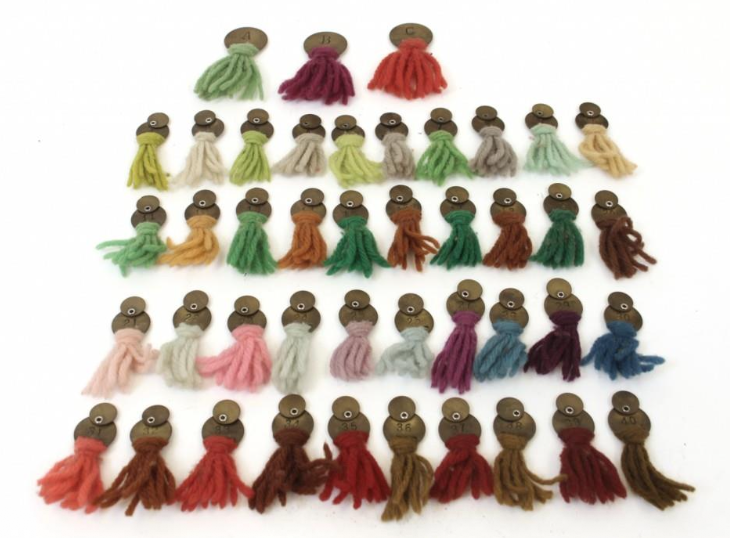
What is the Farnsworth Munsell 100 Hue?
100 caps with colored papers (15 removed to create JND)
Total Error Score (TES) plotted in score sheet
Doesn’t distinguish dichromacy from anomalous trichromacy (i.e Protanope vs Protanomalous)
Time consuming (rarely used)
What does the Error Score (TES) indicate for FM 100 test?
severity of defect
What does the midpoint indicate for FM 100 test?
type of color vision defect
What midpoint values would indicate the different color defects?
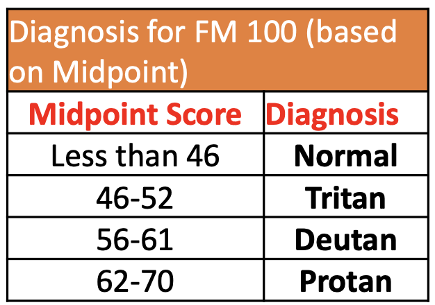
What are the typical axis of confusions in FM 100 Hue test?
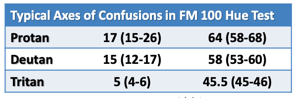
What type of color defect will someone who has diabetic retinopathy have?
Tritan
What type of color defect will someone with Retrobulbar Neuritis associated with MS have?
Recovery from Red-Green defect
What is the function of Farnsworth D-15/Dichotomous D-15/Panel D-15/Regular D-15?
→ distinguish the functionally color blind from the Moderately color defective and the normal” (hence called dichotomous)
used to evaluate Acquired color vision defect
discriminates Protan, Deutan & Tritan defects, but NOT discriminate between Dichromacy and Anomalous trichromacy
has low sensitivity - some subjects with Mild defects (anomalous trichromacy) may pass the test
What is a pass vs fail in Farnsworth D-15?
PASS = ‘mild’ CVD (normals + mildly CVD can complete this test)
FAIL = ‘moderate-to-severe’ CVD (2+ crossings)
True or false - Those who pass the Farnsworth D15 test have a mild deficiency that is unlikely to cause significant handicap in everyday color tasks.
False - they might still have trouble with everyday color tasks
What does the spacing of colors alongs confusion lines allow for?
Makes it easy to:
Pass normal trichromats and many anomalous trichromats
Fail all dichromats
A Young male Fails the Ishihara Color Plate Test but PASSES the Farnsworth D-15 arrangement test. What does this indicate?
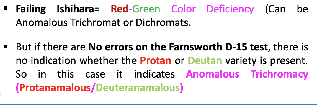
Who typically fails both the Ishihara and Farnsworth D-15 tests?
Protanopes
Deuteranopes
Severe (extreme) anomalous trichromats (nearly dichromatic)
What is the Lanthony Desaturated D15 test?
→ identical to the Farnsworth D-15 test, except the color caps are much less saturated than D-15
detects Anomalous trichromacy (Sensitive test), mild Acquired CVD (e.g.,Cataract or Glaucoma)
needs lots of light (600-800 lux)
What is the Farnsworth Lantern (FaLant)?
2 light lantern for US Navy, with Red, Green and White
No longer commercially available
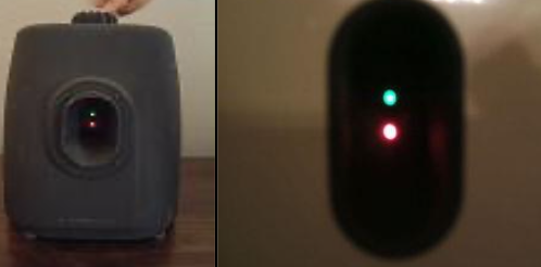
What is the Stereo Optical OPTEC 900?
No longer commercially available
Used in US Military, FAA
What is the Gold Standard for color vision testing for the US Air Force?
Rabin Cone Contrast Test (CCT)
What is the Rabin Cone Contrast Test (CCT)?
1) Detects Cone deficiency severity + Congenital CVD
measures the severity of cone function loss
tracks cone function over time
helps in disease detection + monitor progression
monitor treatment efficacy
2) Detect and monitor Acquired CVD
AMD
TBI
Parkinson’s
GLC
Diabetic retinopathy
MS
Retinal Toxicity due to High Risks meds such as Plaquenil
What is the Nagel Anomaloscope?
→ Only Clinical Instrument that can provide a complete diagnosis of congenital CVD
Anomalous Trichromats from Normal Trichromats
Dichromats from Anomalous trichromat
⭐ standard
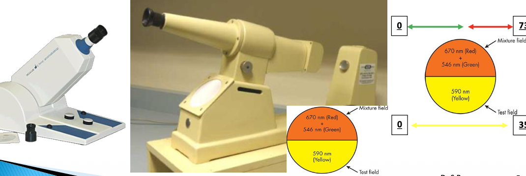
What is the mixture field and test field?
→ observer is asked to match the mixture filed to the test field
.
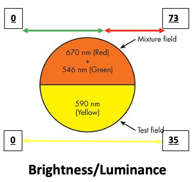
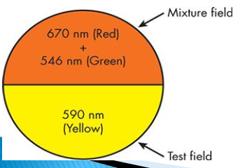
MIXTURE FIELD (upper-half)
RED and GREEN wavelengths
mixture scale setting 0-73 represent various combinations of 546 (Green) to 670 nm (Red)
.
TEST FIELD (lower half):
fixed YELLOW test field (590 nm)
test field knob setting 0-35 varies test field YELLOW Brightness/Luminance
True or False - A person with normal trichromacy can match the test field to the appropriate mixture field?
True - b/c they’re dichromatic
True or False - A person with dichromacy (protanope/deuteranope) can match the test field to the appropriate mixture field?
Yes, no matter what combo of 546 and 670 nm is present in the MIXTURE FIELD
Reason: b/c from approximately 545 to 700 nm, Red–Green Dichromacy pt have only ONE photopigment and manifest MONOCHROMATIC VISION
If the TOP MIXTURE FIELD is set to pure 546 nm, can a patient with Red–Green Dichromacy [Protanope/Deuteranope] adjust the mixture field?

If the TOP MIXTURE FIELD is set to pure 670 nm, can a patient with Red–Green Dichromacy [Protanope/Deuteranope] now adjust the wavelength of the test field so that the two fields appear identical?

How do protanopes and deuteranopes differ on the Nagel anomaloscope?
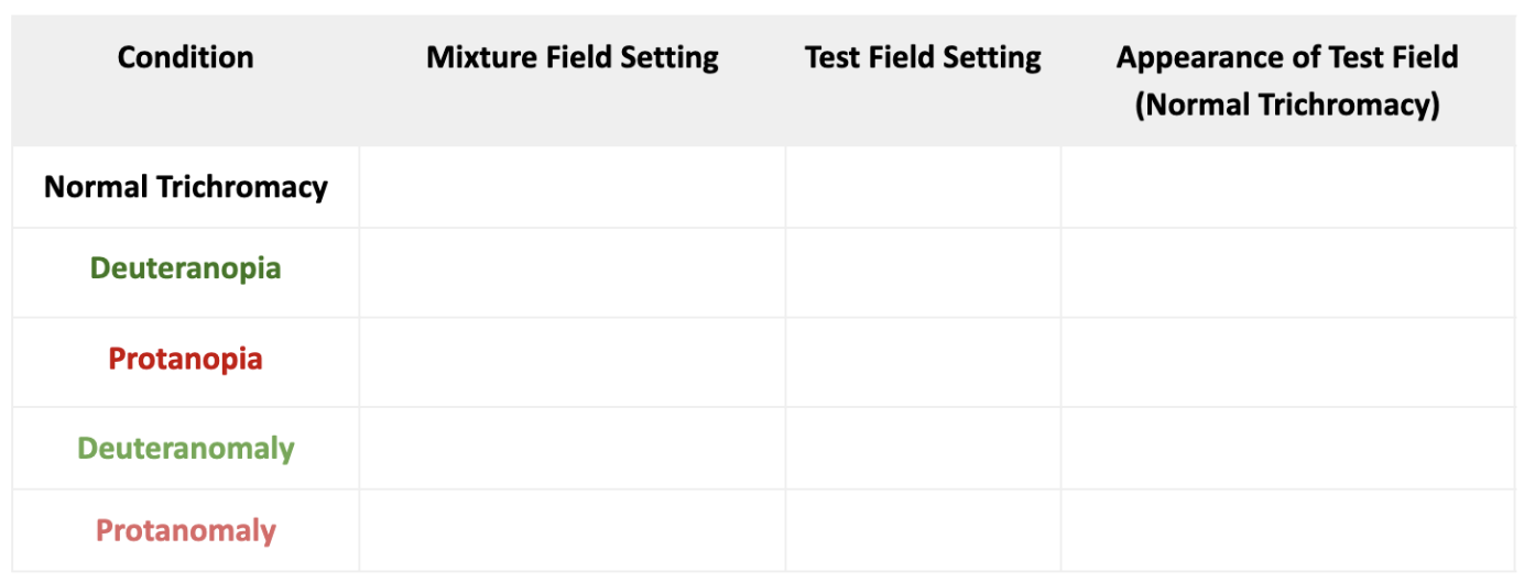
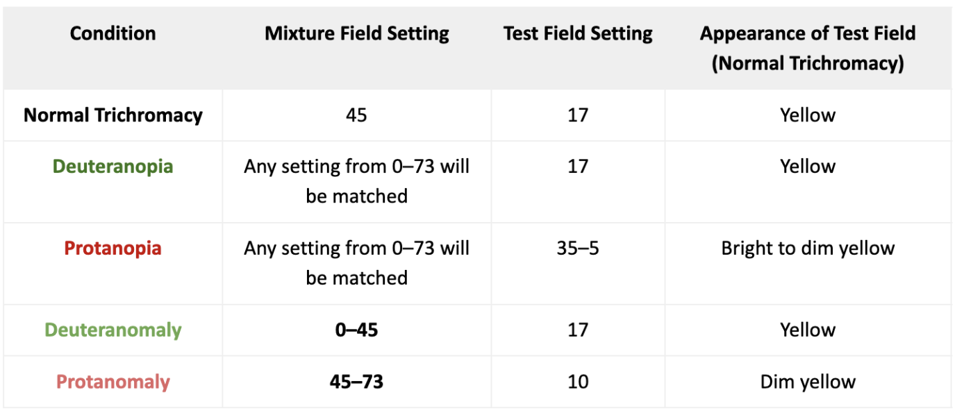
→ Both protanopes and deuteranopes can match any red–green mixture (0–73) to the test field
Deuteranopes: Use the same test field setting because brightness remains constant
Protanopes: Adjust the test field brightness (dimmer for red, brighter for green) because red appears dimmer due to ↓ long-wavelength sensitivity
their luminance function is displaced towards shorter wavelengths
How can the MIXTURE SCALE SETTING distinguish protanomalous from deuteranomalous?
Deuteranomaly - may require more 546 nm (Green) in the MIXTURE FIELD due to displaced M-cone (the mixture looks more red to them) → adjust MIXTURE scale between 0-40
Protanomaly - may require more 670-nm (red) in the MIXTURE FIELD due to displaced L-cone (the mixture looks more green to them) → adjust MIXTURE scale between 50-73
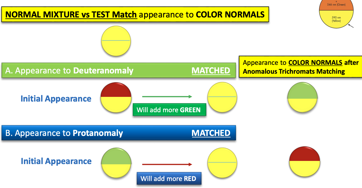
How can the TEST FIELD (BRIGHTNESS) SETTING distinguish protanomalous from deuteranomalous?
Deuteranomaly - has the same luminosity function as normal CVD → adjust TEST FIELD scale to 17
Protanomaly - has the luminosity function similar to protanope → adjust TEST FIELD scale to less than 17 (b/c mixture field already appears dim to them?
How do anomalous trichromats (deutranomolous, protanomalous) perform on color matching tests compared to normal trichromats and dichromats?
Less specific (wider range of matches) than normal trichromats
More specific (narrower range of matches) than dichromats (protanope, deutranope)
What equation is the gold standard ⭐ for diagnosing the FOUR major X-linked color defects?
Rayleigh equation
What is the Rayleigh equation used for?
→ protan/deutan
Red + Green ≡ Yellow
What is the Moreland equation used for?
→ tritan
Blue + Green ≡ Blue-Green
What does trouble with BY defect include?
Macular pigment
Crystalline lens variability
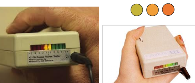
What is the Medmont C-100?
→ Flicker matching (rapid alternation) of Red (650) and Green (560) using dual light emitting diode
Subject controls luminance ratio of Red and Green
Reliably distinguishes
✅ Protans from normal
❌ Deutans from normal
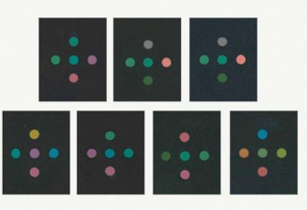
What is the City University Test (CUT)?
→ MATCHTING TEST that invovles matching the Central Hue with one of the Peripheral Hue (which appears similar)
forced choice procedure test
derived from the colors of the D-15
5 hues
1 test hue
1 hue = adjacent D-15 hue
3 hues = Protan, Deutan & Tritan confusion loci
What is the SWAP [Short Wavelength Automated Perimetry]?
→ detects early GLC by isolating S-system function
S cone (blue-yellow pathway) is vulnerable to GLC
Stimulus = Short wavelength (to maximize S cone sensitivity)
Background = yellow (to SUPPRESS (adapting) L & M cones)