Looks like no one added any tags here yet for you.
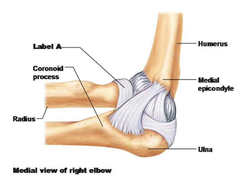
Label A is pointing to the ____.
A. Radial collateral ligament
B. Articular capsule
C. Annular ligament
D. Ulna collateral ligament
C. Annular ligament
What type of synovial joint (based on shape) is the elbow joint?
A. pivot
B. ball and socket
C. plane
D. hinge
D. hinge
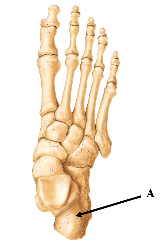
Arrow A is pointing to ____.
A. metacarpal
B. metatarsal
C. talus
D. calcaneus
D. calcaneus
The triceps surae inserts into arrow A via this tendon.
A. central tendon
B. linea alba
C. achilles tendon
D. iliopsoas tendon
C. achilles tendon
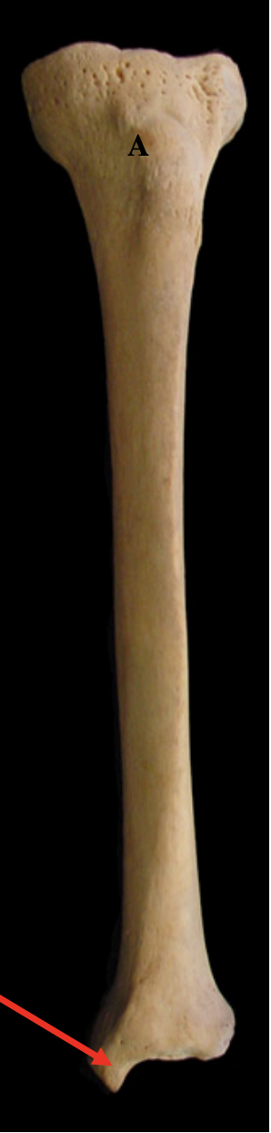
This picture shows the tibia and label A is tibial tuberosity. What structure is the arrow pointing to?
A. lateral condyle
B. lateral malleolus
C. medial condyle
D. medial malleolus
D. medial malleolus
Indicate whether this femur is a left or right bone
A. Left
B. Right
B. Right
The femur will fit into the ____ of the pelvic girdle.
A. Ischial tuberosity
B. Pubic symphysis
C. Auricular surface
D. Acetabulum
D. Acetabulum
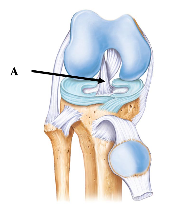
Arrow A is pointing to the ____ on the knee joint (right knee)
A. Fibular collateral ligament
B. Anterior cruciate ligament
C. Posterior cruciate ligament
D. Tibial collateral ligament
B. Anterior cruciate ligament
The function of the ligament in this photo is to
A. Prevent anterior sliding of the tibia
B. Prevent forward sliding of the femur
C. Distribute the compressive load evenly
D. Prevent the leg from moving side to side at the knee
A. Prevent anterior sliding of the tibia
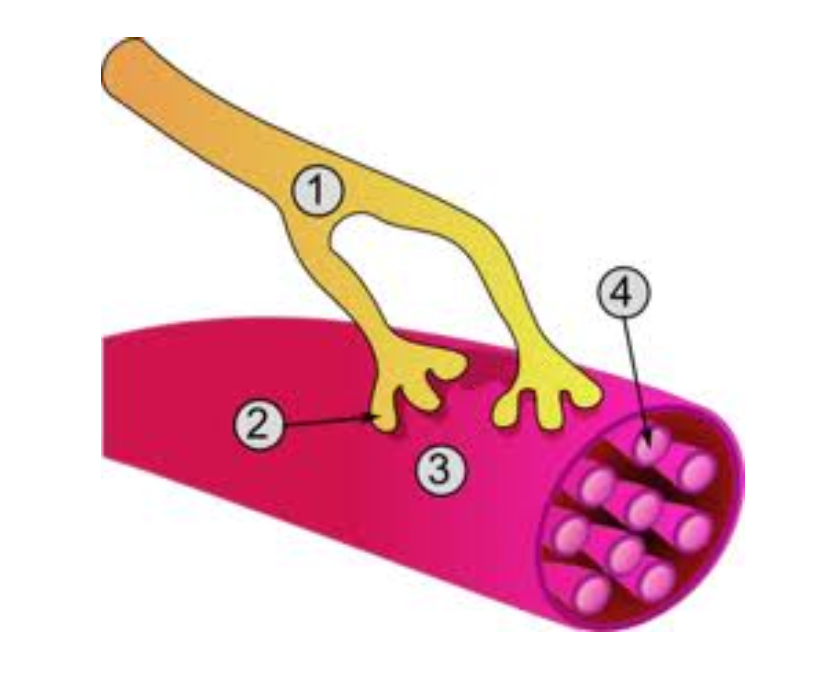
This is a picture of an innervated muscle fiber. Label #2 is pointing to ____ and the point where the neuron meets the muscle fiber is known as the ____.
A. Dendrite, triad
B. Myofibril, synaptic cleft
C. Cell body, sarcolemma
D. Axon terminal, neuromuscular junction
D. Axon terminal, neuromuscular junction
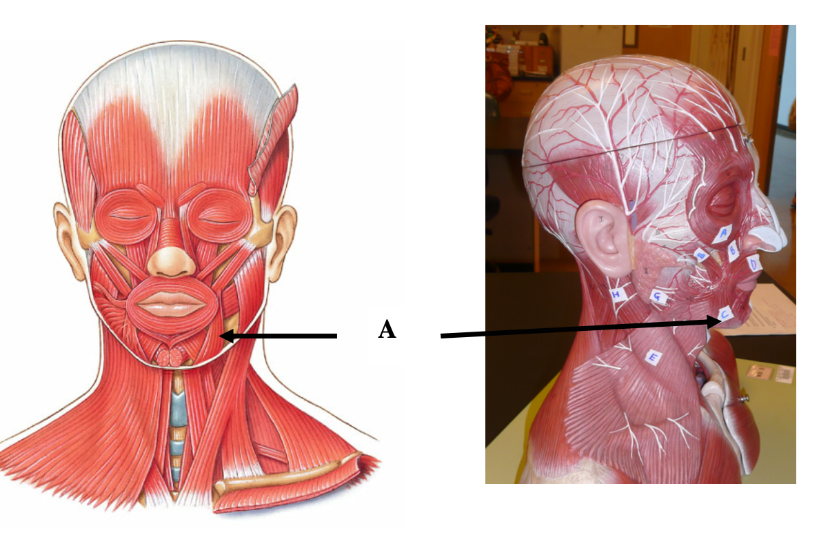
What muscle is arrow A pointing to?
A. risorius
B. orbicularis oculi
C. zygomaticus
D. depressor anguli oris
D. depressor anguli oris
The function of this muscle is to
A. compress the cheek
B. draw corner of the lip laterally
C. draw corners of the mouth downward and laterally
D. raise the lateral corners of the mouth upwards
C. draw corners of the mouth downward and laterally
Identify the muscle labeled E on the model
A. zygomaticus
B. buccinator
C. masseter
D. temporalis
C. masseter
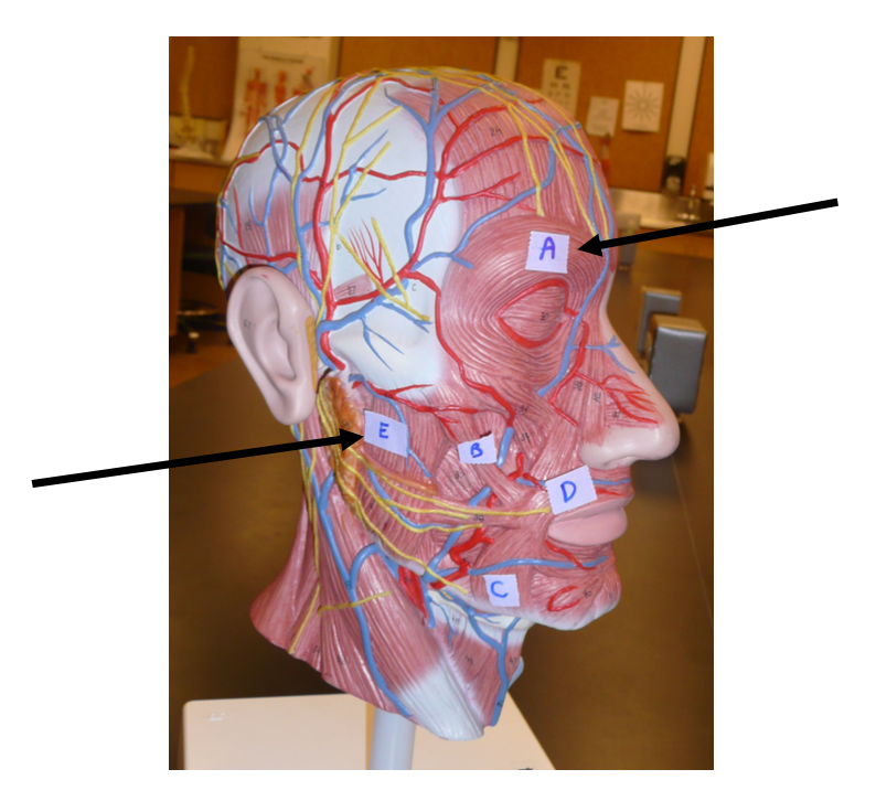
The function of muscle labeled A on the model is to
A. raise eyebrows
B. draw eyebrows together
C. close eye
D. pull scalp posteriorly
C. close eye
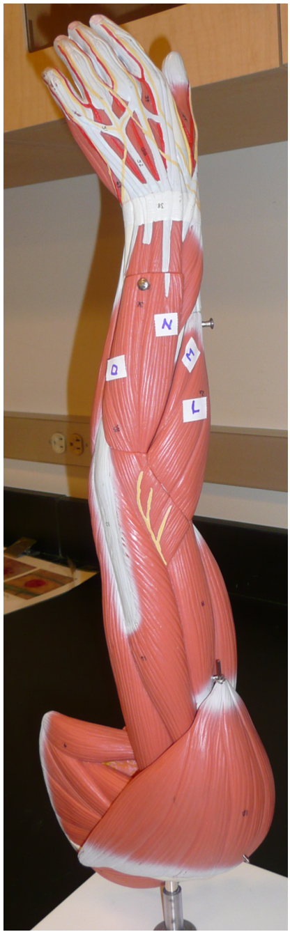
What muscle is label N on the model?
A. brachioradialis
B. flexor carpi ulnaris
C. extensor carpi ulnaris
D. extensor digitorum
D. extensor digitorum
The function of the muscle on label N is to
A. abduct the wrist
B. extend the fingers
C. flex the wrist
D. extend the wrist
B. extend the fingers
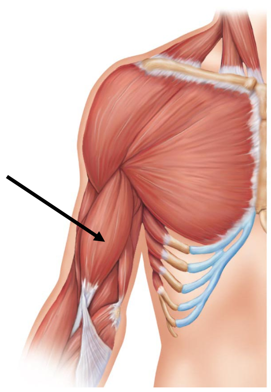
What muscle on the arm is the arrow pointing to?
A. Triceps brachii
B. Biceps brachii
C. Pectoralis major
D. Brachialis
B. Biceps brachii
The insertion of the muscle with the arrow is the
A. ulna
B. radius
C. humerus
D. clavicle
B. radius
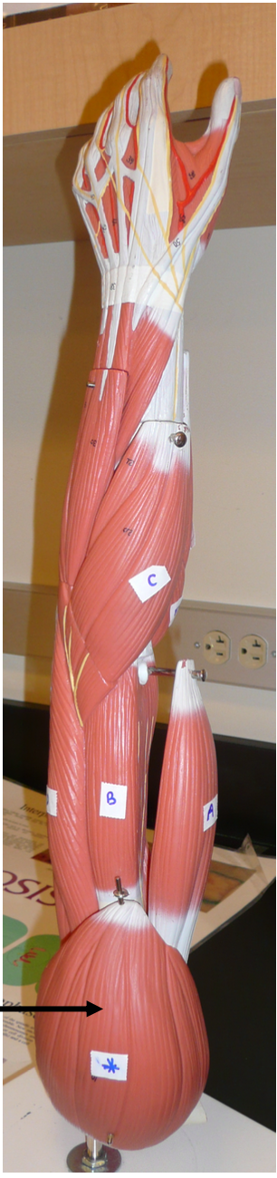
The arrow on the model is pointing to
A. trapezius
B. deltoid
C. levator scapulae
D. brachialis
B. deltoid
The muscle on the arrow is the prime mover of
A. arm flexion
B. arm extension
C. arm adduction
D. arm abduction
D. arm abduction
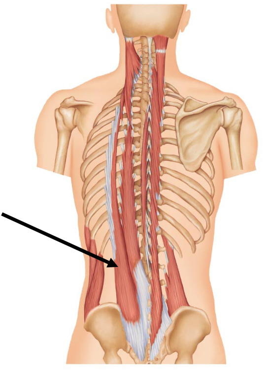
The arrow is pointing to
A. iliocostalis
B. Spinalis
C. Semispinalis
D. Longissimus
A. Iliocostalis
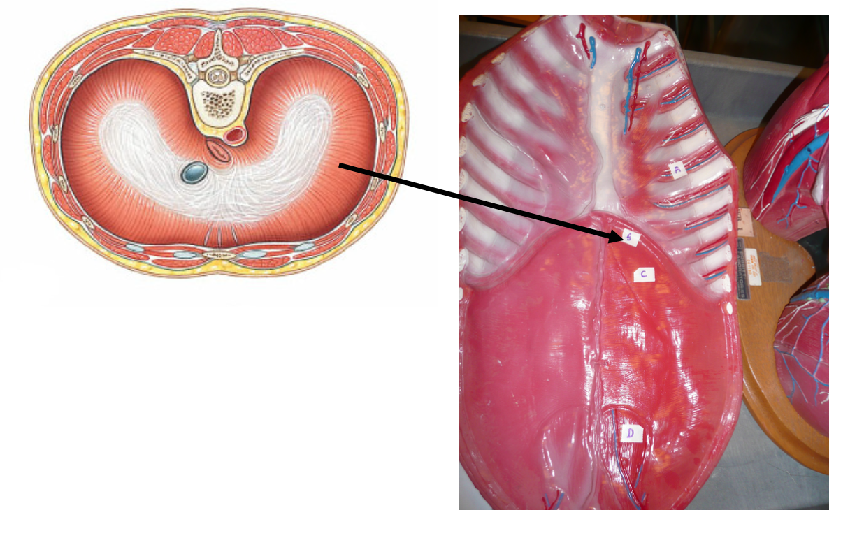
The picture on the left is the muscle labeled B on the torso. What is the muscle?
A. External intercostals
B. Serratus anterior
C. Internal intercostals
D. Diaphragm
D. Diaphragm
The muscle labeled B separates the ____ cavity from the ____ cavity.
A. Abdominal, pelvic
B. Dorsal, ventral
C. Thoracic, abdominopelvic
D. Pleural, pericardial
C. Thoracic, abdominopelvic
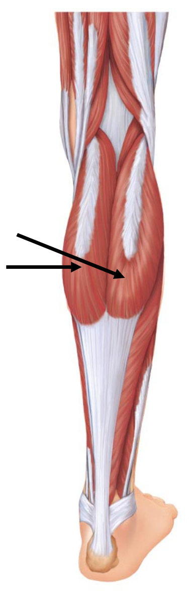
The muscle indicated by the arrows is
A. soleus
B. gastrocnemius
C. popliteus
D. triceps surae
D. triceps surae
The insertion for the muscle indicated by the arrows is the
A. talus
B. metatarsal
C. calcaneus
D. fibula
C. calcaneus
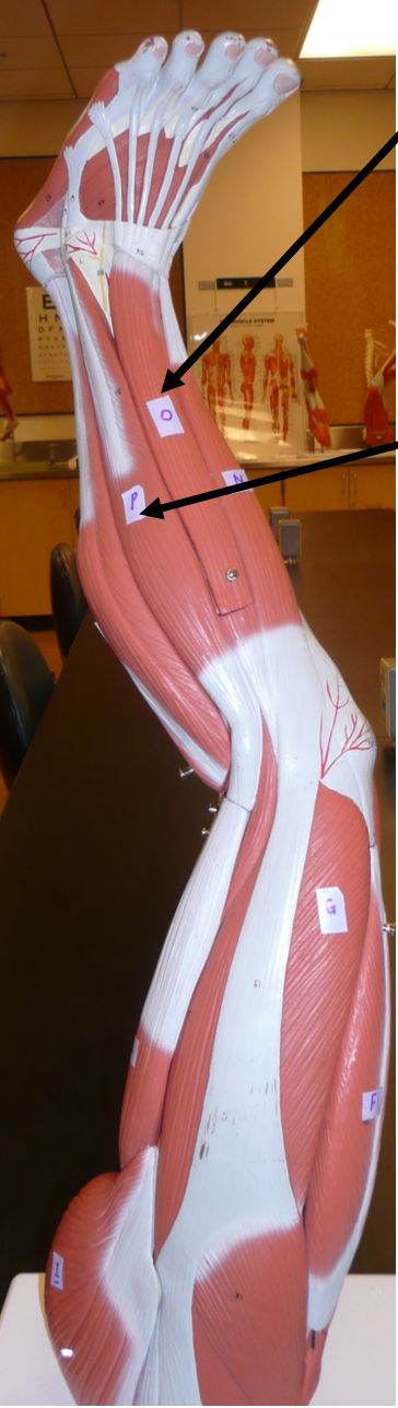
Identify muscle labeled O.
A. tibialis major
B. extensor digitorum longus
C. extensor hallucis longus
D. fibularis longus
B. extensor digitorum longus
Identity muscle labeled P
A. tibialis anterior
B. extensor digitorum longus
C. extensor hallucis longus
D. fibularis longus
D. fibularis longus
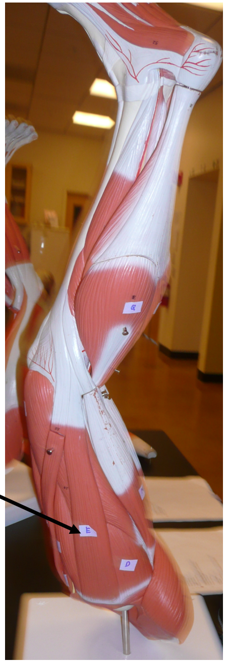
Arrow is pointing to
A. adductor longus
B. adductor magnus
C. sartorius
D. gracilis
D. gracilis
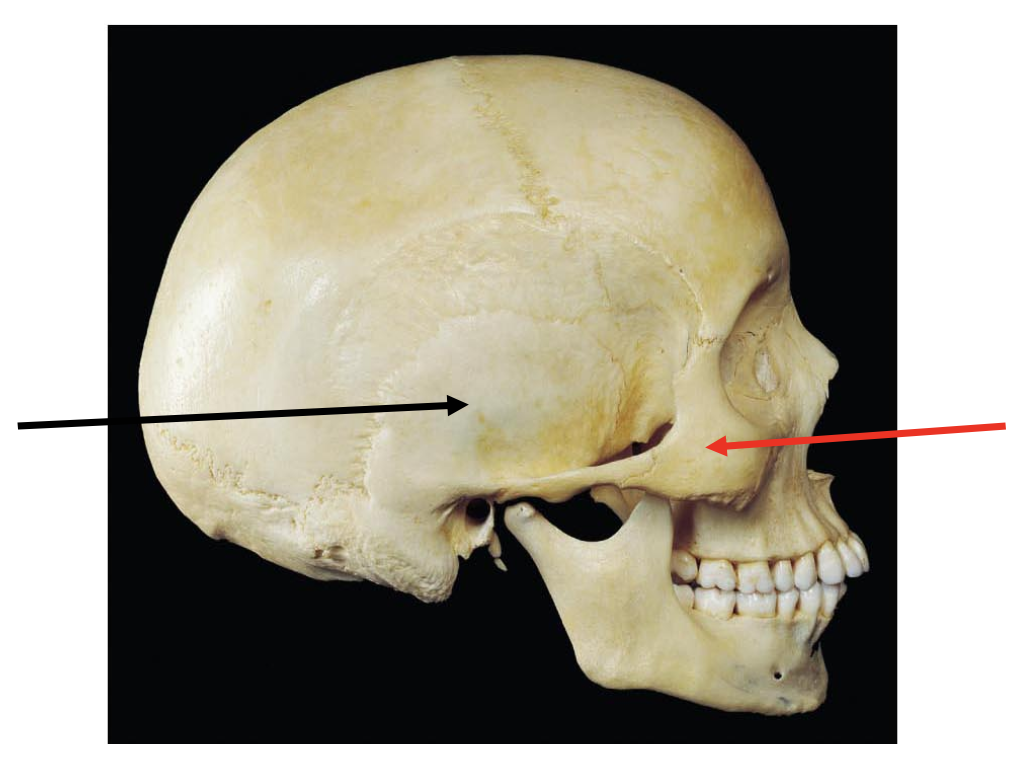
The black arrow is pointing to the
A. parietal bone
B. occipital bone
C. temporal bone
D. frontal bone
C. temporal bone
The red arrow is pointing to the
A. ethmoid bone
B. maxilla
C. zygomatic bone
D. nasal bone
C. zygomatic bone
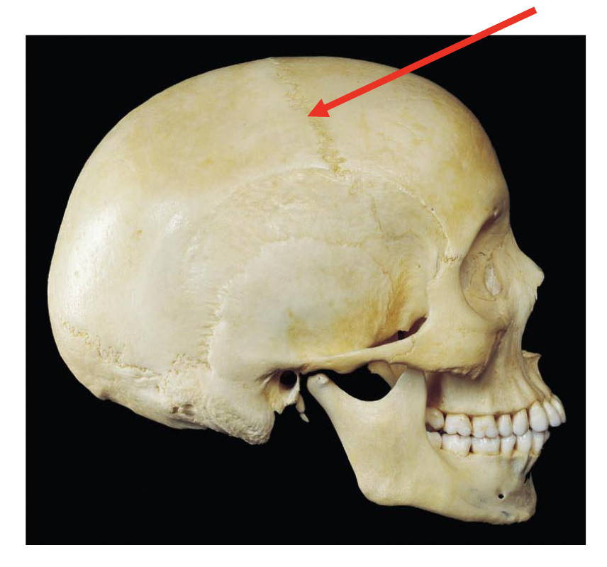
The suture that the red arrow is pointing to is known as the ____ suture and a suture is a type of ____ joint.
A. lambdoid, fibrous
B. Coronal, fibrous
C. Squamous, cartiilaginous
D. Sagittal, synovial
B. Coronal, fibrous
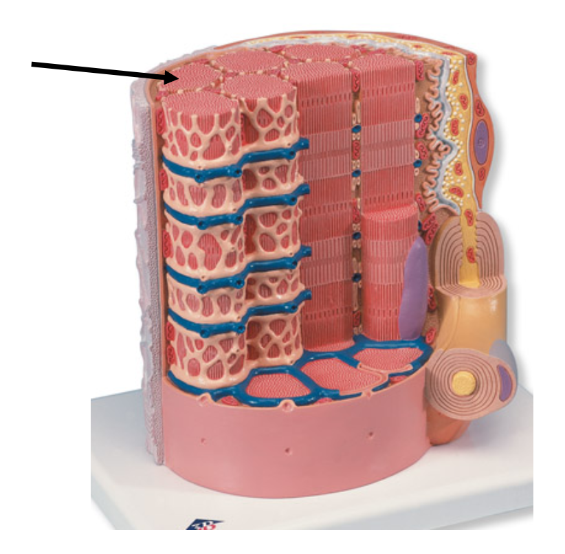
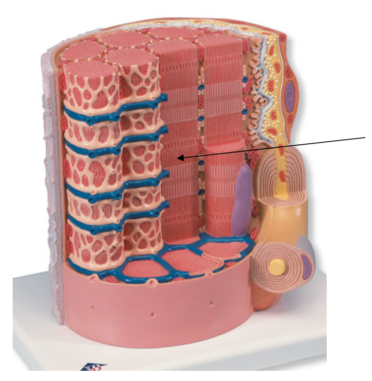
The arrow is pointing to the organelle known as
A. nucleus
B. mitochondrion
C. sarcoplasmic reticulum
D. myofibril
D. myofibril
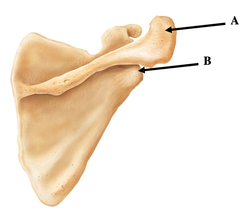
The photo provided is of a scapula. Identify label A.
A. coracoid process
B. glenoid cavity
C. spine
D. acromion
D. acromion
What bone articulates with the part labeled B on the scapula?
A. clavicle
B. humerus
C. radius
D. ulna
B. humerus
Identify the bone to the left.
A. humerus
B. femur
C. clavicle
D. ulna
C. clavicle
The arrow is pointing to the
A. conoid tubercle
B. sternal end
C. coronoid process
D. acromial end
B. sternal end
What vertebra is shown on the left?
A. atlas
B. cervical
C. thoracic
D. lumbar
C. thoracic
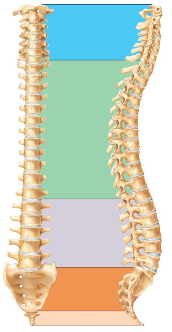
The vertebrae in the blue region are the ____ vertebrae.
A. cervical
B. lumbar
C. thoracic
D. coccygeal
A. cervical
The arrow is pointing to
A. Z disc
B. I band
C. A band
D. M line
C. A band
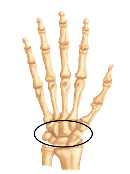
The bones within the oval are known as the
A. phalanges
B. metacarpals
C. carpals
D. proximal phalanges
C. carpals
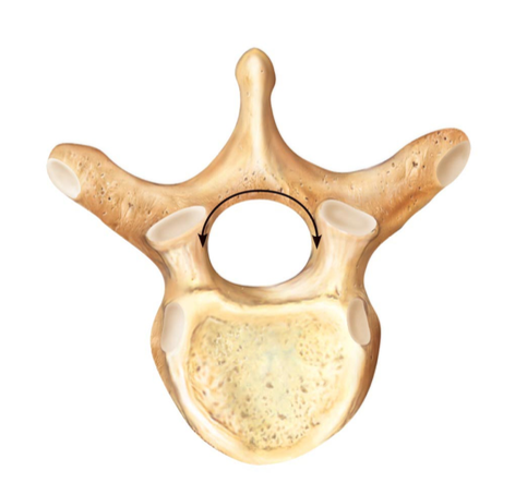
Specimen A is a/an ____ vertebrae.
A. cervical
B. thoracic
C. lumbar
D. atlas
A. cervical
What is found in between vertebrae that function as shock absorbers?
A. Z disc
B. Lingamentum flavum
C. Intervertebral disc
D. Spinous process
C. Intervertebral disc
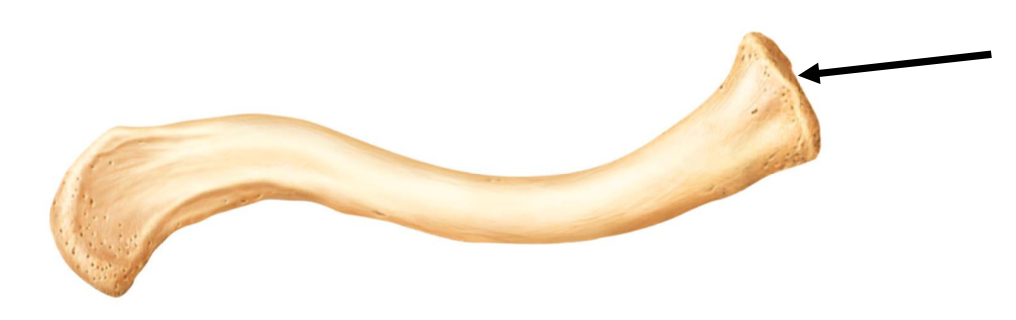
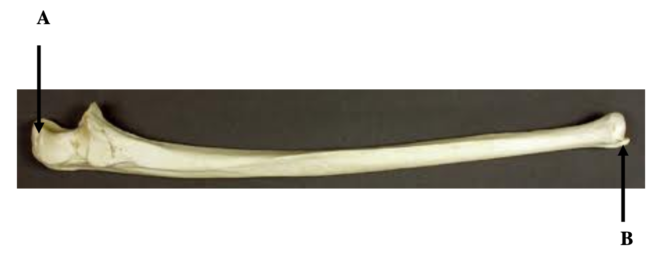
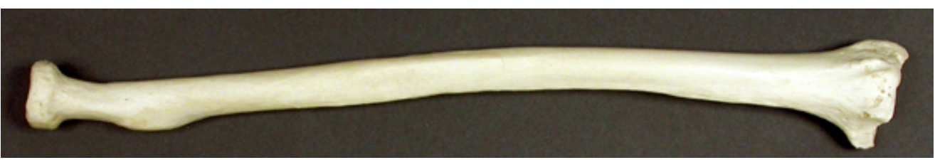
The photo provided is the ulna. Label A is pointing to
A. Coronoid process
B. Corocoid process
C. Olecranon process
D. Trochlea
C. Olecranon process
Label B is pointing to
A. trochlear notch
B. head
C. olecranon process
D. styloid process
D. styloid process
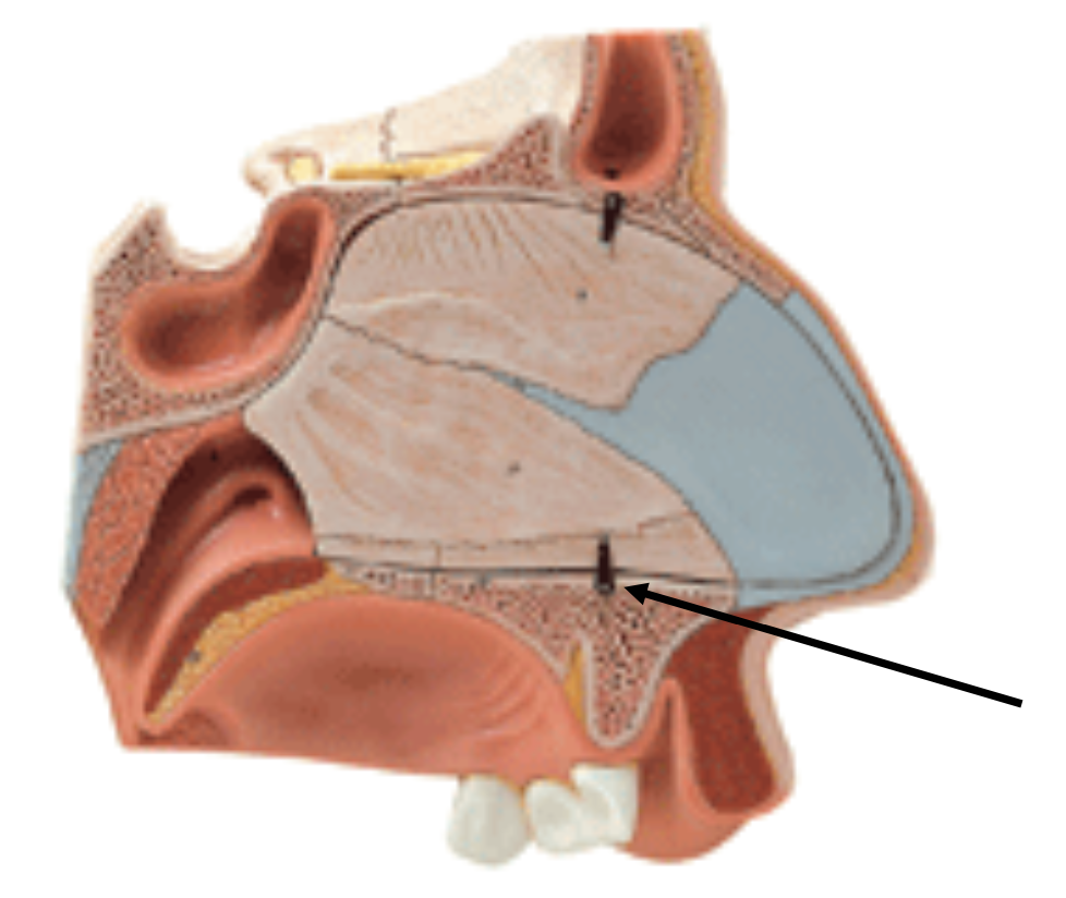
This is a model of the nasal cavity. What bone is the arrow pointing to that forms part of the floor of the nasal cavity that is also the roof of the oral cavity?
A. ethmoid
B. nasal
C. maxilla
D. zygomatic
C. maxilla
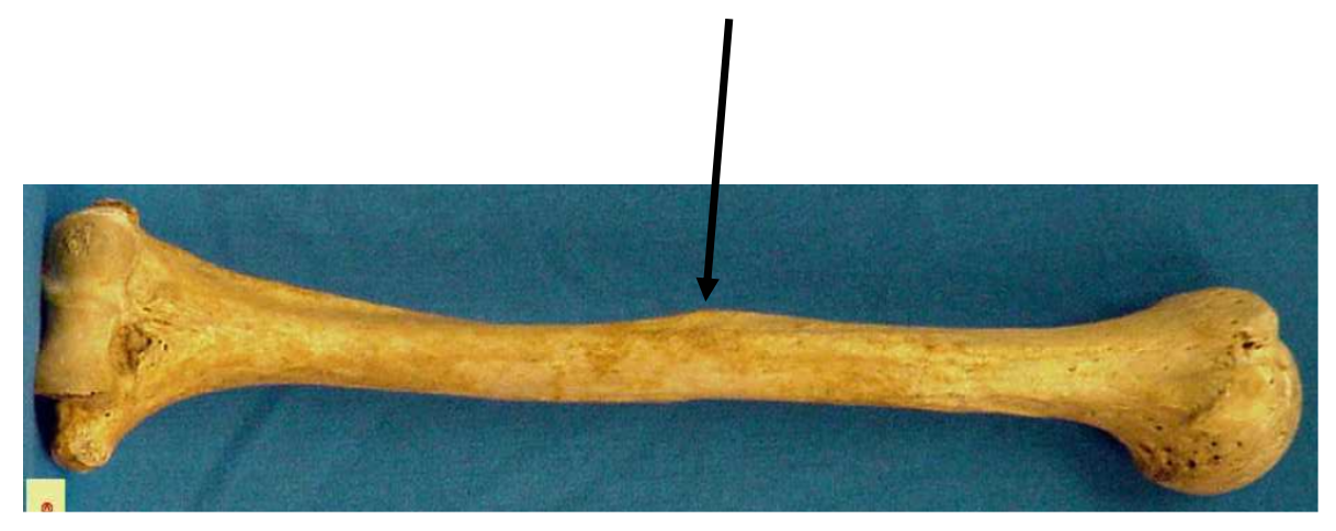
The photo provided shows a humerus. The arrow is pointing to
A. radial groove
B. lesser tubercle
C. deltoid tuberosity
D. capitulum
C. deltoid tuberosity
The muscle that attaches to the structure the arrow points to is
A. biceps brachii
B. triceps brachii
C. brachialis
D. deltoid
D. deltoid
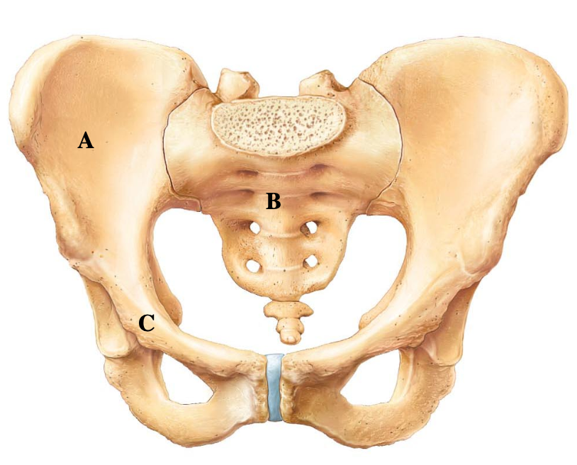
Labels A, B, and C are ____, ____, and ____.
A. ilium, ischium, pubis
B. ilium, sacrum, pubis
C. ischium, coccyx, ilium
D. pubis, sacrum, ischium
B. ilium, sacrum, pubis
The bone in the photo is radius. What structural type of amphiarthrotic joint is formed between the radius and ulna where the interosseous membrane is present?’
A. syndesmosis
B. gomphosis
C. synchondrosis
D. symphysis
A. syndesmosis
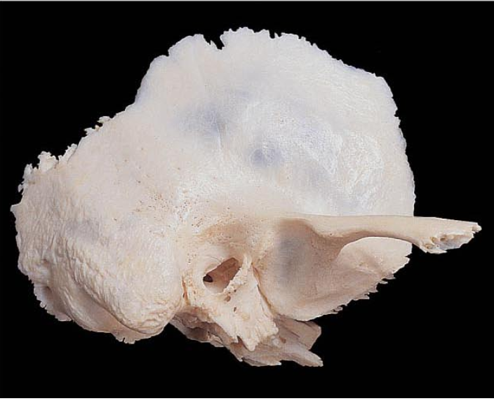
Identify this cranial bone.
A. parietal bone
B. sphenoid bone
C. ethmoid bone
D. temporal bone
D. temporal bone