MicroBio Chapter 1/2
 Call Kai
 Learn
Learn Practice Test
 Spaced Repetition
 Match
Match Flashcards
 Knowt Play
1/74

There's no tags or description
Looks like no tags are added yet.
Name | Mastery | Learn | Test | Matching | Spaced | Call with Kai | Chat |
|---|
No analytics yet
Send a link to your students to track their progress
75 Terms
What is microbiology, and what is a pathogen?
Microbiology is the study of microorganisms/microbes. A pathogen is a microorganism capable of causing disease. Importantly, not all microbes are harmful.
What major groups of microbes/infectious agents does Bricker want you to recognize?
Bacteria, archaea, protozoa, fungi, helminths, algae, viruses, and prions.
Viruses and prions are acellular, meaning they are not cells.
What is the general size order of helminths, protozoa, bacteria, viruses, and prions?
Answer: Largest → smallest:
Helminth → Protozoan → Bacterium → Virus → Prion
Bricker actually used this as a Concept Check, so know it quickly.
What are the four major families of biological macromolecules?
Answer:
Carbohydrates
Lipids
Proteins
Nucleic acids
These molecules perform different jobs in cells, including energy storage, membrane formation, structure, metabolism, and storing genetic information.
Monomer vs. polymer, and dehydration synthesis vs. hydrolysis?
Answer: A monomer is a small building block; a polymer is a larger molecule made by connecting many building blocks.
Dehydration synthesis = builds larger molecules by removing water.
Hydrolysis = breaks molecules apart using water.
Think: build vs. break.
What is a monosaccharide, give a example, and what can it participate in?
Answer: A monosaccharide is one simple sugar unit. Examples include glucose and fructose.
They can participate directly in metabolism or serve as building blocks for larger carbohydrates.
What is a disaccharide, and which examples should you know?
Answer: A disaccharide contains two monosaccharides joined together.
Maltose = glucose + glucose
Lactose = glucose + galactose
Sucrose = glucose + fructose
What is a polysaccharide, give examples, and what is its purpose?
Answer: A polysaccharide is a long chain of monosaccharides.
Examples: starch, cellulose, glycogen.
They can be used for energy/food storage or structural support.
How do you differentiate mono-, di-, and polysaccharides?
Answer:
Mono = one sugar unit
Di = two sugar units
Poly = many sugar units
If an exam describes a long sugar chain, think polysaccharide.
What major lipid types should you recognize, and what can they function in?
Answer: Your material focuses on:
triglycerides
phospholipids
waxes
steroids
Lipids can function in energy storage, cell membranes, and other cellular structures.
What is a triglyceride made of, give examples of some triglycerides and their purpose?
Answer: A triglyceride is made of:
1 glycerol + 3 fatty acids
Fats and oils are triglycerides, and one major purpose is energy storage.
Saturated vs. unsaturated fatty acids?
Answer:
Saturated: no carbon-carbon double bonds; straighter chain.
Unsaturated: one or more carbon-carbon double bonds; often produces bends/kinks.
You should be able to recognize the difference from a diagram.
What is a phospholipid?
Answer: A phospholipid contains glycerol, fatty acids, and a phosphate-containing head.
The head is polar/hydrophilic and interacts with water.
The tails are nonpolar/hydrophobic and avoid water.
What is the purpose of phospholipids, and why do they form a bilayer?
Answer: Phospholipids form the basic structure of cell membranes.
In water:
hydrophilic heads face water
hydrophobic tails point inward toward each other
This naturally produces a phospholipid bilayer.
What should you know about waxes and steroids?
Answer: Mycolic acid is a wax associated with the cell wall of mycobacteria.
Steroids have a ringed structure. Examples include cholesterol and ergosterol.
What is a protein, and what does it do?
Answer: Proteins are chains of amino acids.
Proteins can:
provide structure
act as enzymes
perform metabolic reactions
function as antibodies
be components of membranes, cell walls, and ribosomes
What are amino acids and peptide bonds?
Answer: Amino acids are the building blocks of proteins.
A peptide bond joins one amino acid to another, eventually creating a polypeptide/protein chain.
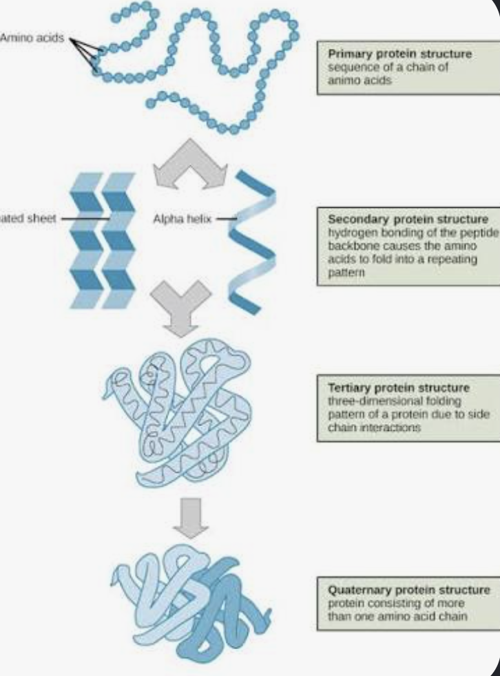
What are the four levels of protein structure?
Answer:
Primary: amino-acid sequence
Secondary: local folding into helices/pleated sheets
Tertiary: overall 3-D shape of one polypeptide
Quaternary: two or more folded polypeptide chains associated together
Memory:
sequence → shapes → 3-D → multiple chains
What is protein denaturation, and why does it matter?
Answer: Denaturation means a protein loses its normal native shape.
Because a protein's function depends on its shape, changing that shape can cause the protein to stop working properly.
What is a nucleotide, and what does it contain?
Answer: A nucleotide is the building block/monomer of DNA and RNA.
Every nucleotide contains:
phosphate
pentose sugar
nitrogenous base
What do you need to know about DNA?
Answer: DNA contains:
deoxyribose sugar
adenine (A)
thymine (T)
cytosine (C)
guanine (G)
DNA is usually double stranded and is involved in storing/transmitting genetic information.
What do you need to know about RNA?
Answer: RNA contains:
ribose sugar
adenine (A)
uracil (U)
cytosine (C)
guanine (G)
RNA is usually single stranded and helps express genetic information, including involvement in protein synthesis.
DNA vs. RNA?
Answer:
DNA: deoxyribose, T, usually double stranded
RNA: ribose, U, usually single stranded
Both have A, C, and G.
Big memory point:
DNA = T
RNA = U
What is ATP composed of and what is its purpose?
Answer: ATP contains:
adenine + ribose + 3 phosphate groups
ATP functions as an important energy-transfer molecule for cellular activities.
What three basic characteristics are common to all cells?
Answer: From your assigned textbook section, all cells contain:
a cell/cytoplasmic membrane
DNA in chromosome(s)
ribosomes for protein synthesis
This directly answers Bricker's learning outcome asking for three characteristics common to all cells.
What are the Five I's, and what is the overall purpose?
Answer:
Inoculation → Incubation → Isolation → Inspection → Identification
It's basically:
put the sample in → grow it → separate it → examine it → determine what it is.
What is inoculation, and what is an inoculum?
Answer: Inoculation is introducing a microbial sample into a growth medium.
The inoculum is the sample containing the microorganisms being introduced.
Think:
inoculum = sample
inoculation = action
What is incubation and why is it necessary?
Answer: Incubation provides the proper conditions for microbial growth, including appropriate temperature and gas conditions.
Purpose: allow microbes to grow and multiply.
What is isolation and why is it important?
Answer: Isolation separates microorganisms so different species can be studied individually.
A patient specimen may contain several organisms. You don't want them all mixed together when trying to identify one particular species.
What is inspection?
Answer: Inspection means examining the culture.
This can involve:
observing colonies with the naked eye
microscopy
staining
What is identification, and how can microbes be identified?
Answer: Identification determines which microorganism is present, often down to species level.
Bricker lists:
microscopy
metabolism/biochemical testing
genetics
immunology
Pure vs. mixed vs. contaminated culture?
Answer:
Pure culture: one microbial species/type.
Mixed culture: multiple microorganisms intentionally/knowingly present.
Contaminated culture: an unwanted microorganism accidentally entered the culture.
What are medium, media, and culture?
Answer:
Medium: material/environment used to grow microorganisms.
Media: plural of medium.
Culture: microorganisms that have been grown in or on the medium.
What are the three physical states of media?
Answer:
Liquid → Semisolid → Solid
Each physical form has different laboratory uses.
When would liquid, semisolid, and solid media be useful?
Answer:
Liquid: organisms grow throughout a fluid; useful for growing large populations.
Semisolid: can help observe patterns such as microbial movement.
Solid: provides a surface where separate colonies can form.
What is agar and why is it useful?
Answer: Agar is a solidifying agent used to make microbiological media firm.
Its major benefit is creating a stable surface where microorganisms can grow into separate visible colonies.
What three properties can be used to classify media?
Answer: Media can be classified by:
physical state
chemical composition
function/purpose
What is chemically defined/defined medium?
Answer: A defined medium has a known exact chemical composition.
You know exactly which chemicals are present and how much is present.
What is complex medium?
Answer: A complex medium contains ingredients whose exact chemical composition isn't fully known, often because they come from biological materials or extracts.
Defined vs. complex media?
Answer:
Defined = exact composition known.
Complex = exact composition not completely known.
If an exam says every chemical and amount is precisely known → defined.
What are general-purpose and enriched media?
Answer: General-purpose media support growth of a broad variety of organisms.
Enriched media contain added nutrients to help organisms with greater nutritional requirements grow.
Blood agar and chocolate agar are examples shown in your material.
What is selective medium and what is its purpose?
Answer: Selective medium allows certain organisms to grow while suppressing others.
Purpose: help isolate a desired microorganism from a mixed sample.
Memory:
Selective = selects WHO grows.
What is differential medium and what is its purpose?
Answer: Differential media allow organisms to grow but make them visibly different based on their reactions, often through color changes.
Memory:
Differential = shows a DIFFERENCE.
Selective vs. differential media?
Answer:
Selective → WHO can grow?
Differential → HOW do the organisms that grow differ?
This distinction is specifically emphasized in Bricker's material.
Why is MacConkey agar both selective and differential?
Answer:
Selective: suppresses many gram-positive organisms.
Differential: produces visible distinctions among organisms that do grow.
Bricker specifically uses MacConkey agar as the example.
What is reducing medium?
Answer: Reducing media contain substances that reduce/remove oxygen.
Purpose: help grow microorganisms that need little or no oxygen.
This is useful textbook depth, but lower priority than selective/differential.
What are fermentation, transport, and enumeration media used for?
Answer:
Fermentation media: detect whether an organism can ferment a carbohydrate; changes may indicate acid/gas production.
Transport media: maintain a specimen while it travels to the laboratory.
Enumeration: used when the goal involves determining the number of microbes.
Know the basic purpose, not every tiny detail.
What is a colony? what does individual bacteria have to do with this
Answer: A colony is a visible mass of microbial cells that grew from a much smaller number of microorganisms.
Individual bacteria are microscopic, but after multiplying into millions of cells, the colony becomes visible.
What are the three isolation techniques you should recognize?
Answer:
Streak plate: progressively streak cells across agar to dilute/separate them.
Loop dilution/pour plate: dilute the sample and place it into agar.
Spread plate: place a sample on the agar surface and spread it across the plate.
All three aim to produce isolated colonies.
Why do microbiologists isolate organisms before identifying them?
Answer: Because a mixed sample can contain multiple microorganisms. Isolation produces separate colonies, allowing one organism to be studied and identified without confusing its characteristics with another organism.
This connects the Five I's:
Isolation happens before Inspection and Identification.
What metric units should you know, largest → smallest?
Answer:
millimeter (mm) → micrometer (μm) → nanometer (nm)
Microorganisms and microscopic structures are commonly measured using these units.
What metric conversions should you know?
Answer:
1 mm = 1,000 μm
1 μm = 1,000 nm
And:
milli = 10⁻³
micro = 10⁻⁶
nano = 10⁻⁹
What are the three elements of good microscopy?
Answer:
Magnification + Resolution + Contrast
Bricker specifically lists knowing these as a learning outcome.
What is magnification?
Answer: Magnification means making the image appear larger than the specimen actually is.
Think:
Magnification = bigger.
What is resolution?
Answer: Resolution, or resolving power, is the ability to distinguish two nearby objects as two separate objects.
Think:
Resolution = detail/separation.
What is contrast?
Answer: Contrast is the difference between the specimen and its background.
Greater contrast makes the specimen easier to see.
Magnification vs. resolution?
Answer:
Magnification = bigger image
Resolution = clearer separation of details
Something can be highly magnified and still look blurry if the resolution is poor.
What resolving-power numbers does Bricker give?
Answer:
Human eye: about 0.2 mm
Light microscope with oil immersion: about 0.2 μm
Why is immersion oil used?
Answer: Immersion oil reduces light scattering, which helps improve resolution.
Think:
less scattered light → better detail.
What basic microscope pathway/parts should you understand?
Answer: Basic light pathway:
Light source → condenser → specimen → objective lens → ocular lens → eye
What is bright-field microscopy?
Answer:
Bright-field is the most widely used light microscopy method.
Light passes through the specimen. It can be used with living unstained specimens or preserved stained specimens.
What is dark-field microscopy?
Answer: Dark-field microscopy produces a bright specimen against a dark background.
It's useful for viewing thin/living organisms that may be difficult to see using ordinary bright-field microscopy.
Textbook-depth card — lower priority than bright-field, TEM, and SEM.
What is phase-contrast microscopy? What does it help internal structures with
Answer: Phase-contrast increases differences within living, unstained cells, helping internal structures become easier to see without staining the specimen.
Textbook-depth card.
What are fluorescence and confocal microscopy?
Answer:
Fluorescence microscopy uses fluorescent substances to make particular cells/structures glow and become easier to identify.
Confocal microscopy obtains sharp images at different depths that can be combined to create detailed three-dimensional views.
Know their general purposes rather than tiny technical details.
Light microscope vs. electron microscope?
Answer:
Light microscopy = uses visible light.
Electron microscopy = uses electrons and can reveal much smaller structures in greater detail.
What is TEM and what is its purpose?
Answer: TEM = Transmission Electron Microscope.
Electrons pass through an extremely thin specimen.
Purpose: see detailed internal structures of cells and viruses.
Memory:
T = Through
What is SEM and what is its purpose?
Answer: SEM = Scanning Electron Microscope.
Electrons scan the surface of the specimen.
Purpose: produce a detailed 3-D-looking surface image.
Memory:
S = Surface
TEM vs. SEM?
Answer:
TEM → THROUGH → internal structures
SEM → SURFACE → 3-D surface appearance
This is one comparison I would know instantly.
Living preparation/hanging drop vs. fixed smear?
Answer: Living preparations allow microorganisms to be viewed alive.
A hanging-drop preparation suspends a drop containing microorganisms from a coverslip, allowing movement/live cells to be observed.
A fixed smear attaches cells to the slide so they can be stained and examined.
Why are stains used, and what is basic vs. acidic dye?
Answer: Cells can be difficult to see because they have little natural contrast. Staining adds color and increases visibility.
Bricker gives:
Basic dyes = positively charged
Acidic dyes = negatively charged
What is a simple stain?
Answer: A simple stain uses one dye to make cells easier to see and observe things like shape and arrangement.
Examples:
crystal violet
methylene blue
Differential vs. special stains?
Answer: A differential stain distinguishes different groups/types of cells.
Examples:
Gram stain
acid-fast stain
A special stain highlights a particular cellular structure.
Examples:
capsule stain
flagellar stain
What are the four Gram-stain steps in order?
Answer:
Crystal violet
Iodine
Alcohol/decolorizer
Safranin
Memorize this order exactly.
What staining results should you recognize?
Answer:
Gram-positive = purple
Gram-negative = pink/red
Acid-fast cells = reddish-purple/red
Non-acid-fast = blue
Endospores = green
Vegetative cells in the endospore stain = pink
How should you classify these stains?
Answer:
Simple stains
crystal violet alone
methylene blue alone
Differential stains
Gram stain
acid-fast stain
Special stains
capsule stain
flagellar stain