Module 1 - Avascular Necrosis & Epiphyseal Disorders
1/40
Earn XP
Description and Tags
Question-and-answer flashcards covering avascular necrosis, Legg-Calvé-Perthes, Osteochondritis Dissecans, Freiberg’s disease, Köhler’s disease, and Kienböck’s disease, including imaging signs and treatments.
Name | Mastery | Learn | Test | Matching | Spaced | Call with Kai |
|---|
No study sessions yet.
41 Terms
What is another name for avascular necrosis?
ischemic necrosis.
What causes bone necrosis in avascular necrosis?
Loss of blood supply to bone.
What are common causes of ischemic necrosis in adults?
Trauma, vascular, metabolic, neoplastic, alcoholism, chronic pancreatitis, radiation, diving, high-dose steroids, or idiopathic (25%).
Which joints are commonly affected by ischemic necrosis in adults?
Hip, shoulder, scaphoid, knee, ankle.
What is the typical age group for adult ischemic necrosis?
30–60 years.
What happens in early vs. later stages of ischemic necrosis?
Early: microfractures. Later: fragmentation, compression, resorption, leading to bone/joint collapse.
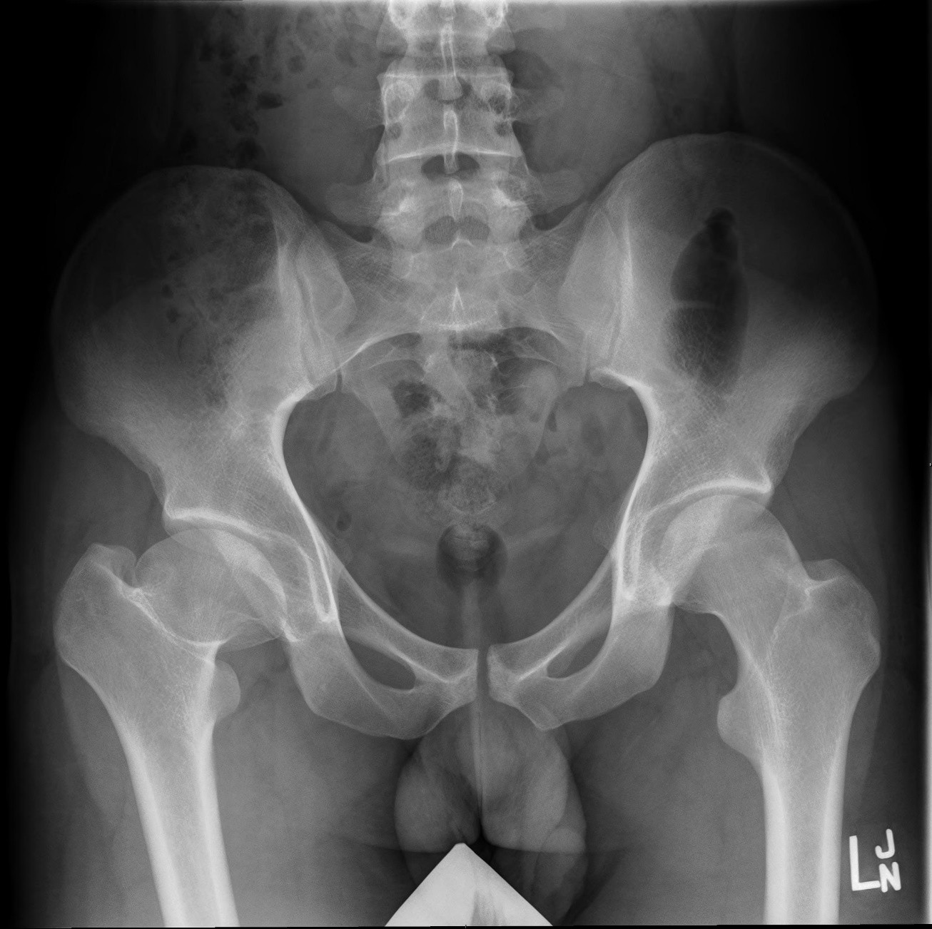
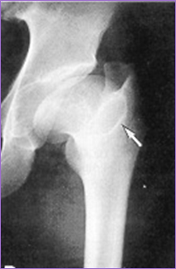
What is the hallmark radiographic sign of hip ischemic necrosis?
Crescent sign – subcortical radiolucent band representing a fracture line.
Which imaging modalities detect early ischemic necrosis best?
MRI, CT, Nuclear Medicine (MRI shows effusion & bone changes; NM shows reduced then increased uptake).
What are common treatments for ischemic necrosis in adults?
NSAIDs, physiotherapy, immobilization, bone grafts, core decompression, joint replacement, stem cell therapy.
What is Legg-Calvé-Perthes disease?
Temporary loss of blood supply to femoral head ossification center before growth plate closure.

What is the typical age and sex distribution of Legg-Perthes disease?
Ages 3–12 years ;MORE common in males 4:1.
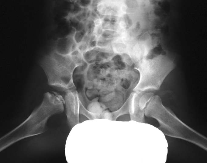
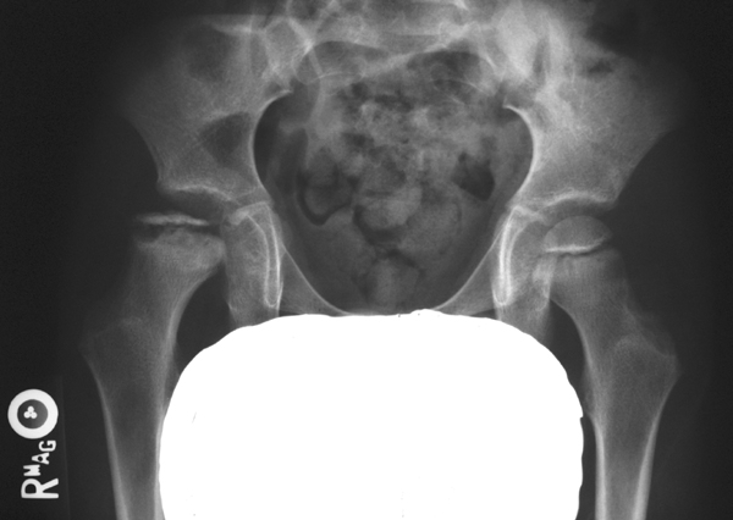
What are the four stages of Legg-Calvé-Perthes disease?
Avascular: ↓ head size, widened joint space. 2. Fragmentation: bone fragments, crescent sign, blurred outline. 3. Repair: revascularization, new bone, wide short neck. 4. Healed/Deformity: flattened, enlarged head, widened neck, enlarged GT.
What symptoms are seen in Legg-Calvé-Perthes disease?
Vague groin pain (may radiate to knee), limp, ↓ hip ROM.
Radiographic features of Legg-Calvé-Perthes disease?
↓ size, flattened femoral head, ↑ density of epiphysis, widened neck, enlarged GT, low MRI signal.

How is Legg-Calvé-Perthes disease treated?
Casting, traction, bed rest, or osteotomy. No cure; deformities often remain.
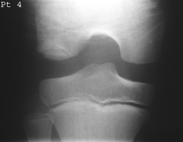
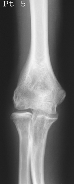
What is Osteochondritis Dissecans?
Avascular necrosis of subchondral bone fragments, often traumatic.
Who is most affected by Osteochondritis Dissecans?
Adolescents (11–20), athletes.

What are 'joint mice'?
Necrotic bone fragments that dislodge into the joint space.
Most common site of Osteochondritis Dissecans?
Medial femoral condyle (75%).

Symptoms of Osteochondritis Dissecans?
Joint effusion, clicking, locking, tenderness.
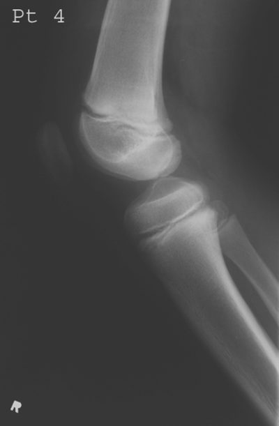
How is it diagnosed radiographically?
MRI/CT for cartilage lesions; X-ray shows fragments and site of origin; tunnel view for knee.
Treatment options for Osteochondritis Dissecans?
Arthroscopy (remove/reattach), drilling, immobilization, protected weight bearing.


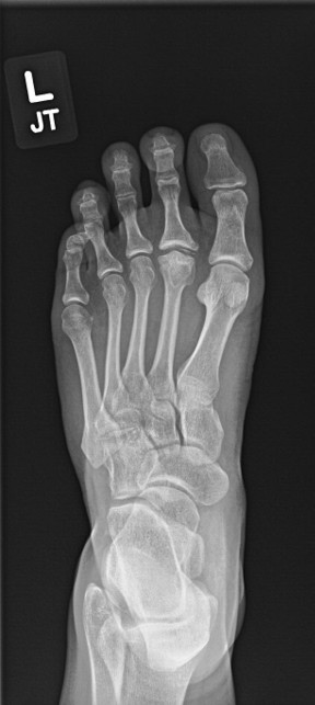
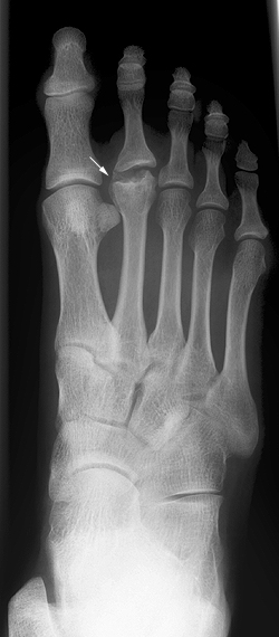
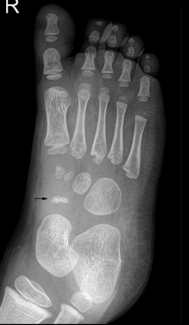
What bone is affected in Freiberg’s disease?
Metatarsal head, usually 2nd (80%), sometimes 3rd.
Who is most affected by Freiberg’s disease?
Teen females (5:1), athletes, high-heel wearers.
Pathogenesis of Freiberg’s disease?
Repeated stress → microfractures at growth plate → necrosis → deformity.
Symptoms of Freiberg’s disease?
Localized forefoot pain, tenderness, worsens with activity.

Radiographic appearance of Freiberg’s disease?
Early: cortex collapse, sclerosis, altered joint space. Late: enlarged, fragmented head.
Treatments for Freiberg’s disease?
Reduced activity, casting, or surgery (excision, transplant).


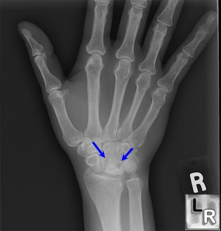
What bone is affected in Köhler’s disease?
Navicular bone (foot).
Typical age group for Köhler’s disease?
Children 3–7, mainly boys.

Signs & symptoms of Köhler’s disease?
Midfoot pain/swelling, limping with weight shifted laterally.

Radiographic findings in Köhler’s disease?
Patchy/homogeneous sclerosis, collapse/fragmentation, eventual revascularization with abnormal shape.
Treatments for Köhler’s disease?
Pain relief, 6–8 weeks casting, arch supports; resolves with time.
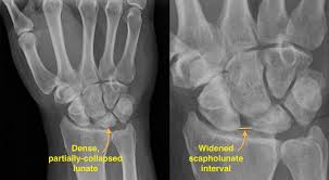
What is Kienböck’s disease?
Avascular necrosis of the lunate bone.
Who is most affected by Kienböck’s disease?
Young adults, often with negative ulnar variance or trauma.

What classification is used for Kienböck’s disease?
Lichtman Stages I–IV (MRI changes → sclerosis → collapse → arthritis).

Symptoms of Kienböck’s disease?
Wrist pain, stiffness, ↓ grip strength.
Radiographic findings in Kienböck’s disease?
Stage I – MRI changes; Stage II – lunate sclerosis; Stage III – collapse ± scaphoid rotation; Stage IV – arthritis.

Treatment options for Kienböck’s disease?
Immobilization, bone grafts, radial shortening/ulnar lengthening, proximal row carpectomy, wrist fusion.
What does the Crescent sign indicate in avascular necrosis of the femoral head?
A crescent-shaped lucent subchondral fissure indicating a fracture line; seen best in abducted position.
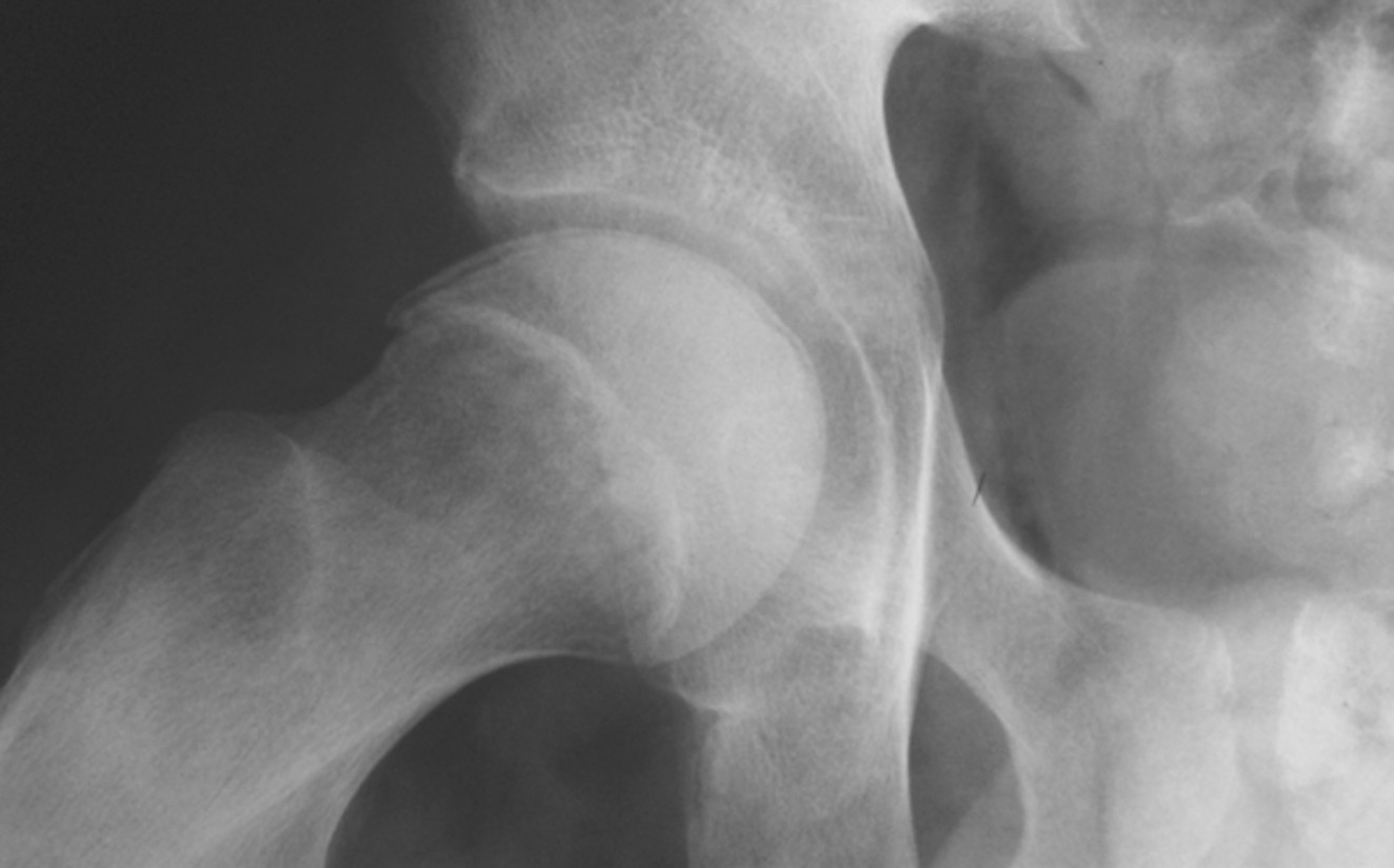
What stage is this disease at?
Stage 4