Practical assessments
1/32
There's no tags or description
Looks like no tags are added yet.
Name | Mastery | Learn | Test | Matching | Spaced | Call with Kai |
|---|
No analytics yet
Send a link to your students to track their progress
33 Terms
Describe GAT procedure
1) Explain procedure to patient
2) Standard hygiene procedures with slit lamp- wipe down chin rest
3) Replace probe with new disposable probe / use a reusable probe that has been prepared correctly
4) Anaesthetic and fluorescein
4) Set up tonometer and illumination
•Cobalt blue light – position illumination system 40 to 60 degrees to one side
•Turn dial to put some weight into the probe
5) 10x to 16x magnification
•Illumination set to 45-60 degrees
•Set tonometer scale to 1.6 grams (16 mmHg)
6) Align probe with centre of cornea
7) Ask patient to blink a few times
•Stare straight ahead
8) Advance probe towards patient’s cornea
9) Watch as the probe touches the cornea
•Advance the probe a little further to applanate the cornea
•Probe arm will be seen to move backwards slightly
10) Look through the eyepieces
•Adjust weight setting by turning dial on tonometer body until mires are correctly aligned
11) Remove probe from cornea & repeat procedure for fellow eye
12) Record result and time of measurement
13) Remove tonometer
14) Check cornea for staining
15) Clean probe if reusable type
•Dispose of probe appropriately if ‘disposable’ type
(See IOP starred flashcards for more information)
Describe the Van Herrick procedure
Set angle of 60 degrees between illumination and observation system of slit lamp
•16x magnification
Optic section: bright, thin beam
Assess relative width of anterior chamber against width of cornea when light just enters anterior chamber
Abduct eye when assessing nasal angle:
•Illumination system locked 60 degrees to the nasal side of the observation system
•Rotate whole system temporally
•Ask patient to look into observation system (eye being measured has to abduct to do this; prevents nose from blocking the slit beam)
(see anterior segment starred flashcards for more information)
Describe the US procedure
Compact handheld ultrasound pachymetry
Procedure:
•Explain procedure to patient
•Topical anaesthetic
•Apply probe tip to to the centre of the patient’s cornea
•Obtain readings according to the manufacturer’s instructions
•Check cornea with slit lamp and fluorescein
(See pachymetry starred flashcards for more information)
Describe how to calibrate GAT
Place tonometer on slit lamp, with probe in place
Insert calibration rod into socket on side of GAT housing
Set rod to the 2 gram setting
•Increase the weight setting on the dial and watch for the probe to rock forwards
•Read off scale to check calibration accuracy
•Decrease weight and watch for probe rocking backwards
Repeat the above, with the rod set to 0 grams and 6 grams
Absolute calibration error and any change in the error between the weight settings should be noted
(See IOP starred flashcards for more information)
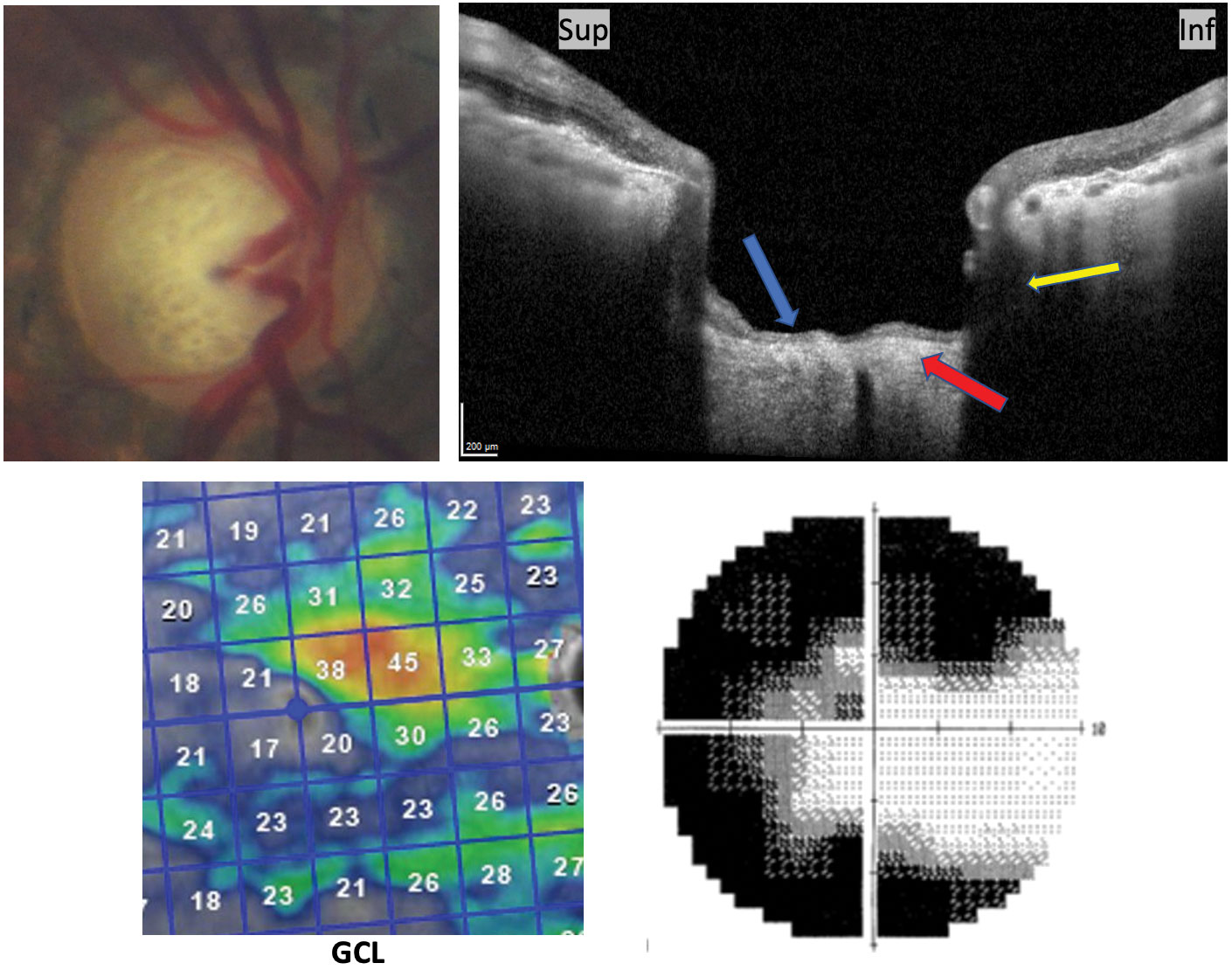
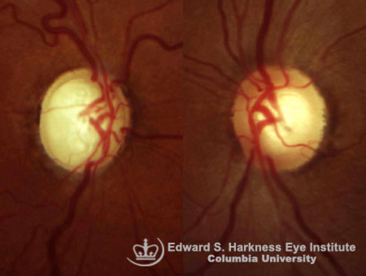
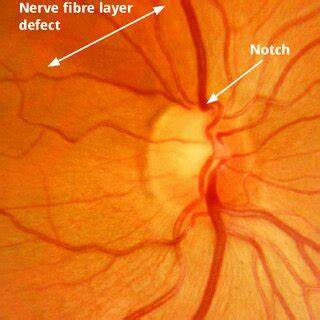
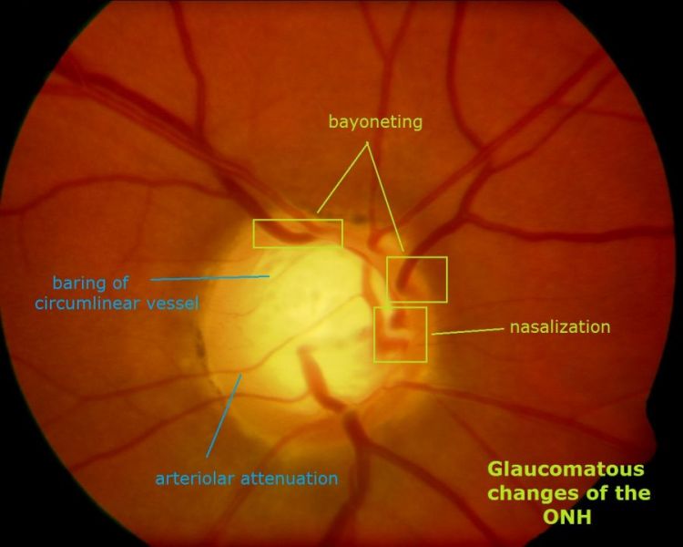
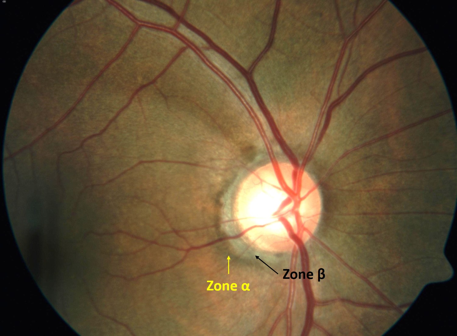
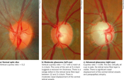
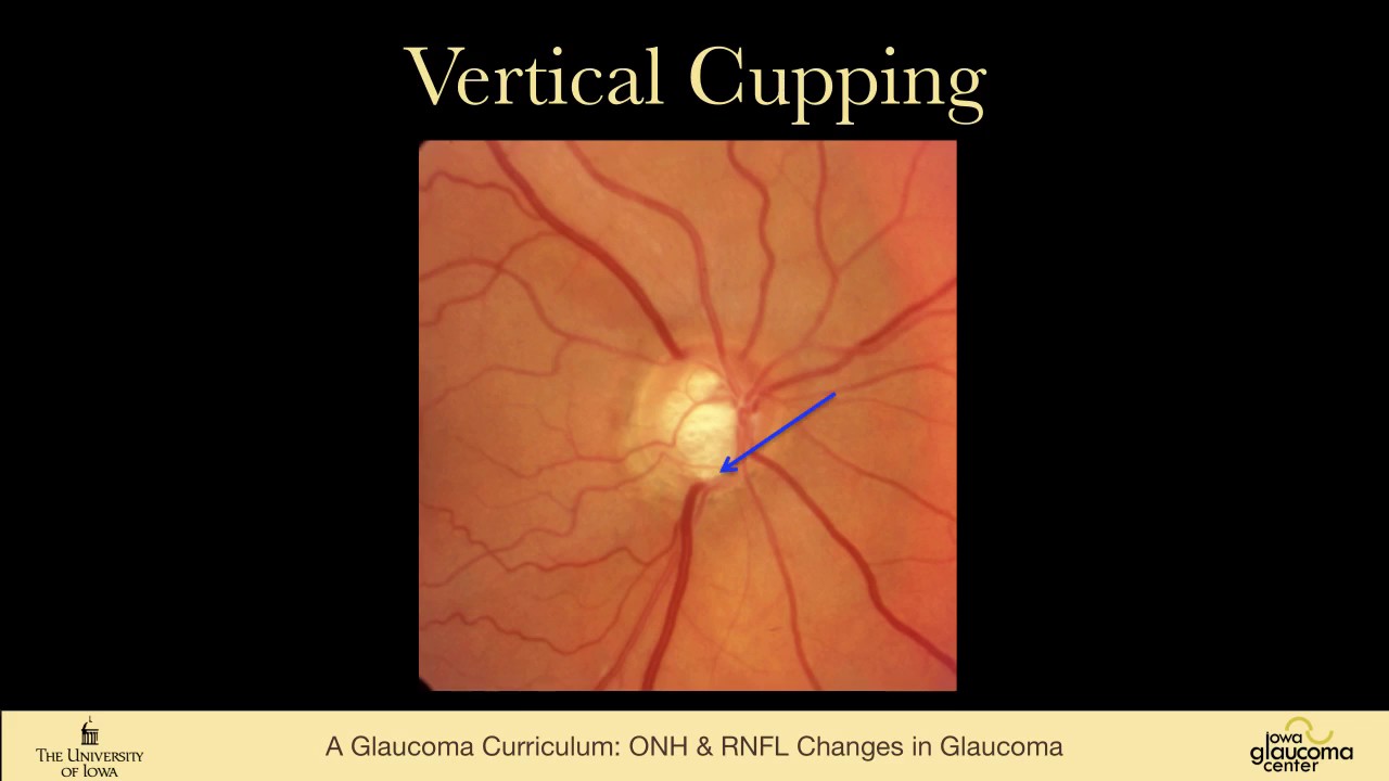
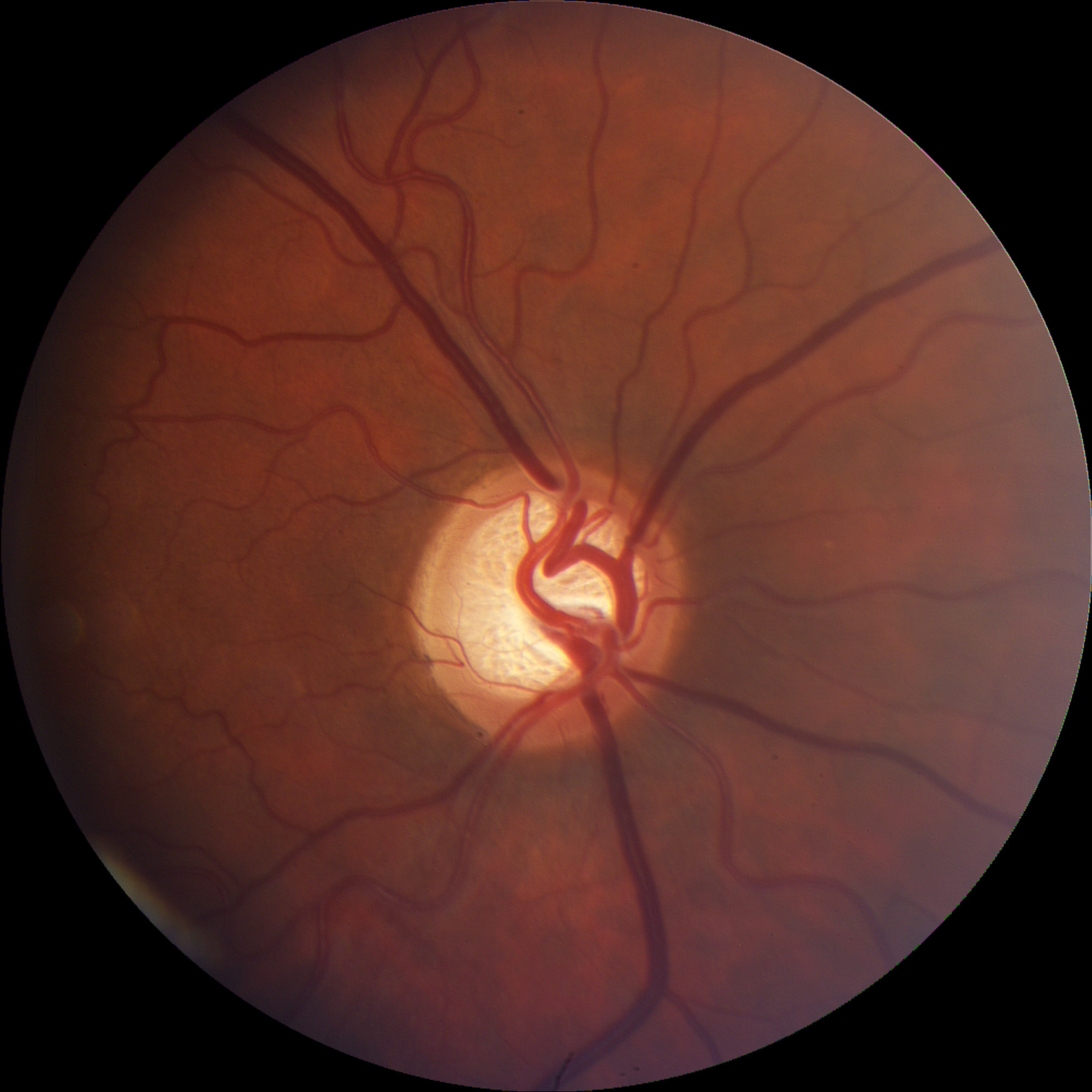
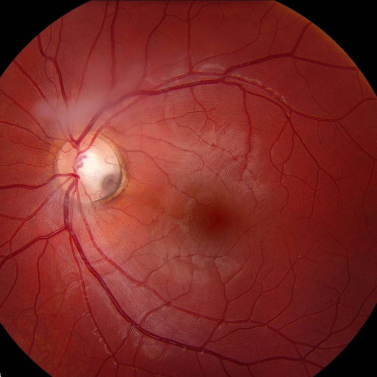
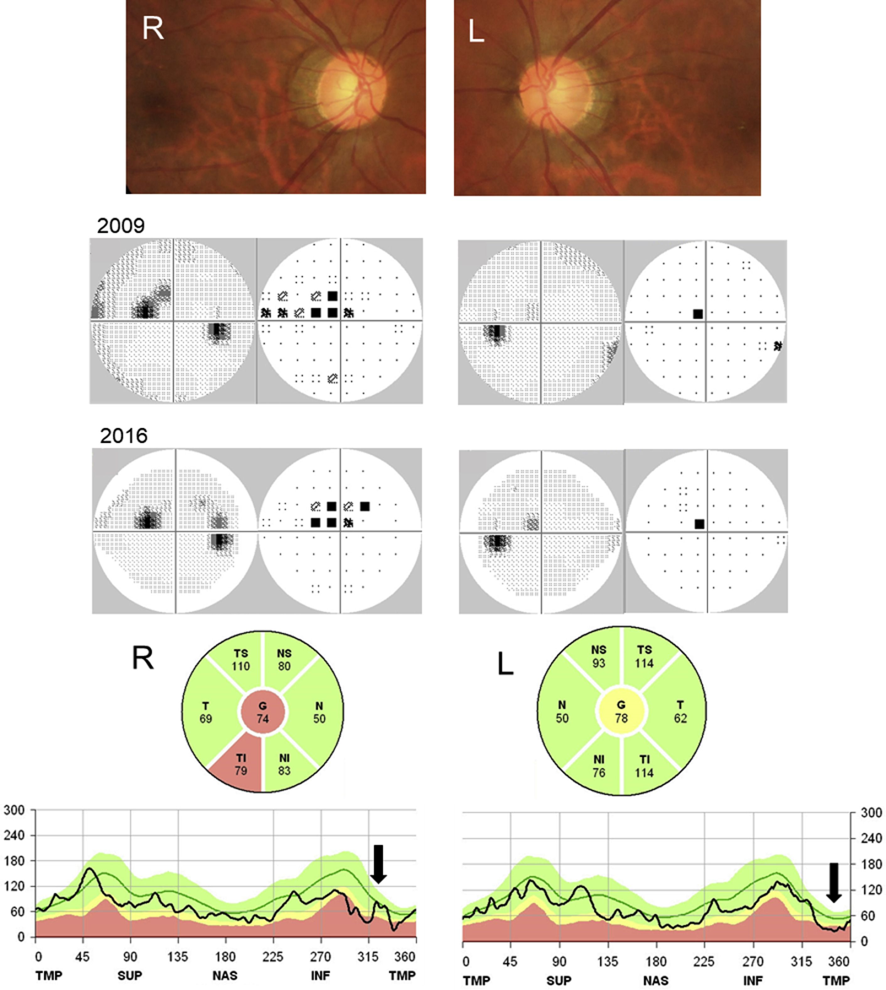
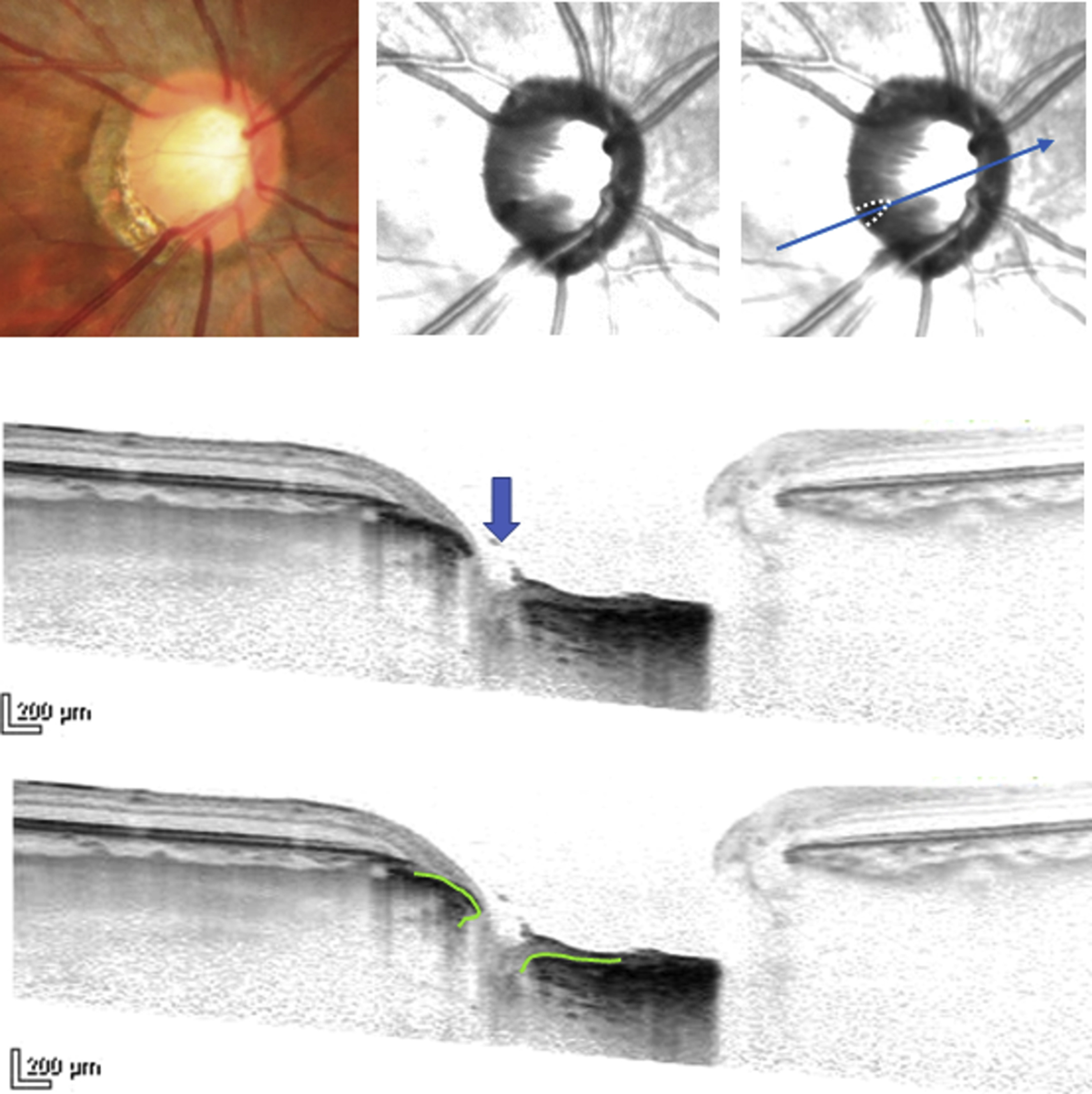
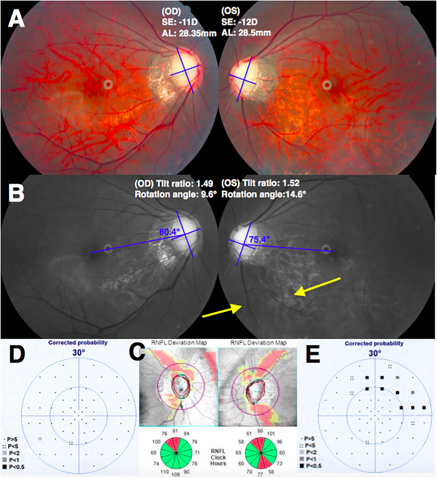
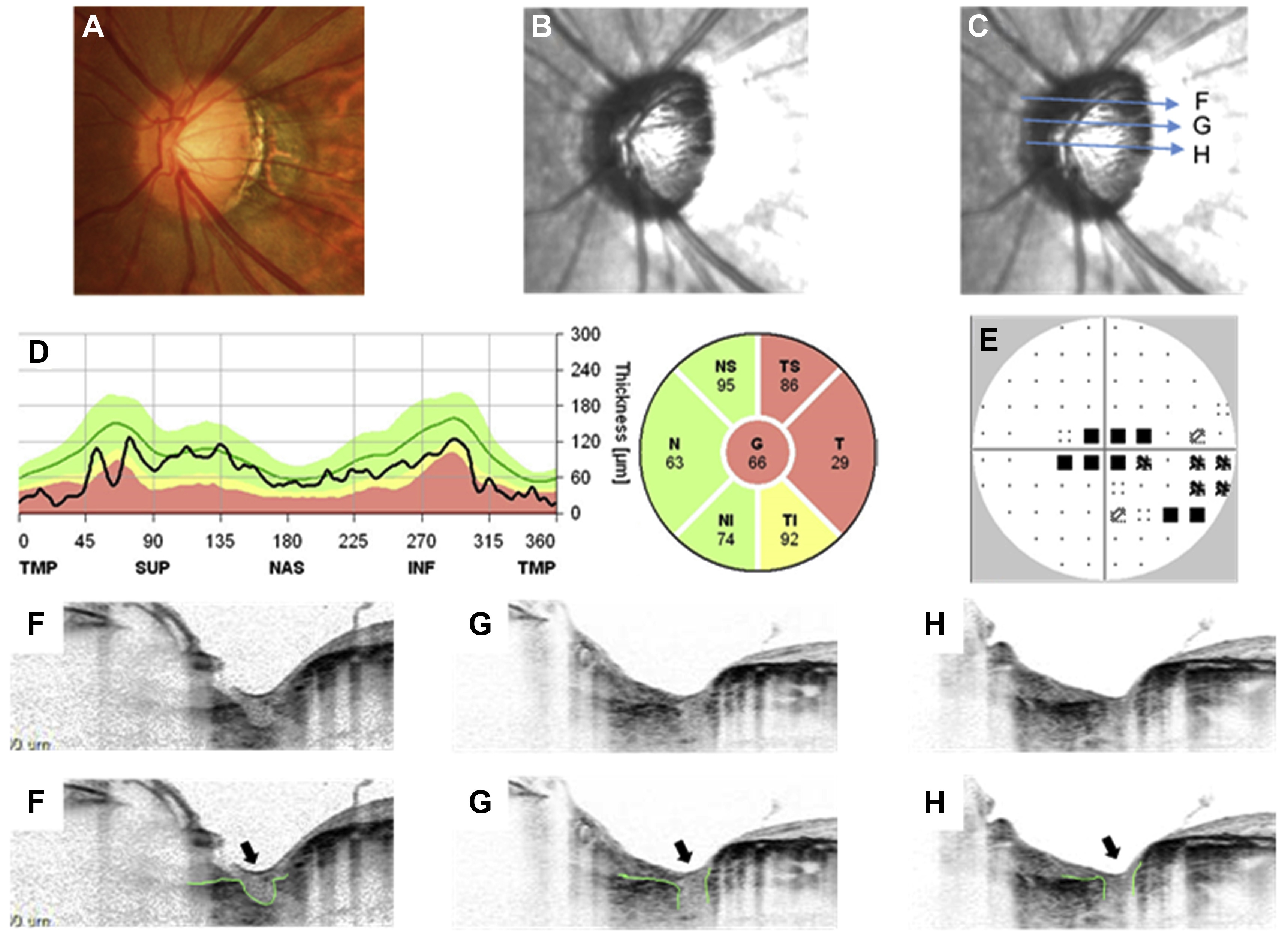
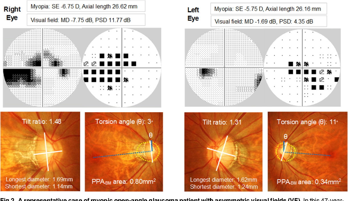
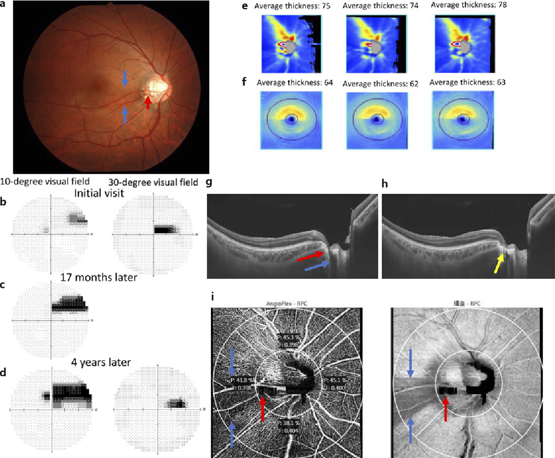
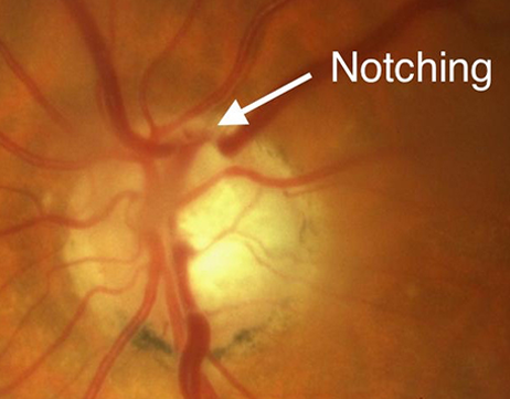
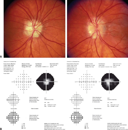
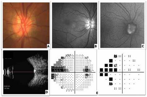
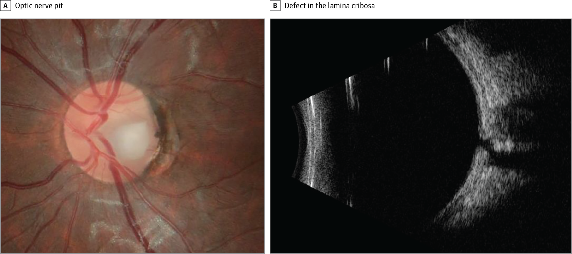
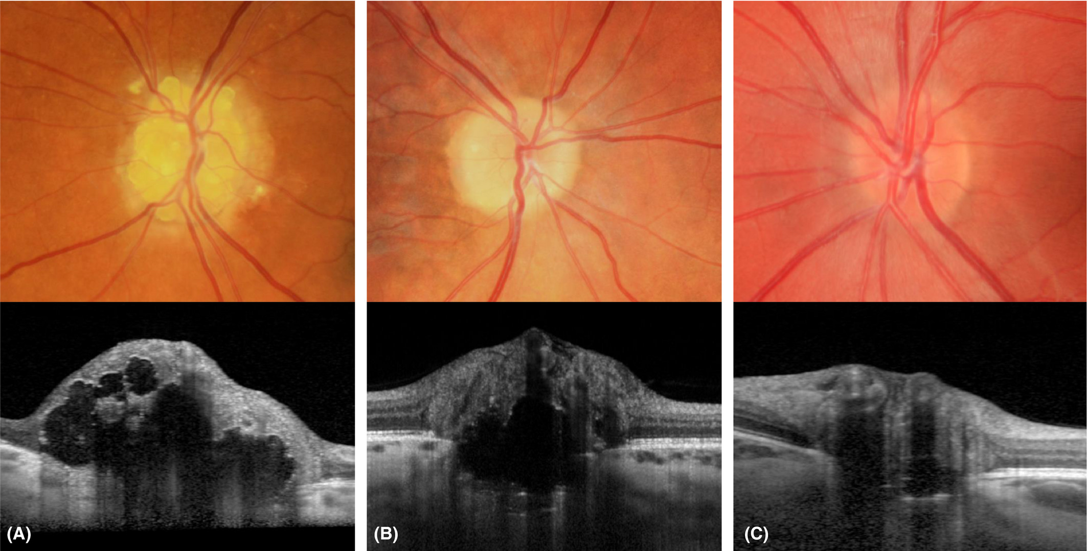
ONH description:
Case scenario case history: