Adaptive Immune Response 8/25
 Call Kai
 Learn
Learn Practice Test
 Spaced Repetition
 Match
Match Flashcards
 Knowt Play
1/106

There's no tags or description
Looks like no tags are added yet.
Name | Mastery | Learn | Test | Matching | Spaced | Call with Kai | Chat |
|---|
No analytics yet
Send a link to your students to track their progress
107 Terms
types of adaptive immunity
humoral and cell-based (cellular)
role of humoral immunity
use antibodies released by B lymphocytes to neutralize/eliminate microbes
role of cellular immunity
use T lymphocytes/effector T cells to regulate immune responses (CD4+ helper T cells) and kill target cells such as virus-infected cells (CD8+ cytotoxic T cells)
antigen
a molecule that could be recognized by lymphocytes (B cells and T cells) or antibodies
immunogens
antigens that are capable of inducing an immune response in the host organism
antigen-presenting cells (APCs)
cells that capture microbial antigens and display them for recognition by T lymphocytes
APC expression
express major histocompatibility complex (MHC molecules);
the majority of T lymphocytes recognize peptide antigens that are bound to and displayed by MHC molecules on APCs
professional APCs
dendritic cells, macrophages, B cells
dendritic cells (DCs)
the most efficient and specialized APCs
macrophages
APCs that engulf microbes and display the microbial antigens to T cells
B cells
APCs that ingest protein antigens and display to helper T cells which enhances antibody secretion
non-professional APCs
all nucleated cells can present antigens to T cells
capture of protein antigens by APC
DCs are present in tissues in contact with external environment so microbes that breach the epithelial barriers and enter the body are captured mainly by DCs
4 step process of capture and display of microbial agents by APC
1. capture antigens (phagocytosis/endocytosis)
2. activation of DC (initiate inflammation and innate immunity)
3. migration of antigen-carrying DC to lymph nodes
4. display antigen to T cells (APC/adaptive immunity)

major histocompatibility complex (MHC)
molecules that are membrane proteins expressed on APCs that display peptide antigens for recognition by T lymphocytes
MHC location
found in all mammals
human MHC proteins
human leukocyte antigens (HLA) that cause rejection after transplantation
MHC locus
contains two sets of highly polymorphic genes (class I and II) and many non-polymorphic genes (class III)
MHC restriction
T lymphocytes of a specific individual can see peptides only when these peptides are displayed by that individual's MHC molecules; one's T cells can only recognize peptide antigens presented by one's own MHC on APC
MHC gene locus
located on chromosome 6; class I and II regions are polymorphic (on each end) and class III region is non-polymorphic (in the middle)

properties of MHC genes
highly polymorphic and codominantly expressed
highly polymorphic nature of MHC genes
many different alleles (10,000 class I MHC alleles and 3000 class II MHC alleles) are present in the populations of human beings
inherited not generated de novo by somatic gene recombination
ensures different individuals are able to present and respond to different microbial peptides
codominant expression of MHC genes
both parental alleles are expressed which increases the number of different MHC molecules that can present peptides to T cells
expression of class I MHC molecules
all nucleated cells (APCs); CD8+ CTLs can kill any type of virus-infected cell
expression of class II MHC molecules
professional APCs (DCs, macrophages, and B lymphocytes); CD4+ helper T lymphocytes interact with dendritic cells, macrophages, and B lymphocytes
class I MHC molecule characteristics
composed of 8-9 amino acids
a1 and a2 domains form peptide-binding cleft and are highly variant (polymorphic)/different among different individuals
a3 domain is invariant/binds coreceptor (CD8) on CD8+ T cells (cytotoxic T cells)
class II MHC molecule characteristics
composed of 10-30 amino acids
a1 and a2 domains form peptide-binding cleft
a1 and B1 domains are highly variant
a2 and B2 domains are invariant/bind coreceptor (CD4) on CD4+ T cells (helper T cells)
6 features of peptide binding to MHC
1. broad specificity (many different peptides can bind to the same MHC molecule)
2. each MHC molecule displays one peptide at a time
3. MHC molecules bind only peptides
4. peptides are acquired during intracellular assembly (class I and II MHC molecules display peptides from different cellular compartments)
5. stable surface expression of MHC molecule requires bound peptide
6. very slow off-rate (MHC molecule displays bound peptide for long enough to be located by T cell)
processing and presentation of protein antigens - class I MHCs
cytosol proteins of any nucleated cells are processed in proteolytic structures called proteasomes and displayed by class I MHC molecules
processing and presentation of protein antigens - class II MHCs
extracellular proteins that are internalized by professional APCs are processed in endosomes and lysosomes and displayed by class II MHC molecules
steps of processing and presentation of cytosol proteins
proteolysis of cytosolic proteins, binding of peptide to class I MHC molecules, transport of peptide-MHC complexes to the cell surface
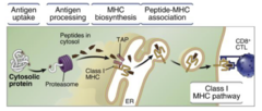
proteolysis of cytosolic proteins
cytosol proteins (intracellular microbes such as viruses) are unfolded, covalently tagged with ubiquitin, and degraded by proteasome peptides
binding of peptide to class I MHC molecules
class I MHC are synthesized in the ER and the transporter associated with antigen processing (TAP) is located in the ER membrane and binds and pumps peptides from cytoplasm to ER
transport of peptide-MHC complexes to the cell surface (MHC-1)
class I MHC bind peptide, become stabilized, and transported to cell surface then recognized by CD8+ T cells; class I MHC empty, unstable, degraded
steps of processing and presentation of extracellular proteins
internalization and digestion of antigens, binding of peptides to class II MHC molecules, transport of peptide-MHC complexes to cell surface
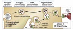
internalization and digestion of antigens
extracellular microbes (such as bacteria and fungi) or microbial proteins are internalized by APCs (phagocytosis or endocytosis), enter endosomes/phagosomes, fuse with lysosomes, and are broken down into peptides
binding of peptides to class II MHC molecules
class II MHC are synthesized in ER, carry invariant chain, and fuse with endosomal vesicle
transport of peptide-MHC complexes to cell surface (MHC-2)
class II MHC bind a peptide, stable, delivered to cell surface, recognized by CD4+ T cells; class II is empty, unstable, degraded
significance of MHC complex-associated antigen presentation
the restriction of T cell recognition to MHC-associated peptides ensures that T cells see and respond only to cell-associated antigens; express on cell membrane and require intracellular biosynthetic and assembly steps
significance of segregating the class I and class II pathways of antigen processing
immune system is able to respond to extracellular and intracellular microbes in different ways that are best able to combat these microbes
class I MHC (CD8+ T cell) antigen processing
all nucleated cells infected by intracellular microbes
class II MHC (CD4+ T cell) antigen processing
macrophages and DCs ingest bacteria and fungi (extracellular microbes) and B cells ingest microbial protein (extracellular microbes)
functions of APCs
display peptides for recognition by T cells ("first signal") and express "second signal" for T cell activation
"first signal" of APCs
APC express MHC-peptide complex and T cells express T cell receptor and CD4/CD*
"second signal" of APCs
APC express B7 and T cells express CD28
types of APCs
class I MHC (all nucleated cells) and class II MHC (DCs, macrophages, B cells)
structure of class I MHC molecules
variant a1 and a2 domains that bind antigen peptides and invariant a3 domain that binds CD8
structure of class II MHC molecules
variant a1 and B1 domains that bind antigen peptides and invariant a2 and B2 domains that bind CD4
properties of peptides binding to MHC molecules
broad specificity, one at a time, only peptides, intracellular assembly machinery, stable when bound to peptides, slow off-rate
processing and presentation of protein antigens in cytosol
class I MHC, CD8+ T cells, kill antigen-expressing target cells
processing and presentation of protein antigens extracellularly
class II MHC, CD4+ T cells, activate macrophages and DCs to kill microbes, help B cells secrete antibodies
types of antigen receptors
B cell receptors (BCR) and T cell receptors (TCR)
features of antigen receptors
BCRs and TCRs recognize chemically different structures, contain different regions, and do not deliver intracellular signals by themselves
chemical structures recognized by BCRs
shapes or conformations of macromolecules including proteins, lipids, carbohydrates, nucleic acid, and small chemical groups, and diverse microbes and toxins in their native forms
chemical structures recognized by TCRs
peptides presented by APCs = cell-associated antigens
regions on BCR/TCR
variable (V) regions that recognize antigens and vary between clones of lymphocytes
constant (C) regions that have structure integrity and effector functions conserved among all clones
BCR/TCR complexes
do not deliver intracellular signals by themselves and are instead associated with invariant molecules that deliver intracellular signals after antigen recognition
antibodies
aka immunoglobulins (Igs) that have two forms - membrane-bound BCR and secreted proteins
membrane-bound BCR antibody form
recognize antigens and initiate the responses
secreted protein antibody form
neutralize and eliminate microbes and their toxins
structure of antibodies
two identical heavy (H) chains that each contains a variable (V) region and 3-4 constant (C) regions and two identical light (L) chains that each contains a variable (V) region and a constant (C) region
Ig domain
a characteristic three-dimensional shape of two layers of a B sheet held together by a disulfide bridge
CDRs (complementarity determining region) role
antigen recognition and binding
binding of antigens by antibodies
antigen-binding site of an antibody is composed of the V regions of both the heavy and light chains and the core antibody structure contains two identical antigen binding sites; each V region contains three hypervariable regions (CDRs)
5 isotypes of antibodies
IgA, IgD, IgE, IgG, IgM
IgA function/secreted form
mucosal immunity; mainly dimer but also monomer and trimer
IgD function/secreted form
naive B cell antigen receptor; monomer
IgE function/secreted form
defense against helminthic parasites and immediate hypersensitivity
monomer
IgG function/secreted form
opsonization, complement activation, antibody-dependent cell-mediated cytotoxicity, neonatal immunity, feedback inhibition of B cells; monomer
IgM function/secreted form
naive B cell antigen receptor (monomeric form), complement activation; pentamer
features of antibodies
five classes (isotopes), differ in C regions, heavy-chain class (isotope) switching, capable of binding a wide variety of antigens (macromolecules and small chemicals), epitopes, affinity, BCR complex
epitopes of antibodies
determinants that are the part of the antigens that are recognized by antibodies; can be sequences of linear epitopes or shapes of conformational epitopes
affinity of antibodies
the strength of antibody binds to one epitope of an antigen
affinity maturation
affinity increases in secondary immune response
BCR complex of antibodies
Ig attach to Iga and IgB that transmit signals to the interior of B cells leading to B cell activation
T cell receptors for antigens structure
membrane-bound heterodimeric protein that contains an a chain (one V and one C region), a B chain (one V and one C region), and CDRs (three hypervariable complementarity-determining regions)

antigen recognition by TCR
the V region of the a and B chains of the TCR participate in specific recognition of antigen peptides displayed by MHC molecules; each TCR recognizes as few as one to three residues of the MHC-associated peptides
TCR complex
TCR, CD3, and zeta proteins that transmit signals; TCR does not transmit signals to T cells
production of diverse antigen receptors
antigen receptor genes are inherited, a collection of diverse BCR/TCR is called lymphocyte repertoires/immune repertoires, diverse antigen receptors are generated through random recombination or junctional diversity
comparison of TLR4, MHC, and BCR receptors
TLR4 is inherited and identical across the population, MHC is inherited as one single expression but there are different types across the population, BCR is inherited and expressed differently in the cell and throughout the population

inherited antigen receptor genes
Ig H/L-chain loci and TCR a/B-chain loci contain 30-45 variable (V) region gene segments, one or a few constant (C) region gene segments, diversity (D) segments (only Ig H-chain and TCR B-chain loci), and joining (J) gene segments
mechanism of V(D)L recombination
random selection of gene segments (random selection or somatic recombination) during B cell development

random/somatic recombination
generation of diverse antigen receptors of gene segments that code for the V regions of BCR/TCR
combinational diversity
different combinations of V, D, and J gene segments
mechanisms of producing junctional diversity
exonucleases may remove nucleotides from V, D, and J; terminal deoxynucleotidyl transferase (TdT) catalyzes the random addition of nucleotides to the junctions between V/D segments and D/J segments; overhanging DNA sequences may be generated
maturation and selection of B cells
stem cell > pro-B > pre-B (pre-BCR and u) > immature B (IgM) > mature B (IgM and IgD)
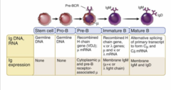
early steps in B cell maturation
recombination of heavy-chain loci occurs in pre-B cells and pre-B cells express u heavy-chain, surrogate light chains, and Iga and IgB, forming the pre-B cell receptor (pre-BCR) complex
completion of B cell maturation
pre-BCR complex triggers recombination of light chain loci leading to coexpression of IgD and IgM
selection of mature B cells
positive selection (survival) or negative selection (apoptosis)
positive selection of mature B cells
survive - express functional BCR AND do not recognize self-antigens
negative selection of mature B cells
apoptosis - fail to express functional BCR OR recognize self-antigens
outcomes of maturation and selection of B cells
generate a collection of mature B cells that are able to recognize any microbial antigens
maturation and selection of T cells
stem cell > double negative (CD4-CD8-) pro-T cell > pre-T cell > double positive (CD4+CD8+) immature T cell > mature CD4+ or CD8+ T cell (or apoptosis)

early steps in T cell maturation
thymus, pro-T cells (CD4-CD8-) undergo TCR gene recombination, pre-T cells with pre-TCR complex triggers TCR a gene recombination, CD4+CD8+ immature T cells express complete aB TCR
selection of mature T cells
positive selection (survive and express CD4+CD8+ and TCR) or negative selection (apoptosis)
positive selection of mature T cells
survive - weakly recognize self MHC with self peptides resulting in expression of either CD4+ or CD8+ (single-positive T cells)
negative selection of mature T cells
apoptosis - do not recognize self MHC with self-peptides (such as fail to express functional CD4+CD8+ or TCR) or strongly recognize self MHC with self-peptides
outcomes of maturation and selection of T cells
generate a collection of mature T cells that are able to recognize any foreign antigen peptides
recognized by antigen receptors
BCR (diverse microbes and toxins in their native form) and TCR (peptides presented by APCs)
BCR structure
H chains, L chains, V region (CDRs), C region