Embryology 4 | Organogenesis: Weeks 4-8
1/28
Earn XP
Description and Tags
https://youtu.be/LtMztjJwH5E?t=922 / https://youtu.be/VBuCiIQQNss
Name | Mastery | Learn | Test | Matching | Spaced |
|---|
No study sessions yet.
29 Terms
What causes head and tail folding in the embryo?
Head and tail folding in the embryo happens because the brain and spinal cord grow quickly. The front (head) end folds because the brain is getting bigger. The tail end folds because the spinal cord is getting longer. These folds help form the forebrain and hindgut.
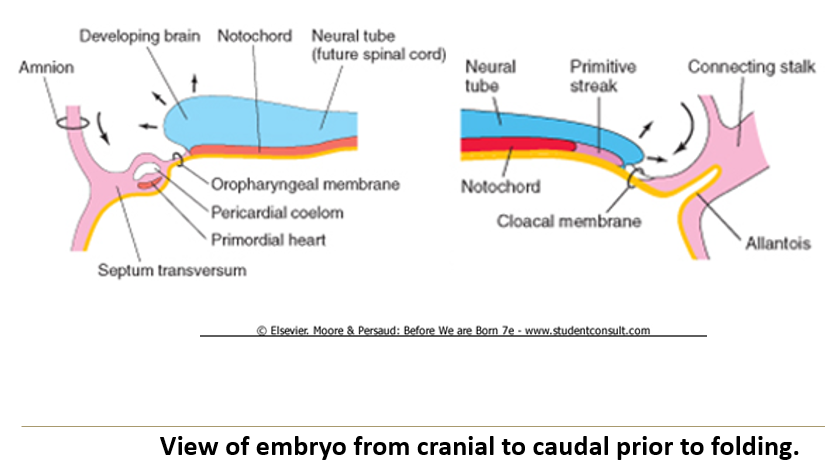
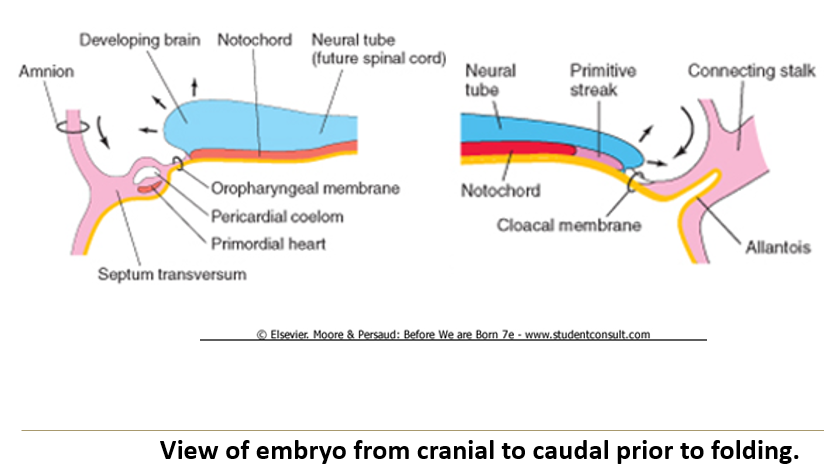
What is the anatomical order visible in a 3-week embryo from caudal to cranial?
Connecting stalk (with allantois)
Cloacal membrane
Caudal neural tube
Mid neural tube
Developing brain
Oropharyngeal membrane
Cardiogenic region
Septum transversum.
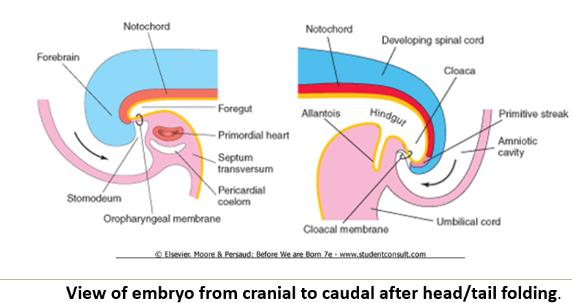
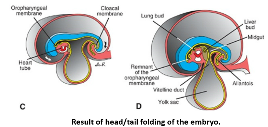
What are the results of cranial folding?
The oropharyngeal membrane and cardiogenic region move to a ventral position, with the oropharyngeal membrane now cranial to the heart.
The foregut forms by internal incorporation of part of the yolk sac.
The stomodeum forms, marking the primordium of the mouth.
What causes caudal folding?
Caudal folding happens because the spinal cord grows longer. As it stretches out, it pulls the tail end of the embryo inward and curves it under the body. This helps shape the lower part of the body, like the abdomen and pelvis.
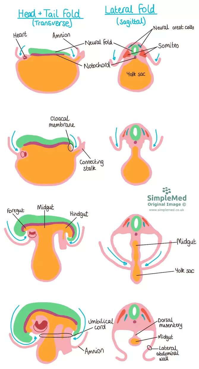
What are the results of caudal folding?
The hindgut forms by internal incorporation of the yolk sac, and its terminal portion dilates into the cloaca.
The cloacal membrane and connecting stalk are repositioned to the ventral side of the embryo.
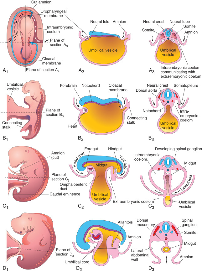
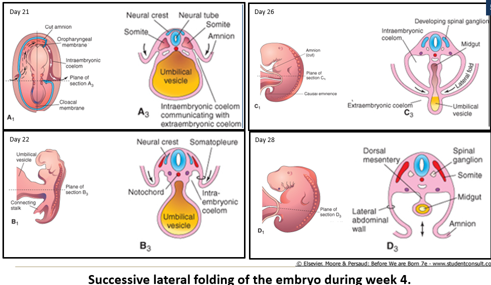
What causes lateral folds in the embryo?
The rapid growth of the spinal cord and somites, causing the body wall to fold toward the median plane.

What are the results of lateral folding?
A portion of the yolk sac is incorporated as the midgut, suspended by the dorsal mesentery.
The umbilical cord begins forming from the connecting stalk, covered by amniotic epithelium, and incorporating the yolk stalk (later called the vitelline duct).
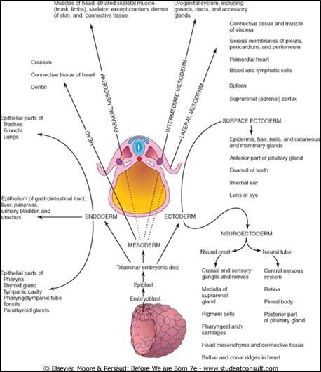
What does specialization of the ectoderm form?
The neural tube and neural crest, which form the CNS, PNS, and outer coverings.
The remaining surface ectoderm gives rise to the epidermis, derivatives of the skin, and melanocytes.
What are some ectodermal derivatives?
Cornea and lens of the eye.
Epithelium of the oral and nasal cavities, paranasal sinuses, and anal canal.
Epithelium of the pineal and pituitary glands.
Some cranial bones and pharyngeal (branchial) cartilages from the neural crest.

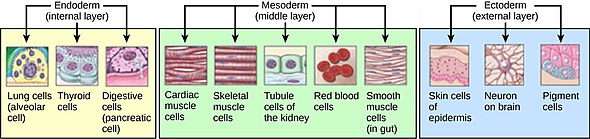
What is the primary role of the endoderm in development?
The endoderm forms the lining of the primitive gut, which becomes the lining of the GI tract.

How do organs and glands form from the endoderm?
By outpocketing from the primitive gut, leading to endothelial linings or secretory cells.
What are some foregut derivatives from the endoderm?
Thyroid and parathyroid glands
Thymus
Respiratory tract
Auditory tube and cavities of middle and inner ear
Liver
Pancreas
What are some hindgut derivatives?
The epithelial portions of the reproductive ducts and glands, and the urethra and bladder.
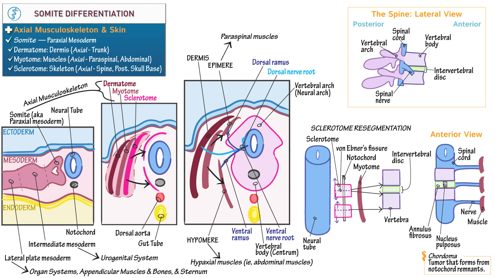
What are the three main derivatives of the somites (paraxial mesoderm)?
Sclerotome: forms vertebrae and ribs
Dermatome: forms the dermis of the dorsal skin
Myotome: forms skeletal muscles
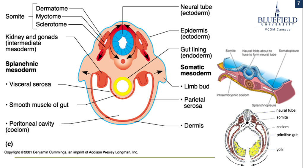
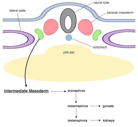
What does the intermediate mesoderm form?
The gonads, kidney, adrenal cortex, and ureter

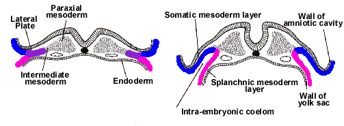
What are the divisions of lateral mesoderm and what do they form?
Somatic mesoderm forms the ventral skin dermis, serosa of the ventral body cavity, and contributes to limb buds (bones, ligaments, dermis)
Splanchnic mesoderm forms smooth muscle of the gut, visceral serosa, heart, blood and lymphatic vessels, and bone marrow/lymphoid tissues
How is gestational age clinically determined?
By time since the last normal menstrual period or conception. Normal gestation is 40 weeks last normal menstrual period or 38 weeks post-conception.
Why might patient history not be reliable for determining gestational age?
Irregular cycles after oral contraceptives, pregnancy, or breastfeeding
Oligomenorrhea (irregular menstrual cycles)
Confusion between implantation spotting and menstruation
Why is knowing gestational age clinically important?
It helps guide clinical decisions, such as timing invasive procedures or assessing risks from teratogen exposure.
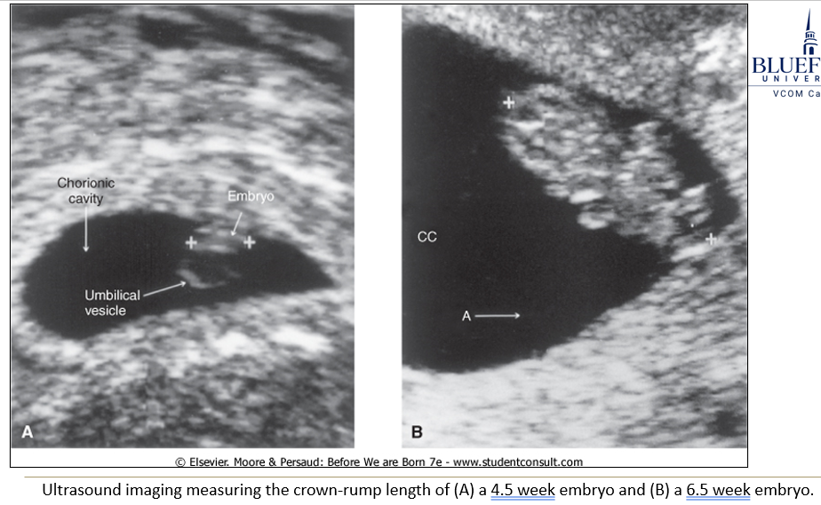
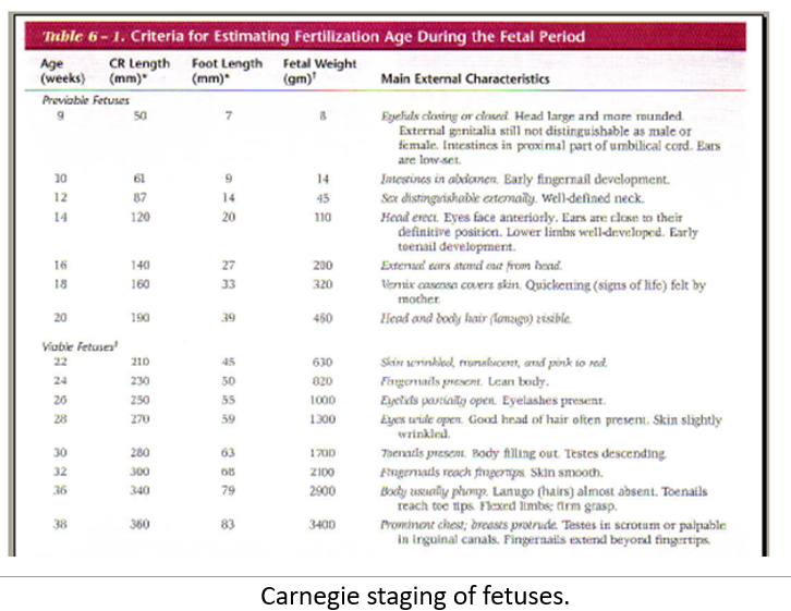
How is gestational age clinically assessed via ultrasound?
The crown-rump length is the most common measurement, with others like foot length used later in pregnancy. Measurements correlate to gestational age.
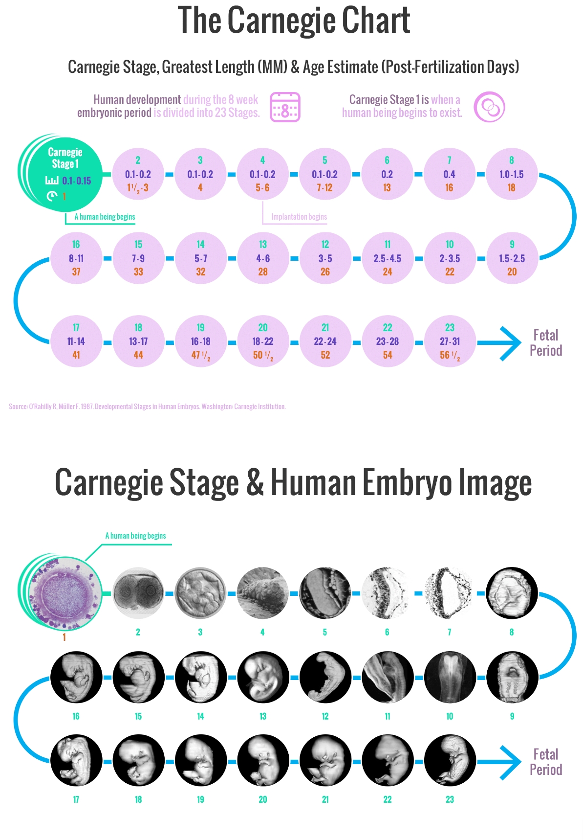
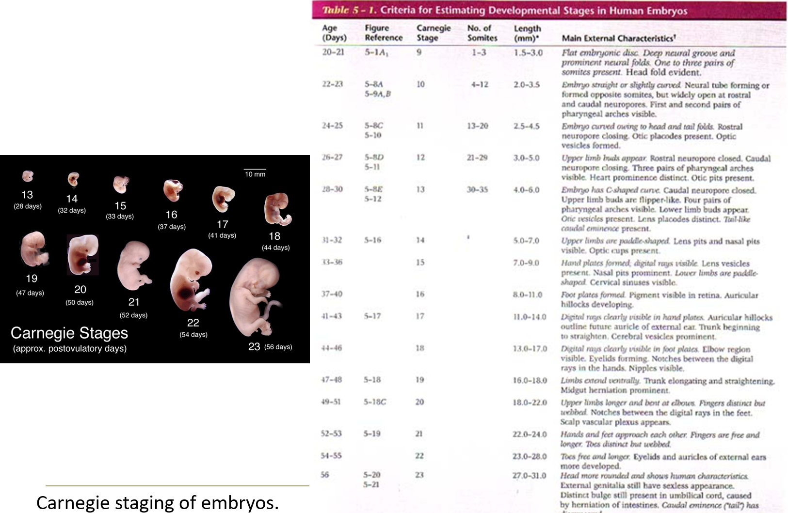
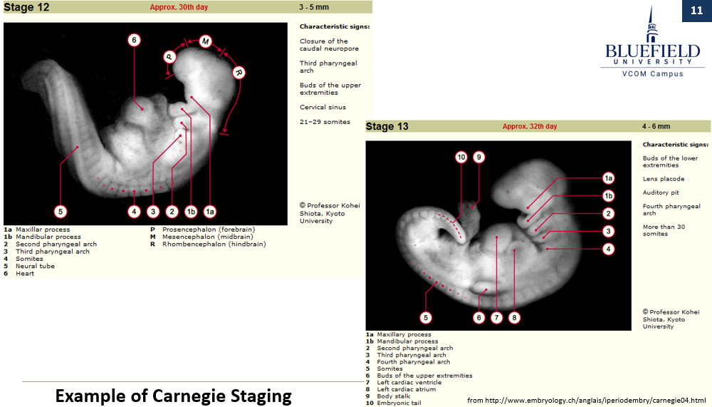
What system is used to assess developmental stage in miscarried embryos?
The Carnegie system, which uses standardized morphological features to stage development and assess causes like growth retardation.

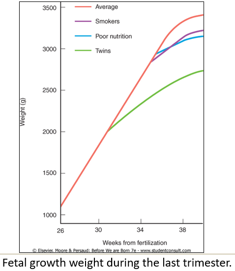
Does neonatal length and weight always correlate with gestational age?
No, there is considerable variation due to genetic and environmental factors.
Is there a specific weight or gestational age that guarantees fetal viability?
No, there is no magic threshold; viability depends largely on lung and nervous system development.

What is the role of insulin-like growth factor I?
IGF-1 helps the body grow and develop. It encourages cells to divide (mitogenic) and build new tissues (anabolic), especially during childhood and puberty.
Where is IGF-1 produced and how is it linked to growth?
Produced by the fetus, and serum IGF-1 levels correlate with fetal growth.
What can mutations in IGF-1 cause?
Mutations in IGF-1 can result in intrauterine growth retardation
When does growth hormone become more important than IGF-1?
GH becomes the primary growth hormone after birth.
What is intrauterine growth retardation?
Refers to a fetus that is at or below the 10th percentile of expected birth weight for its gestational age.
What are some causative factors for intrauterine growth retardation?
Teratogens (chemicals causing abnormal development)
Congenital infections
Poor maternal health (e.g., hypertension, renal/cardiac disease)
Maternal nutritional status
Maternal drug use (cigarettes, alcohol, social drugs)
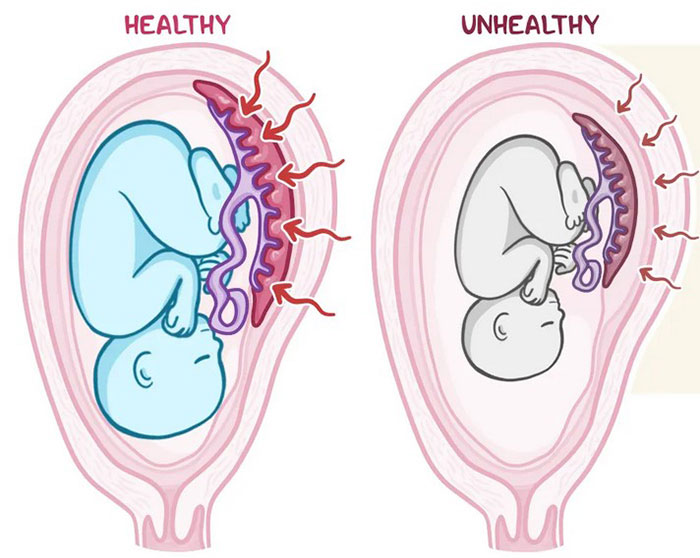
Placental insufficiency
Multiple births