A&P II: Exam 2 | Lymphatics, Respiratory, Digestion, and Metabolism
1/118
There's no tags or description
Looks like no tags are added yet.
Name | Mastery | Learn | Test | Matching | Spaced | Call with Kai | Chat |
|---|
No analytics yet
Send a link to your students to track their progress
119 Terms
nonspecific (innate) immunity
no specific recognition of invaders, no memory component. This is the first and second lines of defense
specific (adaptive) immunity
specific recognition of invaders with a memory component
lymphatic vessels
begin as lymphatic capillaries and they are closed at one end. They then unite to form large lymphatic vessels. These vessels then pass through the lymph nodes. The lymph nodes are located in areas with encapsulated organs with masses and B and T cells. These function as lymph filters.
myeloid tissue
bone marrow: site for the production of blood cells in the medullary cavities. is also known as red bone marrow
how lymphatics differ and compare to the circulatory system
Lymph vessels travel one-way, back to the heart, venous route; has similar structure to veins including tunics and valves (?); structural difference includes the lymph vessel's need to diffuse bigger molecules; lymph transport slower and has lower pressure and speed than veins
Blood and nutrients travel through blood vessels (arteries, veins, and capillaries). The lymphatic system is responsible for draining excess fluid from the tissues and returning it to the circulatory system.
distribution of lymphatic vessels
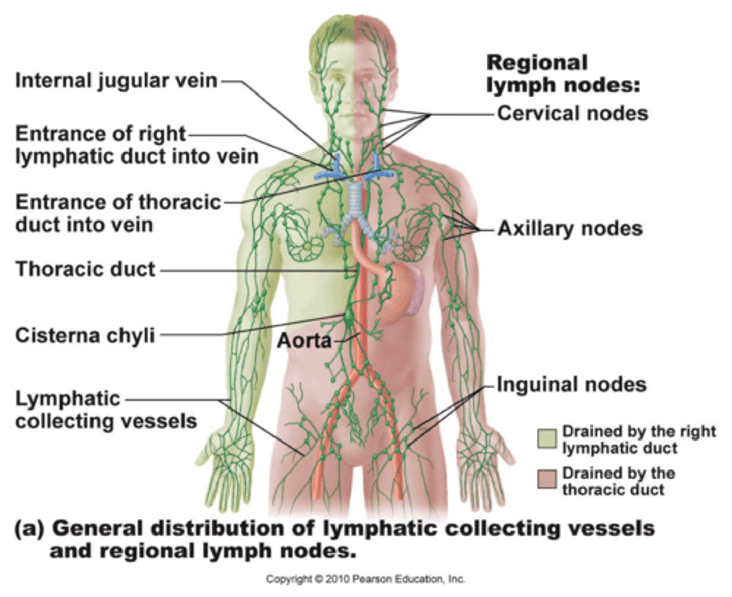
lymphatic capillaries
have a larger diameter than blood capillaries. They have a unique one-way structure in order to permit interstitial fluid to flow in but not out.
how veins and vessels are similar
These large lymphatic vessels resemble veins in structure, but the vessels have thinner walls and more valves than veins.
LEUKOCYTES
F. LEUKOCYTES [WBCs] Myeloid derived
1. Monocyte-Macrophages - fixed and free big eaters [Reticuloendothelial System]
Fixed: Langerhan Cells, Kupffer Cells, Microglea, Osteoclasts etc. Present
antigens; Clean up after microphage battles
2. Microphages - kamikaze little eaters
a. Neutrophils - invading bacteria & cellular debris
b. Eosinophils - target foreign cpds or pathogens coated with antibodies
3. Basophils and Mast Cells - Inflammatory response Histamine, Heparin,
Prostaglandins
primary lymphatic organs
sites where stem cells divide and become immunocompetent (the ability of the body to produce a normal immune response following exposure to an antigen). These are formed in the red bone marrow and thymus.
secondary lymphatic organs
sites where most immune response occurs. These are the lymph nodes, spleen, and lymphatic nodules.
Major Histocompatibility Complex
The function of MHC molecules is to bind peptide fragments derived from pathogens and display them on the cell surface for recognition by the appropriate T cells.
Major histocompatibility complex (MHC), group of genes that code for proteins found on the surfaces of cells that help the immune system recognize foreign substances. MHC proteins are found in all higher vertebrates. In human beings the complex is also called the human leukocyte antigen (HLA) system.
MHC Class I
- found on all nucleated cells
- built into all body cells except RBCs
MHC Class II
- found on antigen presenting cells and lymphocytes
- only on antigen presenting cells
immune system response
an antigen triggers an immune response -> activates t cells and b cells -> t cells attack the antigen and stimulate the b cells -> activated b cells mature and produce antibodies -> antibodies attack the antigen
origins of t-cells , how they develop, competent or not
t-cells migrate to the thymus from red bone marrow. dendritic cells and specialized epithelial cells help educated and mature t-cells. after the negative/positive selection process, immunocompetent t-cells go to the inner medulla.
interleukin
- a type of cytokine
- interleukin 1 is released by active macrophages
>> triggers the release of ACTH resulting in glucocorticoid release, moderates the immune response, lowers resistance to disease
- interleukin 2 is needed for virtually all immune responses
b-cells, how they develop, competent or not
Produced in the bone marrow, B cells migrate to the spleen and other secondary lymphoid tissues where they mature and differentiate into immunocompetent B cells. Part of the adaptive immune system, B cells are responsible for generating antibodies to specific antigens, which they bind via B cell receptors (BCR).
properties of immunity
1) specificity: activated by and responds to a specific antigen
2) versatility: is ready to confront any antigen at any time
3) memory: "remember" any antigen it has encountered
4) tolerance: responds to foreign substances but ignores normal tissues
exogenous antigens
present in fluid outside body cells
Antigen-presenting cells (APCs) include dendritic cells, macrophages and B cells
Ingest antigen, process, place next to MHC-II molecule in plasma membrane, and present to T cells
endogenous antigens
antigens inside body cells
Infected cell displays antigen next to MHC-I
cell mediated (cellular)
- lymphocytes act against target cell
>> DIRECTLY - by killing infected cells
>> INDIRECTLY - by releasing chemicals that enhance inflammatory response
- Cellular immunity has cellular targets
T LYMPHOCYTES
cytotoxic t-cells directly attack invading antigens. particularly effective against intracellular pathogens, some cancer cells and foreign tissue transplants.
antibody mediated (humoral)
- antibodies, produced by lymphocytes, circulating freely in body fluids
- bind temporarily to target cell
>> temporarily inactive
>> mark for destruction by phagocytes or complement
B LYMPHOCYTES
b-cells transform into plasma cells making antibodies (Abs) or immunoglobulins. work against extracellular pathogens in fluids outside cells.
cell mediated and antibody mediated both...
- have helper t-cells aid in both processes
- both types work together to defend the body
innate immunity
- genetically determined
- present at birth
acquired immunity
- not present at birth
- achieved by exposure to antigen
active immunity
natural active example: exposure to chicken pox
artificial active example: immunization against polio
passive immunity
natural passive example:
maternal antibodies crossing the placenta
artificial passive example:
vaccination against the effects of a snake bite
suppressor t-cells
(Ts) inhibit the activation of t cells and b cells
helper t-cells
(Th) activate other t cells and b cells
memory t-cells
have previously encountered and responded to their related antigen
b-cell differentiation
A single B cell or a clone of cells with shared specificity, upon encountering its specific antigen, divides to produce many B cells. Most of such B cells differentiate into plasma cells that secrete antibodies into blood that bind the same epitope that elicited proliferation in the first place.
???????
delayed hypersinsitivity
Delayed hypersensitivity is a major mechanism of defense against various intracellular pathogens, including mycobacteria, fungi, and certain parasites, and it occurs in transplant rejection and tumor immunity.
it is not antibody mediated but rather is a type of cell-mediated response. CD4+ helper T cells recognize antigen in a complex with Class 2 major histocompatibility complex.
autoimmune disorders
immune response mistakenly targets normal cells
immunodeficiency diseases
immune system does not develop properly or is blocked
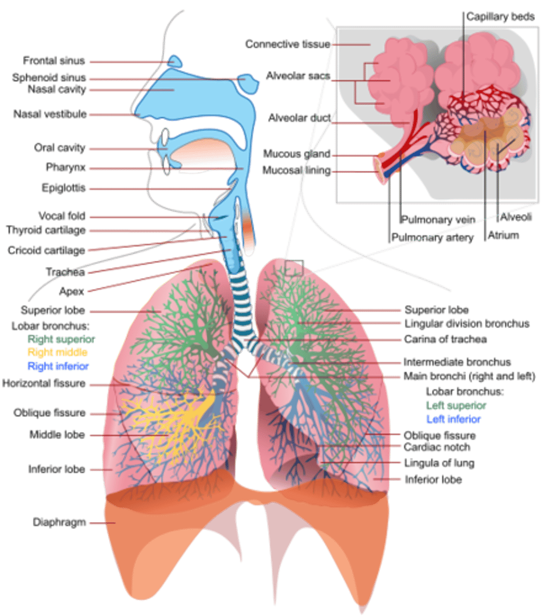
upper respiratory vs lower respiratory
the upper respiratory consists of the: nose, pharynx, and associated structures
the lower respiratory system consists of the: larynx, trachea, bronchi, and lungs
anatomy of the respiratory system
conducting zone vs respiratory zone
the conducting zone conducts air to the lungs - while the respiratory zone is the main site of gas exchange
respiratory tree order
1. trachea
2. primary bronchi
3. secondary bronchi
4. tertiary bronchi
5. bronchioles
6. terminal bronchioles
differences between the right and left lung
left lung:
>two lobes; superior & inferior.
>It is longer and lighter than the right lung
> has a cardiac notch at its mediastinal/anterior border.
right lung:
>is divided into three lobes; superior, middle, and inferior.
chronic pulmonary diseases
bronchitis and emphysema
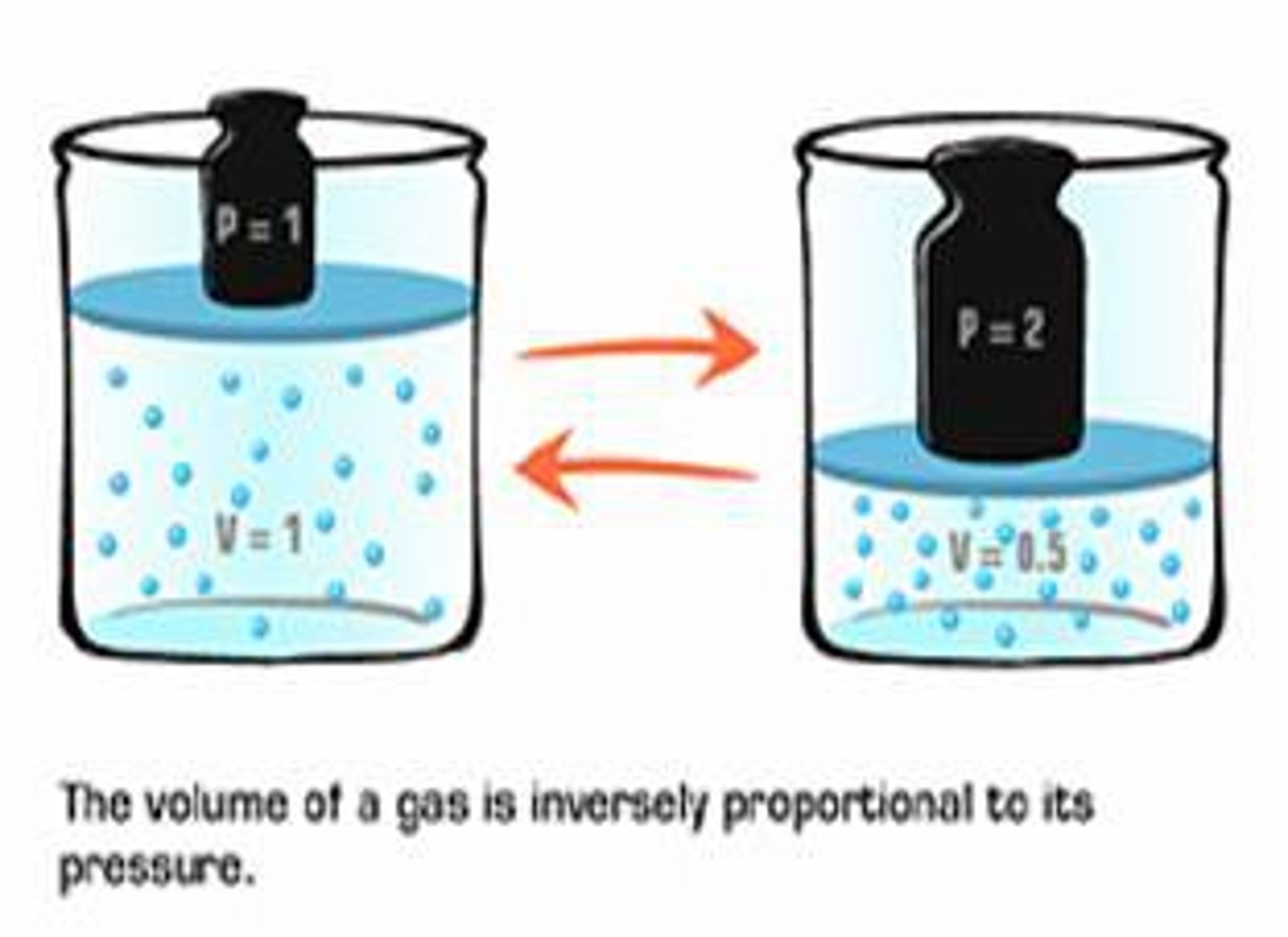
Boyle's Law
pressure of a gas in a closed container is inversely proportional to the volume of the container
example: if the pressure is 2 atm, then the volume is 0.5 liters
Henry's Law
The more pressure that is put on a solution, the more gas that can be dissolved in a liquid. Lessening the pressure causes that gas to come out of the liquid.
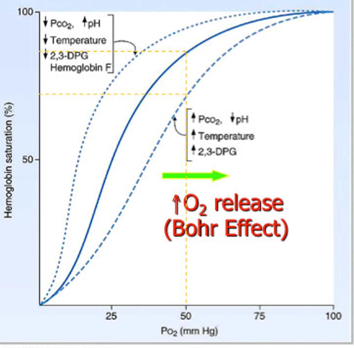
Bohr Effect
As acidity increases (pH decreases), affinity of Hb for O2 decreases
Decreased Attraction of Hb to O2, Right shift caused by an increase in PCO2.
Hb loads more O2 in lungs where pH is high and unloads more in tissues where pH is low
quiet vs active respiration
Inhalation (always active) and exhalation (passive or active); 2 types of inhalation = quiet and forced; 2 types of exhalation = quiet and forced
how respiratory chemical regulation works
Homeostasis is maintained by the respiratory system in two ways: gas exchange and regulation of blood pH. Gas exchange is performed by the lungs by eliminating carbon dioxide, a waste product given off by cellular respiration.
Oxygen-Hemoglobin Dissociation Curve
Other factors affecting affinity of hemoglobin for oxygen
Each makes sense if you keep in mind that metabolically active tissues need O2, and produce acids, CO2, and heat as wastes
Acidity
PCO2
Temperature
This curve is an important tool for understanding how our blood carries and releases oxygen.
"hemoglobin affinity for oxygen"; that is, how readily hemoglobin acquires and releases oxygen molecules into the fluid that surrounds it.
chloride shift
HCO3- accumulates inside RBCs as they pick up carbon dioxide
Some diffuses out into plasma
To balance the loss of negative ions, chloride (Cl-) moves into RBCs from plasma
Reverse happens in lungs - Cl- moves out as moves back into RBCs
anatomy of the digestive system
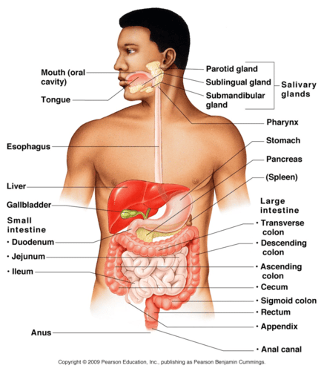
gastrointestinal (GI) tract organs
mouth, most of pharynx, esophagus, stomach, small intestine, and large intestine
accessory digestive organs
teeth, tongue, salivary glands, liver, gallbladder, and pancreas
digestive processes
ingestion, secretion, mixing + propulsion, digestion, absorption, and defecation
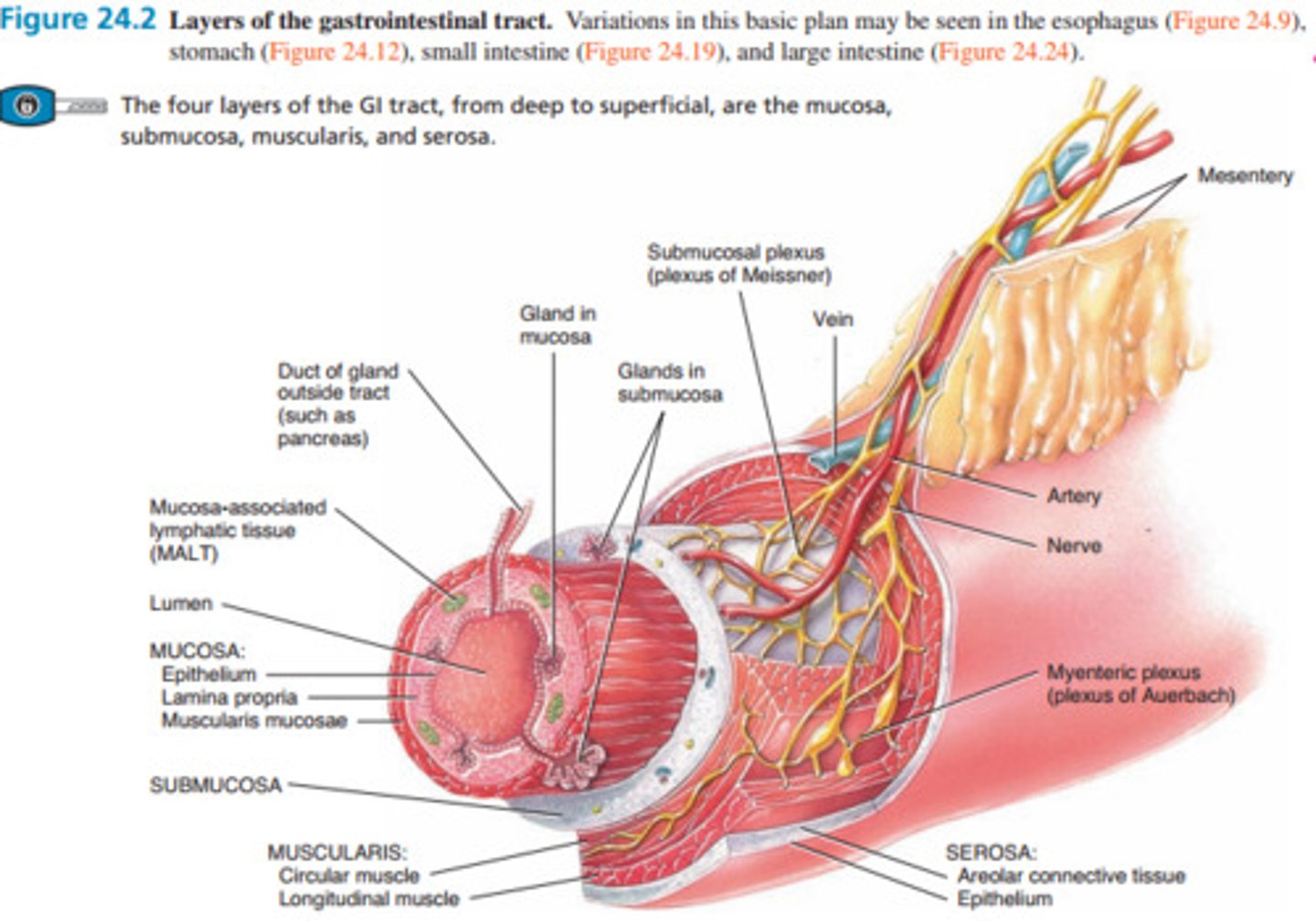
layers of the GI tract
from in to out
mucosa, submucosa, muscularis, serosa
mucosa
inner lining of the GI tract, MALT, contains muscularis mucosae which is a thin layer of smooth muscle making folds to increase surface area
submucosa
connective tissue that binds the mucosa to the muscularis, contains many blood and lymphatic vessels, and the submucosal plexus
muscularis
voluntary skeletal muscle found in mouth, pharynx, upper esophagus, and anal sphincter
serosa
outermost covering of organs in the abdominal cavity, also called visceral peritoneum
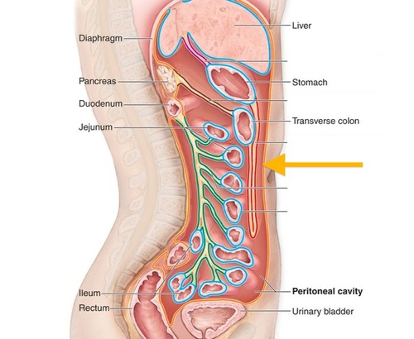
peritoneum
- largest serous membrane of the body
- parietal and visceral peritoneum
- major peritoneal folds: greater omentum, falciform ligament, lesser omentum, mesentery, and mesocolon
--- weave between the viscera and bind organs together
peritoneum
layers of the gastrointestinal tract
oral/buccal cavity
- formed by cheeks, hard and soft palates, and the tongue
- a space that extends from gums and teeth to fauces
- in this cavity, the salivary glands will release saliva
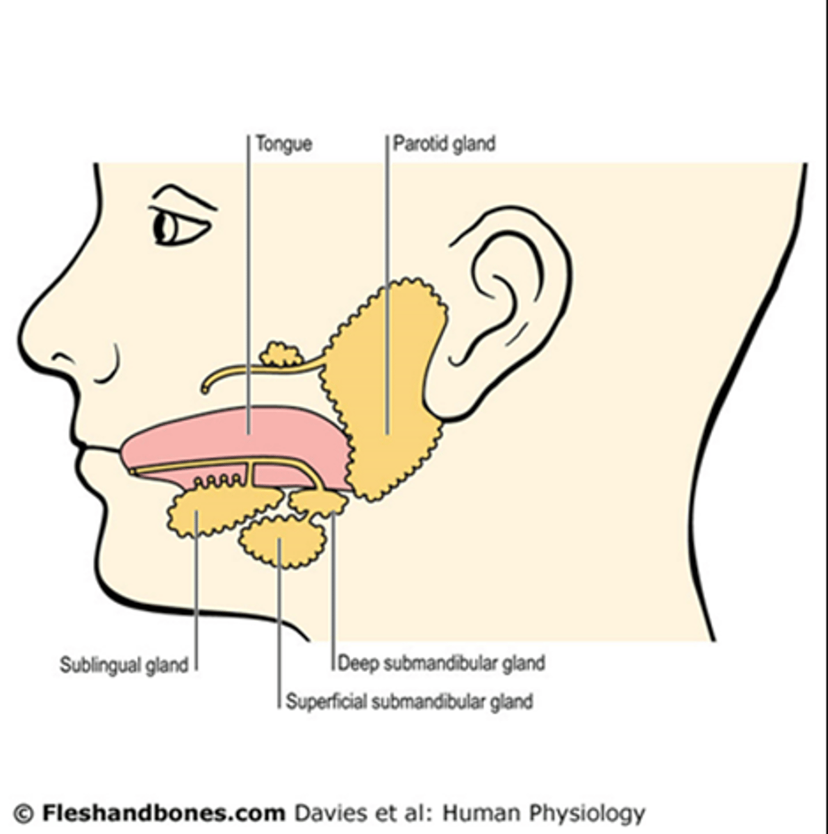
salivary glands
- release saliva
- when food enters the mouth, secretion increases to: lubricate, dissolve, and begin chemical digestion
- parotid, submandibular, and sublingual
what are the three pairs of major salivary glands that secrete most of the saliva?
parotid, submandibular, and sublingual
tongue
accessory digestive organ, maneuvers food for chewing, and lingual glands secrete salivary lipase
teeth/dentes
accessory digestive organ, emulsify food with mechanical digestion
adult dentition
4 incisors, 2 canines (cuspids), 4 premolars (bicuspids), and 4 molars
child dentition
2 incisors, 1 canine (cuspid), 2 molars. have no premolars, but 'first' molars.
saliva
- mostly water 99.5%, the rest 0.5% are ions, dissolved gases, urea, uric acid, muscus, etc
- not all salivary glands produce the same saliva
- salivation is controlled by the ANS
>> parasympathetic: promotes secretion of saliva
>> sympathetic: decreases salivation
mechanical digestion in the mouth
chewing/mastication, food manipulated by tongue->ground by teeth->mixed with saliva, and forms a bolus
chemical digestion in the mouth
the release of salivary amylase (acts on starches) and lingual lipase (acts on triglycerides)
pharynx
passes from nasal cavity into esophagus
> the nasopharynx only functions in respiration. the oropharynx and laryngopharynx participate in digestive and respiratory functions.
-- consists of the: nasopharynx, oropharynx, and laryngopharynx
esophagus
function: secretes mucous, transports food. does not produce any enzymes or participate in absorption.
- made of mucosa to protect against wear and tear. contains two sphincters to regulate movement into the esophagus (UES) and movement into the stomach (LES). contains adventitia.
stomach
function: serves as a mixing chamber and holding reservoir. contains the gastric pits. rugae moves food along. peristaltic movements with rugae.
cells in the gastric pits
Goblet, parietal, chief, and entero-endocrine cells (G-Cells)
gastric pits
These structures are lined with cells that produce chemicals that help the stomach digest food.
what do the gastric pits produce
contain many exocrine cells that secrete digestive enzymes and hydrochloric acid into the lumen, or hollow region, of the stomach. Mucous cells found throughout the stomach lining and gastric pits secrete mucus to protect the stomach from its own digestive secretions.
mechanical digestion
- mixing waves
- gentle, ripping peristaltic movements
- creates chyme
chemical digestion
salivary amylase: digestion continues until inactivated by acidic gastric juice
lingual lipase: acidic gastric juice activates lingual lipase. digest triglycerides into fatty acids and diglycerides.
pepsin
- secreted by cheif cells
- secreted as inactive pepsinogen
- digests proteins
what are the phases of gastric secretion?
cephalic, gastric, and intestinal
the gastric phase
function: enhance secretion started in cephalic stage, acidify chyme, initiate digestion of proteins by PEPSIN
duration: long (3-4) hours
actions: increased acid and pepsinogen production; increased motility and initiation of mixing waves
the intestinal phase
function: control rate of chyme entry into duodenum
duration: long (hours)
actions: feedback inhibition of gastric acid and pepsinogen production; reduction in gastric motility
the cephalic phase
function: prepares stomach for the arrival of food.
duration: short
actions: stimulation of mucus, enzyme, and acid production which leads to an increased volume of gastric juice ---> stimulation of gastrin released by G cells
nutrient pool
fuels the cell's metabolic activities both catabolic and anabolic
pancreas
gland that secretes pancreatic juice into the duodenum, where it mixes with bile to digest food
pancreatic juice
- mostly water
- sodium bicarbonate: buffers acidic stomach chyme
- ENZYMES: pancreatic amylase, proteolytic enzymes-- trypsin, chymotrypsin, carboxypeptidase, elastase--- pancreatic lipase, ribonuclease and deoxyribonuclease
exogenous and endogenous cells in the pancreas
exogenous: in pancreatic acini
endogenous: in pancreatic islet
liver
- is the heaviest gland of the body
- composed of: hepatocytes, bile canaliculi, and hepatic sinusoids
liver products
Processing of hemoglobin for use of its iron content (the liver stores iron) Conversion of poisonous ammonia to urea (urea is an end product of protein metabolism and is excreted in the urine) Clearing the blood of drugs and other poisonous substances. Regulating blood clotting.
gallbladder
function: to store and concentrate bile produced by the liver until it is needed in the small intestine
- absorbs water and ions to concentrate bile up to ten-fold
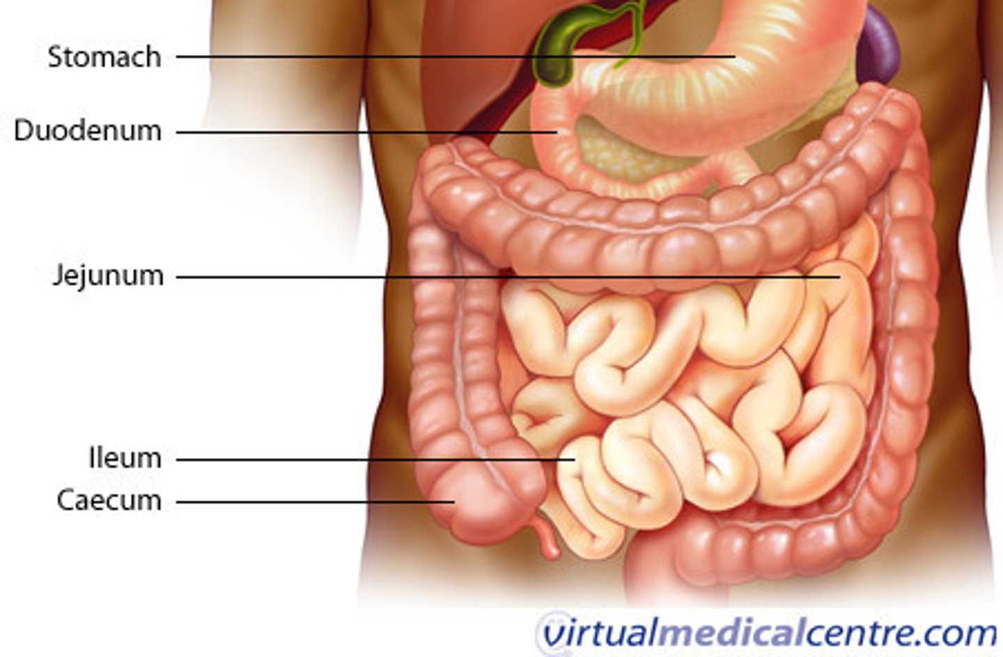
divisions of the small intestine
regions: duodenum, jejunum, and ileum
histology: Absorptive cells (digest and absorb), goblet cells (mucus), intestinal glands (intestinal juice), Paneth cells (lysozyme), and enteroendocrine cells
Abundance of MALT
small intestine structure for larger surface area
> circular folds: permanent ridges of mucosa and submucosa, cause chyme to spiral
> villi: fingerlike projections of mucosa
> microvilli: fingerlike projections of apical membrane of absorptive cells -- brush border with brush border enzymes
small intestine anatomy
intestinal juice
provides itself as a liquid medium for aiding in absorption
brush border enzymes
enzymatic digestion occurring on the surface rather than just in lumen -- breaks down enzymes into glucoses
alpha enzymes include: sucrase, lactase, and maltase
large intestine anatomy
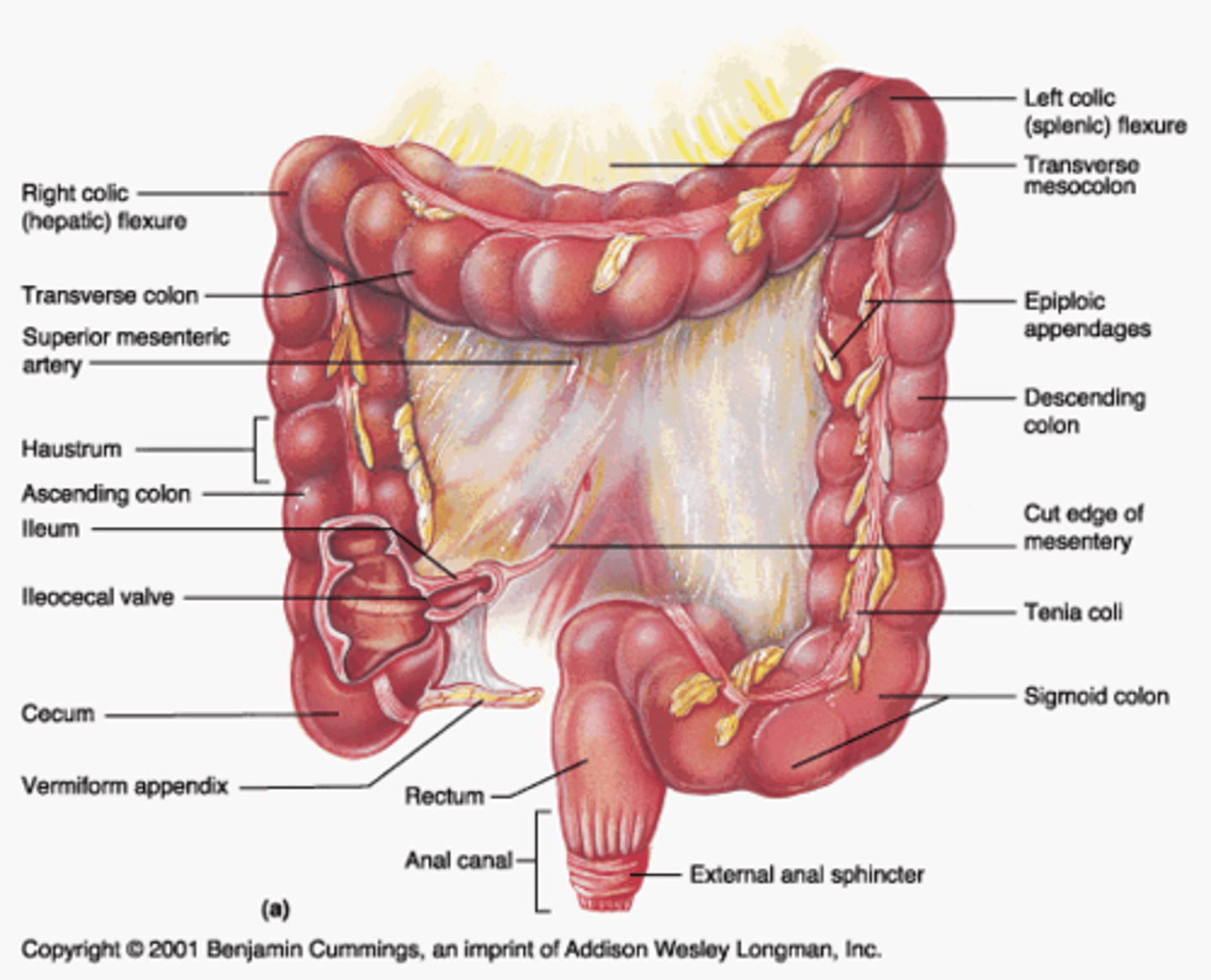
large intestine
to complete absorption, produce certain vitamins, and form/expel feces
digestion in the large intestine
mechanical: haustral churning, peristalsis, mass peristalsis (drives contents) of colon toward rectum
chemical: final stage of digestion through bacterial action, fermentation of carbohydrates, produce some B/K vitamins, mucus but no enzymes
remaining water absorbed along with ions and some vitamins
zymogen cascade
trypsinogen ---(enteropeptidase)---> tripsin --->
procarboxypeptidase ---> carboxypeptidase --->
chymotrypsinogen --> chymotrypsin
zymogen
inactive form of an enzyme