Lab exam 1
 Call Kai
 Learn
Learn Practice Test
 Spaced Repetition
 Match
Match Flashcards
 Knowt Play
1/67

There's no tags or description
Looks like no tags are added yet.
Name | Mastery | Learn | Test | Matching | Spaced | Call with Kai | Chat |
|---|
No analytics yet
Send a link to your students to track their progress
68 Terms

Frontal
A plane that divides the body into anterior (front) and posterior (back) portions.
pleural cavity
each surrounds a lung, the serous membrane of each plural cavity is the pleura
Mediastinum
anatomical portion of the thoracic cavity between the medial walls of the pleural cavities, that contains the heart, trachea, esophagus, and major blood vessels.
Abdominal cavity
contains stomach, spleen, liver, gallbladder, small intestine, and most of the large intestine. Serious membrane of the abdominal cavity is the peritoneum.

Right and left hypochondriac region

Epigastric region

Right and left inguinal (iliac)
Parietal layer
Lines the walls of the cavities
Visceral layer
Covers the viscera within the cavities
Serous membrane
Double layered fluid filled sack that surround organs like the heart and the lungs
Serous fluid
Between the two layers reduces friction and allows the viscera to slide somewhat during movements
Visceral pleura
Clings to the surface of the lungs
Parietal pleura
The outer layer serous pleural membrane
Peritoneum cavity
Serous membrane of the abdominal cavity
Visceral peritoneum
Covers the abdominal viscera
Parietal peritoneum
Lines the abdominal wall

Trachea
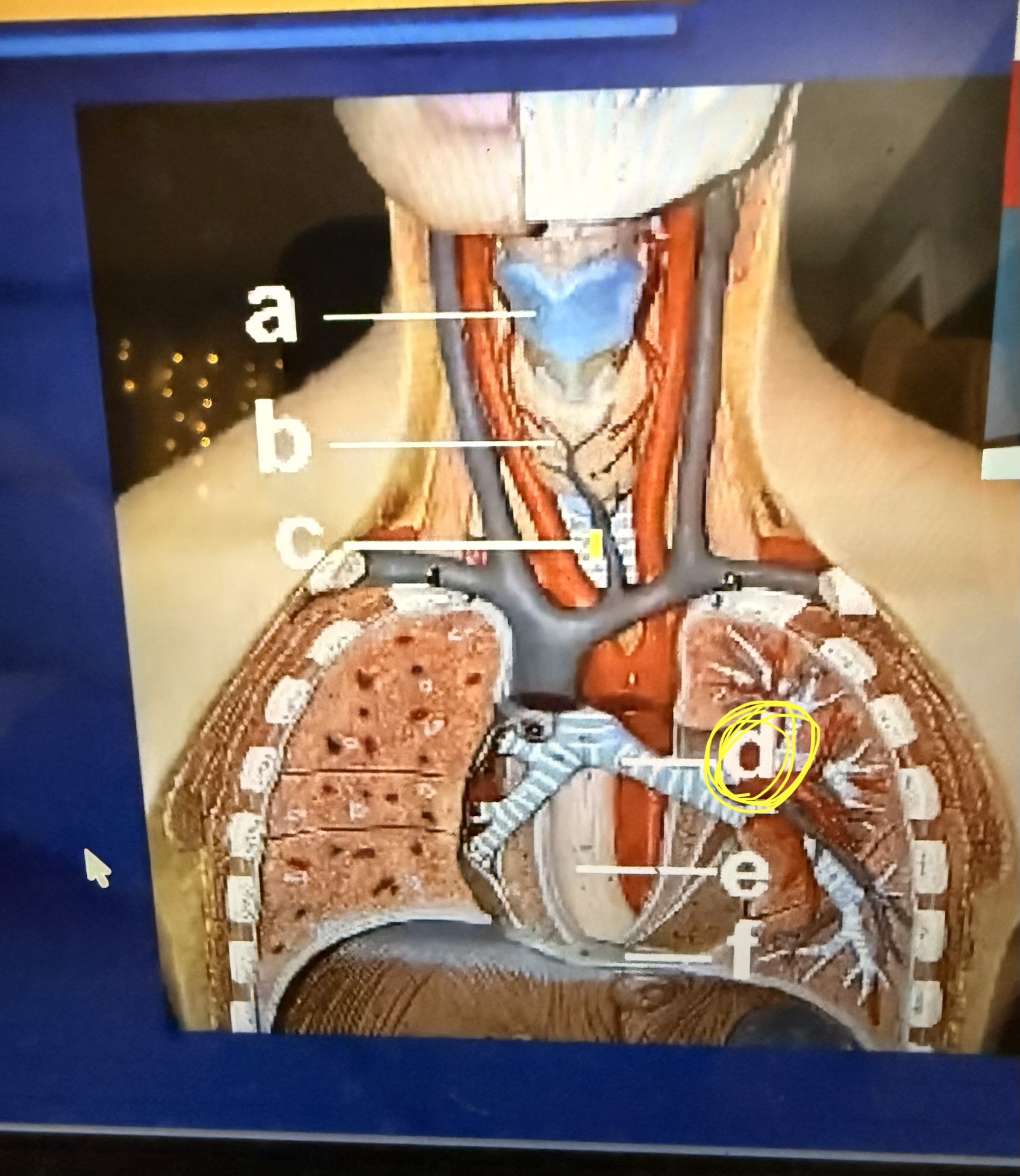
Left primary bronchus
Left lung is differed by
Left superior lube of the lung and then left inferior lobe lung


Esophageal hiatus

Right adrenal gland

right ureter

Fundus region
greater curvature


cardiac sphincter

pyloric sphincter
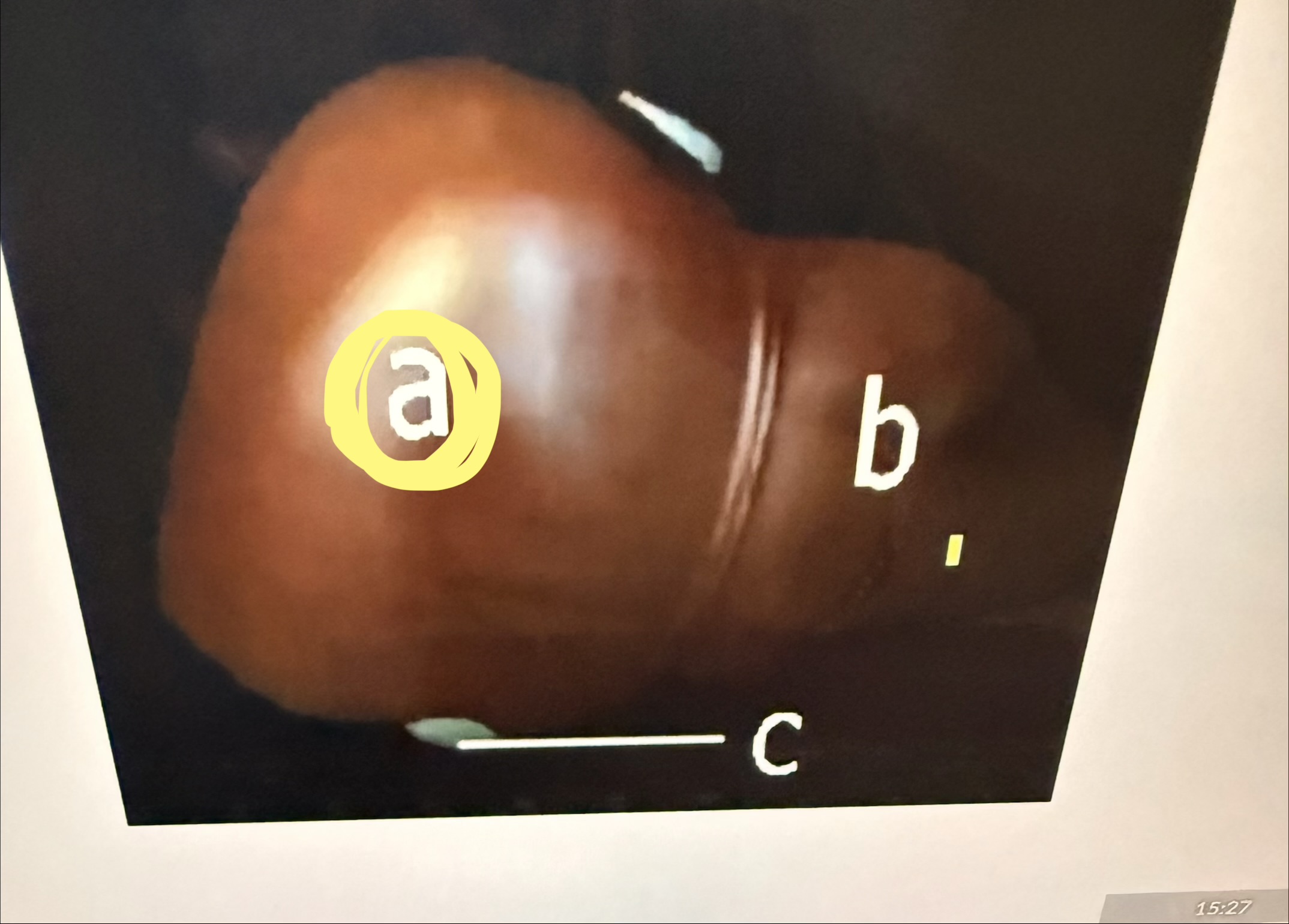
Right lobe of liver
Greater omentum

Cecum of the large intestines


sigmoid colon

haustra

Taeniae coli
Sagittal
A plane that divides the body into left and right portions.

Transverse
A plane that divides the body into superior (upper) and inferior (lower) portions.

spleen

stomach


esophagus
thyroid glands


right adrenal glands

right ureter

rugae

gallbladder

cecum
Scanning lens
40x (4x objective \(\times \) 10x ocular)
Low dry lens
100x (10x objective \(\times \) 10x ocular)
High dry lens
400x (40x objective \(\times \) 10x ocular)
G1 phase
The cell grows larger, copies organelles, and performs normal daily functions.
S phase
The cell replicates its entire DNA genome so each new cell gets an exact copy.
G2 phase
The cell makes final growth spurts and manufactures proteins needed for division.
G0 phase
A resting or quiescent state where a cell exits g1 cycle and stops dividing while remaining metabolically active
Mitotic cell cycle
An orderly sequence of growth and division that produces two genetically identical daughter cells
Interphase cell cycle
Cell grows, duplicates it DNA, and prepares for division
The steps of mitosis
Prophase
Metaphase
Anaphase
Telophase
Stages of cell division
Interphase
Prophase
Metaphase
Anaphase
Telophase
Interphase
DNA duplicated
Growth
Preparation for cell division
Prophase
Chromosomes condense
Metaphase
Chromosomes( consisting of two chromatids) line up at equatorial plate
Anaphase
The attachments between sister chromatids break
Telephase
New nuclei form each with two complete sets of chromosomes
Cytokinesis divide cell into two daughter cells
Resolving power
Magnification of object to distinguish fine detail
Golgi complex
Organelle that packages and processes proteins before secretion.
SER (Smooth Endoplasmic Reticulum)
Organelle that synthesizes lipids and steroids.
Mitochondria
Organelles that produce ATP (energy) for cell function.
Microvilli
Cellular structures that increase surface area for maximum absorption.
Lysosomes
Organelles containing enzymes that destroy foreign bacteria.
the light source on a microscope can be adjusted by
raising or lowering the condenser, or by opening or closing the diaphragm

hypogastric region
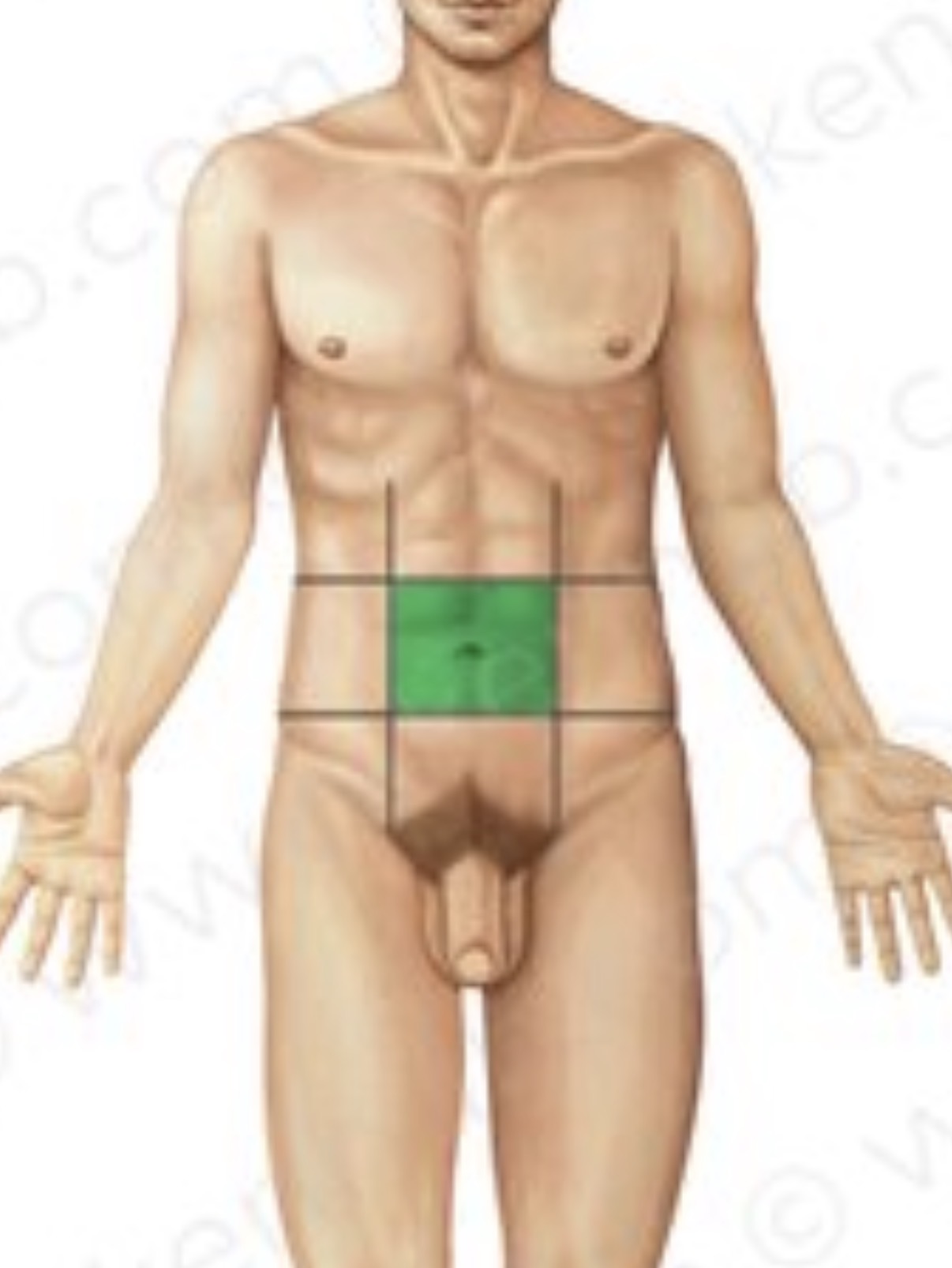
umbilical region