APEX Respiratory Physiology with PLQ, Quiz, & RE
 Call Kai
 Learn
Learn Practice Test
 Spaced Repetition
 Match
Match Flashcards
 Knowt Play
1/214

There's no tags or description
Looks like no tags are added yet.
Name | Mastery | Learn | Test | Matching | Spaced | Call with Kai | Chat |
|---|
No analytics yet
Send a link to your students to track their progress
215 Terms
Anatomic Deadspace begins in the mouth and ends
small airways
terminal bronchioles
respiratory bronchioles
alveolar ducts
terminal bronchioles
what is ventilation
is the process of exchanging gas between the atmosphere and the lungs
2 functions:
O2 is delivered to HGB to support aerobic metabolism
CO2 is the primary end products of aerobic metabolism and is eliminated from the blood
What are the muscles of inspiration
Primary: Diaphragm (tidal breathing and increases the superior-inferior dimension of the chest)
External intercostals (tidal breathing, increase the anterior-posterior diameter)
Sternocleidomastoid (accessory)
Scalene muscles (Anterior, middle & posterior scalene) (accessory)
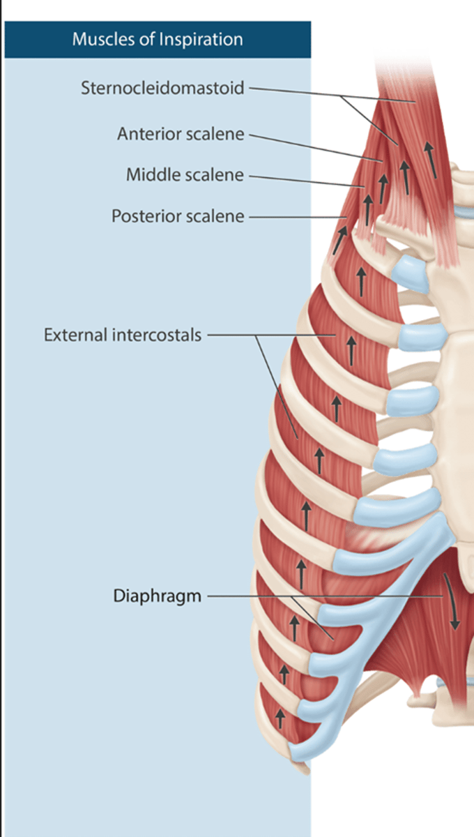
What is inspiration
Contraction of inspiratory muscles reduces thoracic pressure and increases thoracic volume
Example of Boyle's law
The diaphragm and external intercostals contract during inspiration (tidal breathing)
The diaphragm increases the superior-inferior dimension of the chest
The external intercostals increase the anterior-posterior diameters
What are the muscles of expiration
Internal intercostals
Rectus abdominis
External oblique
Internal Oblique
Transverse Abdominis
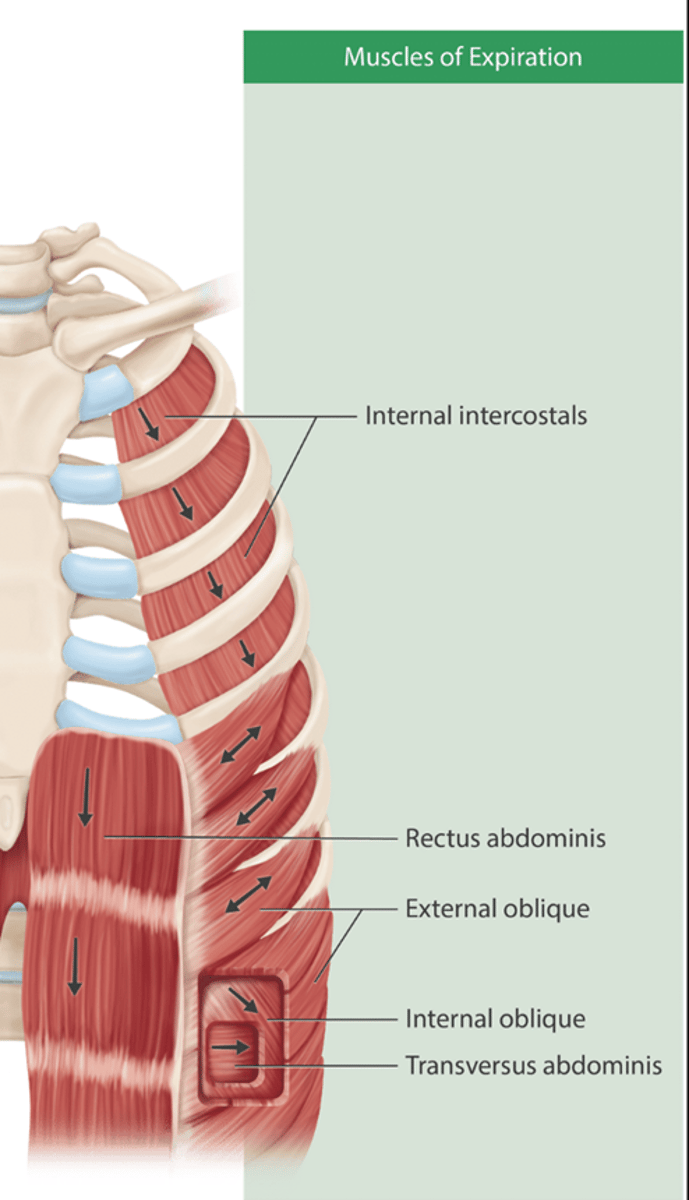
What is exhalation
Exhalation is usually passive and is driven by the recoil of the chest wall
Active exhalation is carried out by the abdominal muscles (Think TIREs- transverse abdominis, internal oblique, rectus abdominis, external oblique)
Internal intercostals play a secondary role in active exhalation
Exhalation is active when minute ventilation increases or in pts with lung disease such as COPD
A forced exhalation is required to cough and clear the airway of secretions; VC of at least 15ml/kg is required for effective cough
Divisions of airway
Airway is a conduit to transfer gas between the atmosphere and the blood and follows the pressure gradient
divided into 3 zones
Conducting (1-4), transitional (17), respiratory (20-23)
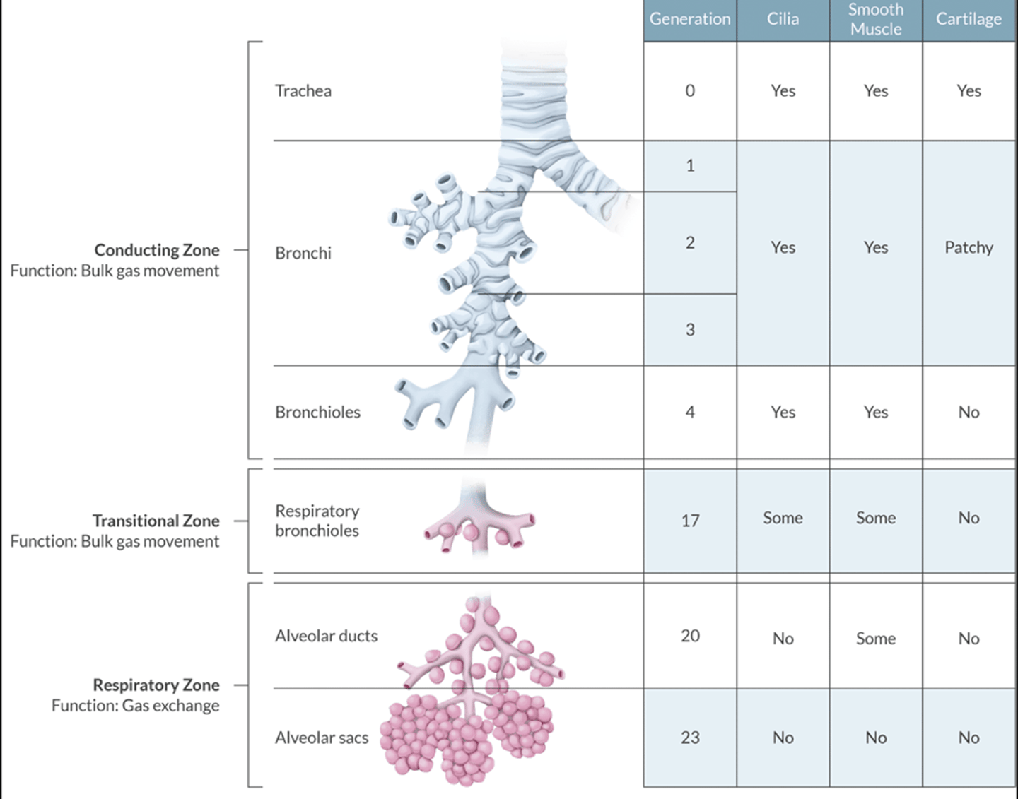
What is the conducting zone & function
function: bulk gas movement
DOES NOT participate in gas exchange= anatomic deadspace
the conducting zone begins at the nares and mouth ends with the terminal bronchioles
the terminal bronchioles are the last structures perfused by the bronchial ciiculation
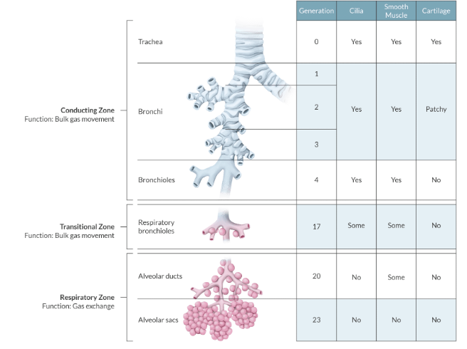
What is the transitional zone
Function: bulk gas movement
The region that contains the respiratory bronchioles
these structures serve as dual function of air conduit and gas exchange
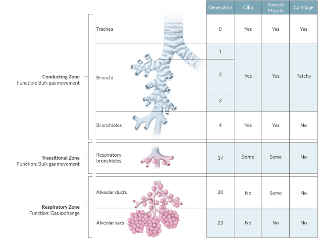
What is the respiratory zone
function: gas exchange
this is where gas exchange takes place
the respiratory zone beings at the alveolar ducts and extends to alveolar sacs
The respiratory bronchioles mark the beginning of this zone (controversial per Apex)
Gas exchange occurs the falt type 1 pneumocytes by diffusion
Airway patency
Airways must remain patent for air movement & gas exchange to occur.
Since bronchioles and alveolar ducts don't contain cartilage= compressible
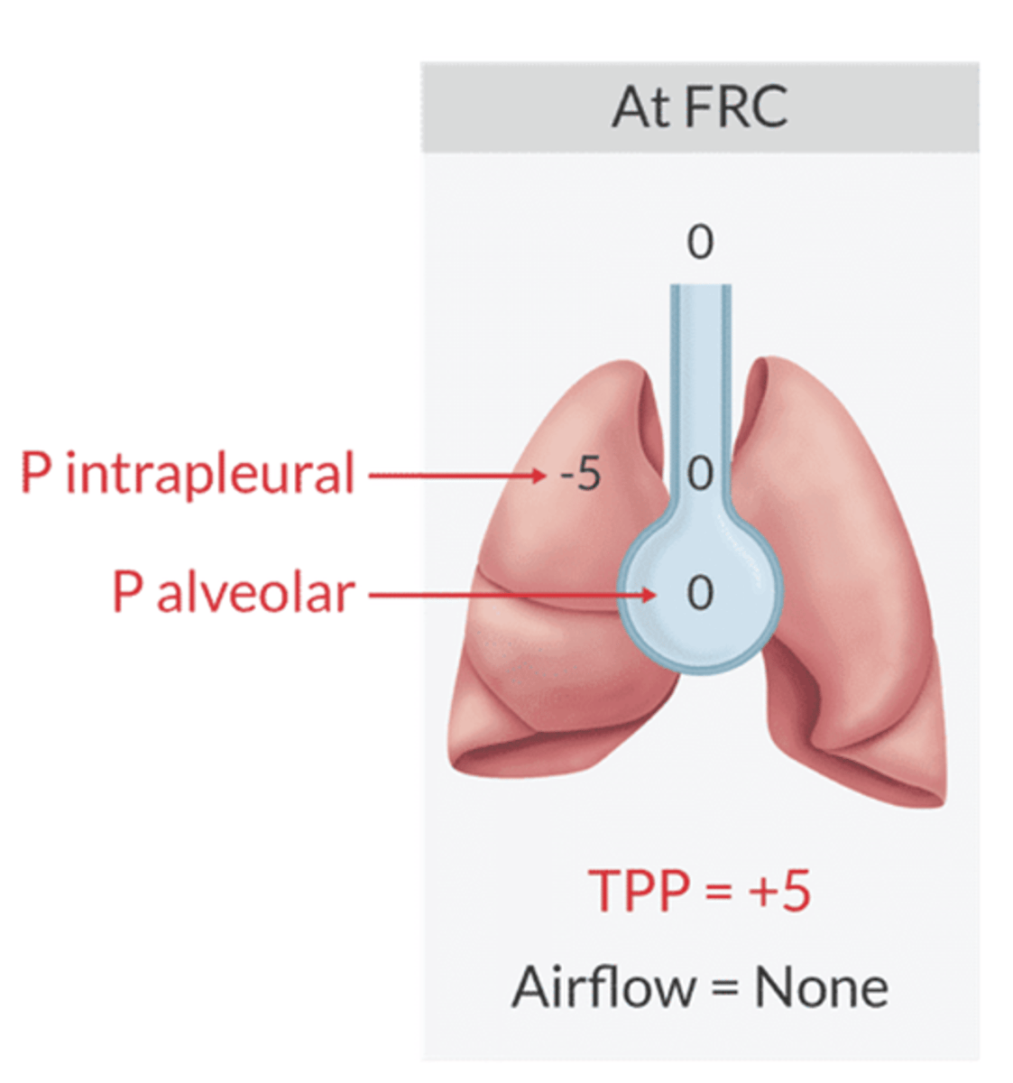
To prevent collapse the pressure inside the airway must be greater than the pressure outside the airway
alveolar pressure is the pressure in the airway
intrapleural pressure is the pressure outside the airway
Transpulmonary pressure is the difference between the 2.
TPP= Alveolar Pressure- Intrapleural pressure
if positive= airway open
If negative= airway collapses
What happens during tidal breathing
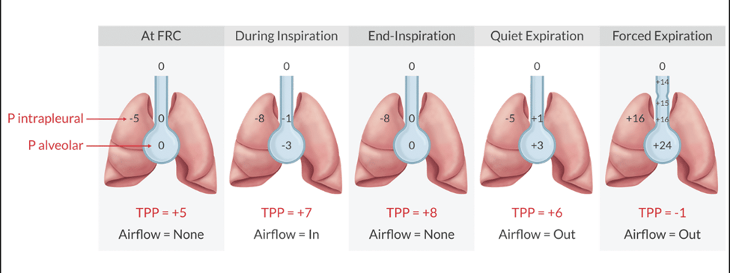
TPP is always positive= airway open
Intrapleural pressure is always negative= keeps lungs inflated
Alveolar pressure becomes slightly negatice during inspiration and slightly positive during expiration
There is no airflow in end-inspiration aka FRC
aside from pathological processes (pneumothorax) the ONLY time that intrapleural pressure becomes positive is during forced expiration
Math of Tidal Breathing
What is the primary determinate of CO2 elimination
Minute ventilation
Tidal volume
alveolar ventilation
RR
alveolar ventilation
What happens in tidal breathing with gas exchange
Vt is the gas that is inhaled and exhaled during a breath.
normal= 6-8ml/kg
When you take a breath, part of the Vt is delivered to the respiratory zone (gas exchange), the remainder of the Vt sits in the conducting airways (deadspace)
dead space (Vd)= 2ml/kg
When you exhale the conducting zone is the first to be removed
this is followed by the exhalation of respiratory zone gas
any condition that increases Vd makes it more difficult to eliminate expiratory gases from the lungs
Increased Vd widens the Paco2- EtCo2 gradient causes CO2 retention
What is the ventilation rate
Ventilation rate is the air removed into and out of the lungs in a given period of time.
What is Minute ventilation
aka Ve
the amount of air in single breath (Vt) multiplied by the number of breaths per minute
MV= Vt X RR
Normal = 5L/min
What is alveolar ventilation
aka Va
only measures the fraction of Ve that is avaliable for gas exchange
it removes deadspace gas from the minute ventilation
Alveolar ventilation= (Vt- Vd) X RR
Alveolar ventilation= CO2 production / PaCo2
Va is directly proportional to CO production and inverse to PaCO2
Normal 3.5L/min
Which conditions will MOST likely increase the PaCo2 to EtCo2 gradient? 3
PPV
LMA
hTN
ETT
neck flexion
atropine
PPV
hTN
Atropine
Overview of definitions of deadspace
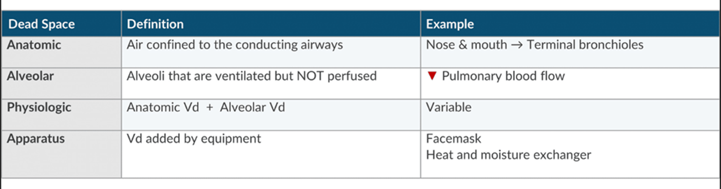
What is the Vd to Vt ratio
this is the fraction of Vt that contributes to deadspace.
factors that alter Vd or V/Q will affect Vd/Vt ratio
Vd/Vt in a normal 70kg pt= 0.33 (150/450)
Mechanical ventilation increases alveolar pressure and increases ventilation relative to perfusion
mechanical ventilation increases Vd/Vt to 50%
****the most common cause of increases Vd/Vt under GA is reduction in CO. ETCo2 acutely decreases, think hTN before considering other causes
What airway device reduces deadspace
ETT decreases Vd (bypasses most of the conducting zone)
LMA reduces Vd by bypassing anatomic deadspace like the ETT
Neck flexion decreases Vd
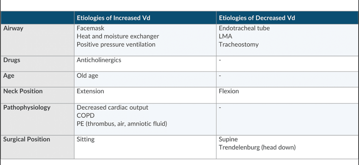
What increases deadspace
facemask
neck extension (opens the hypopharynx and increases deadspace
atropine increases Vd because it is a bronchodilator and increases volume of the conducting airway
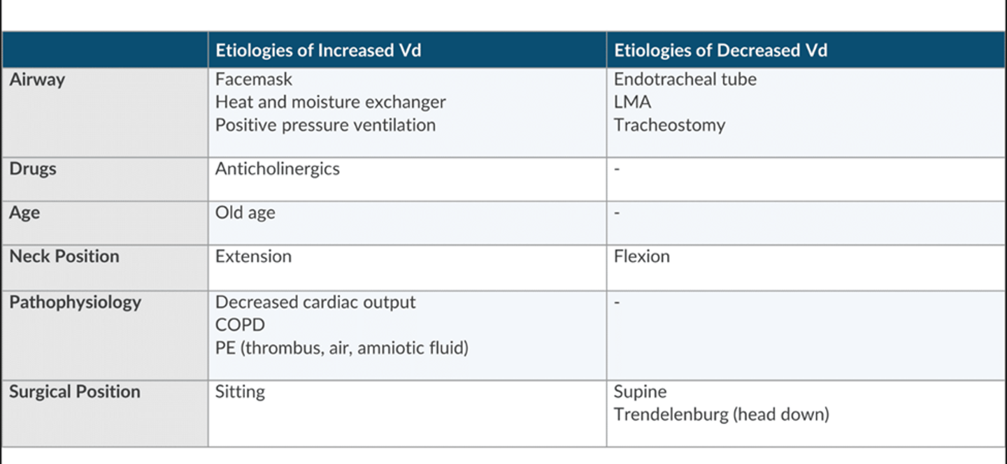
In deadspace increases what happens to minute ventialtion
must increase RR, Vt, or both to maintain a constant PaCo2.
Where does deadspace begin in the circle ssytem
at the y piece
anything proximal to the y piece does not influcen the deadspace nor does increasing the length of the circuit.
only exception is an incompetent valve (incompetent valve makes the entire limb apparatus deadspace)
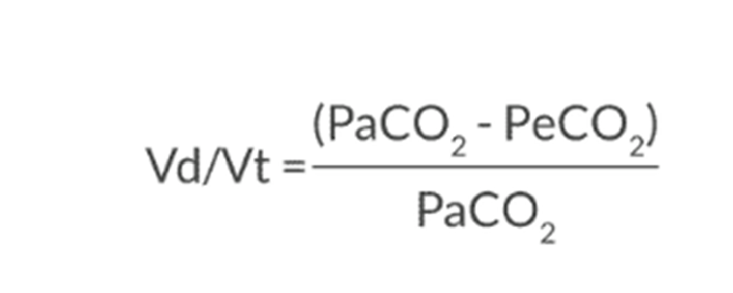
How to use the Bohr Equation
To calculate the physiologic deadspace
compares the partial pressure of the CO2 in the blood vs. the partial pressure of the Co2 in the exhaled
The greater the difference the greater the amount of deadspace
many clinicals use the difference between PaCO2 and EtCo2 as a gross estimation of deadspace but this does not determine the cause of deadspace but only that the amount has changed
A pt in the sitting position. When compared to the apex of the lung, which of the following are higher in the base? (select 2)
BF
PP of alveolar gas
V/Q ratio
PP of alveolar CO2
Blood flow
PP of CO2
What is alveolar compliance (V)
this is a change in alveolar volume for a given change in pressure
not all alveoli are ventilated equally. gravitational forces are largely responsible for this d/t the effect on TTP
At end expiration the apex of the lungs is largest (higher TPP) and the alveoli at the base is the smallest (TPP is lowest)
The alveoli that has a greater degree of volumetric change (compliance) is going to be better ventilated and have better gas exchange.
** understand that the alveolar compliance represents a normal lung. At lung volume below FRC (low lung volume)= compliance is less.
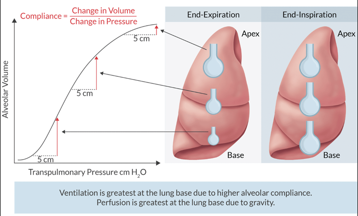
Alveoli in the Apex compliance
has the poorest ventilation because they have the least compliance
the slope of the curve is less steeps and has smaller volumetric change throughout the respiratory cycle
Alveoli in the Bases Compliance
have the greatest ventilation, because have the greatest compliance
The slope of the curve is steeper in this region and larger volumetric changes through the respiratory changes
Alveolar Perfusion (Q)
gravity and hydrostatic pressure affects the distribution of BF through the lungs
when standing upright= less BF to apex and more to bases (V/Q higher in apex and less in bases)
Basic Consequences of V/Q and position changes
Understand the picture.
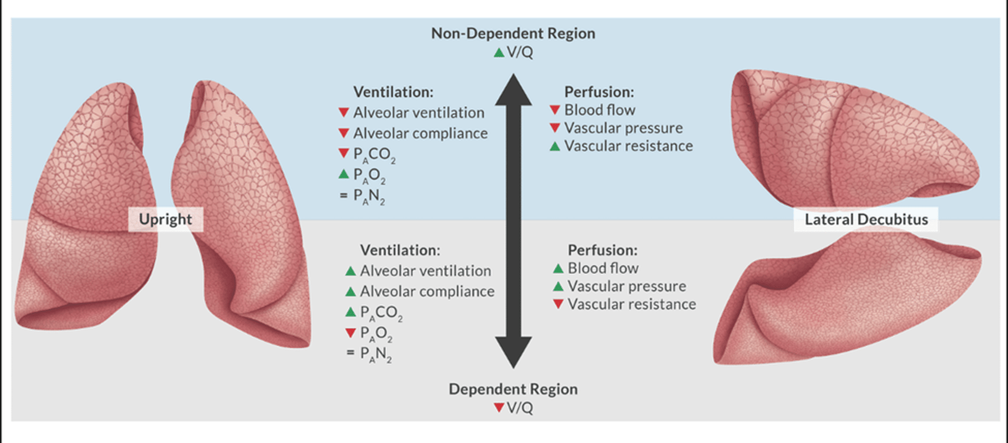
Popular question: if the V/Q is greater in the non-dependent lung region why is ventilation greater in the dependent region?
alveoli with the greatest ventilation undergo the greatest volumetric change through respiratory cycle.
in the wake pt these alveoli are on the steepest slope of the curve= better compliance=better ventilation
Different Surgical positions and V/Q
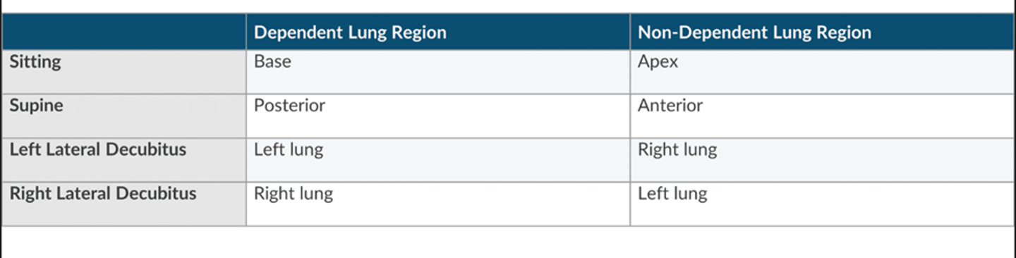
Identify the statement that represent of MOST accurate understanding of V/Q mismatch (select 2)
Bronchioles constrict to minimize zone 1
The A-a gradient is small
Blood passing through under-ventilated alveoli tend to retain CO2
HPV minimizes deadspace
Bronchioles constrict to minimize zone 1
Blood passing through under-ventilated alveoli tend to retain CO2
V/Q mismatch theory
normal V/Q= 0.8
V/Q=1 is perfect match
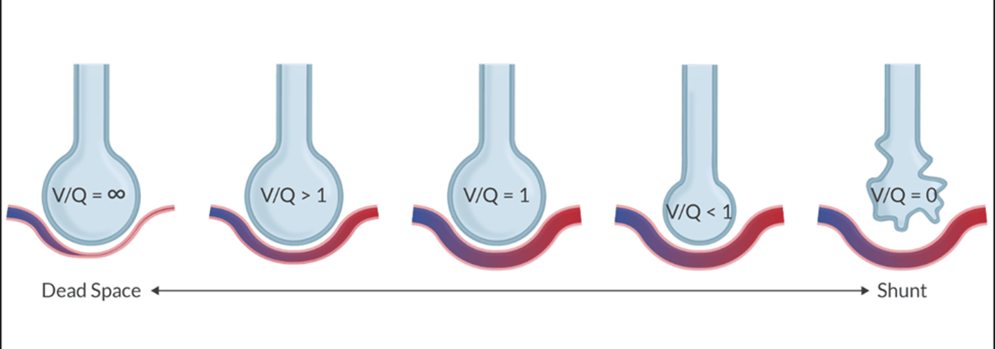
V/Q mismatch in practice
Assume the image is of a pt in the sitting position
V/Q are perfectly matched at intersection
Apex V>Q
Base V
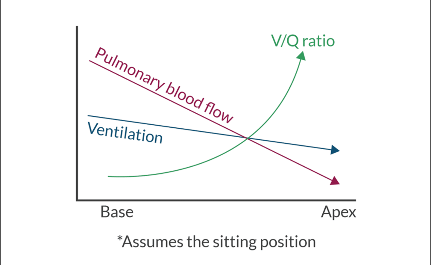
How does this apply to clinical
V/Q mismatch (atelectasis) is the most common cause of hypoxia in the PACU
FRC is smaller d/t GA and surgery= less radial traction to hold the airway open= atelectasis and right to left shunt, V/Q mismatch, and hypoxemia
Treatment= humidified O2 and maneuvers to open the airway (coughing, mobility, Incentive spirometry)
What happens to under ventilated alveoli
blood passing through under ventilated alveoli retain CO2 and is unable to take in enough O2
What happens to overventilated Alveoli
BF gives off excess amount of CO2 (CO2 diffuses 20 times faster than O2)
even though blood can get rid of alot of CO2 it cannot proportionally take up O2 in the same way. (Ex: oxyHGB curve, cannot exceed 100mmHg)
What happens in the whole lung
a lung with V/Q mismatch eliminates CO2 from overventilated alveoli to compensate for the under-ventilated alveoli. PACO2 and PaCO2 gradient is small with V/Q mismatch but CO2 retention= failure of compensatory mechanism
V/Q mismatch cannot absorb more O2 from overventilated alveoli to compensate for the underventiated alveoli. PAO2-PaO2 gradient is large in V/Q mismatch
Compensation of V/Q mismatch
body responds by attempting to match ventilation to perfusion
to combat deadspace (zone 1) the bronchioles constrict to minimize ventilation of poorly perfused alveoli
to combat shunt (zone 3) HV reduces PBF to poorly ventilated alveoli
variables described by the law of la place include the following except
tension
pressure
radius
density
density
Law of la place
the relationship of pressure, radius, and wall tension
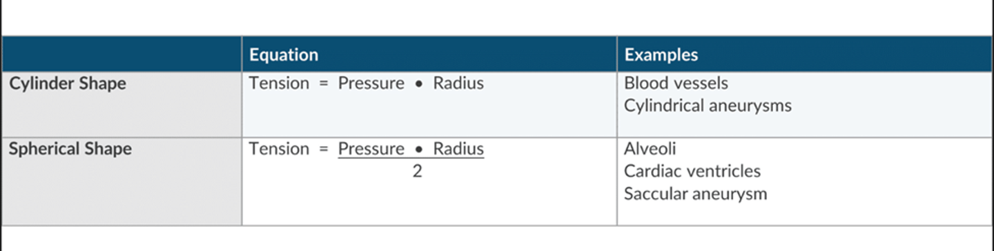
Surfactant (brief overview we have seen this a million times)
according to la place can determine the tendency of alveoli to collapse
Directley proportional to surface tension (more tension=more collapse)
inverse to alveolar radius (small radius= collapse)
produced by type 2 pneumocytes
each alveoli contain the same amount of surfactant
large alveoli= smaller concentration
small alveoli= larger concentration
as radius changes so does the concentration of surfactant
type 2 pneumocytes begin producing surfactant by 22-26 weeks and peak at 35-36 weeks. (preemie= no surfactant= give betamethasone)
Select the correct statement regarding the west zones of the lungs (select 3)
in zone 3 alveolar pressure exceeds venous pressure
in zone 1 alveolar pressure in higher than arterial pressure
in zone 2 venous pressure is higher than alveolar pressure
in zone 2 ventilation is greater than perfusion
in zone 1 there is no pulmonary BF
In zone 3 PBF is proportional to arterial to venous pressure gradient
in zone 1 alveolar pressure in higher than arterial pressure
in zone 1 there is no pulmonary BF
In zone 3 PBF is proportional to arterial to venous pressure gradient
West zones
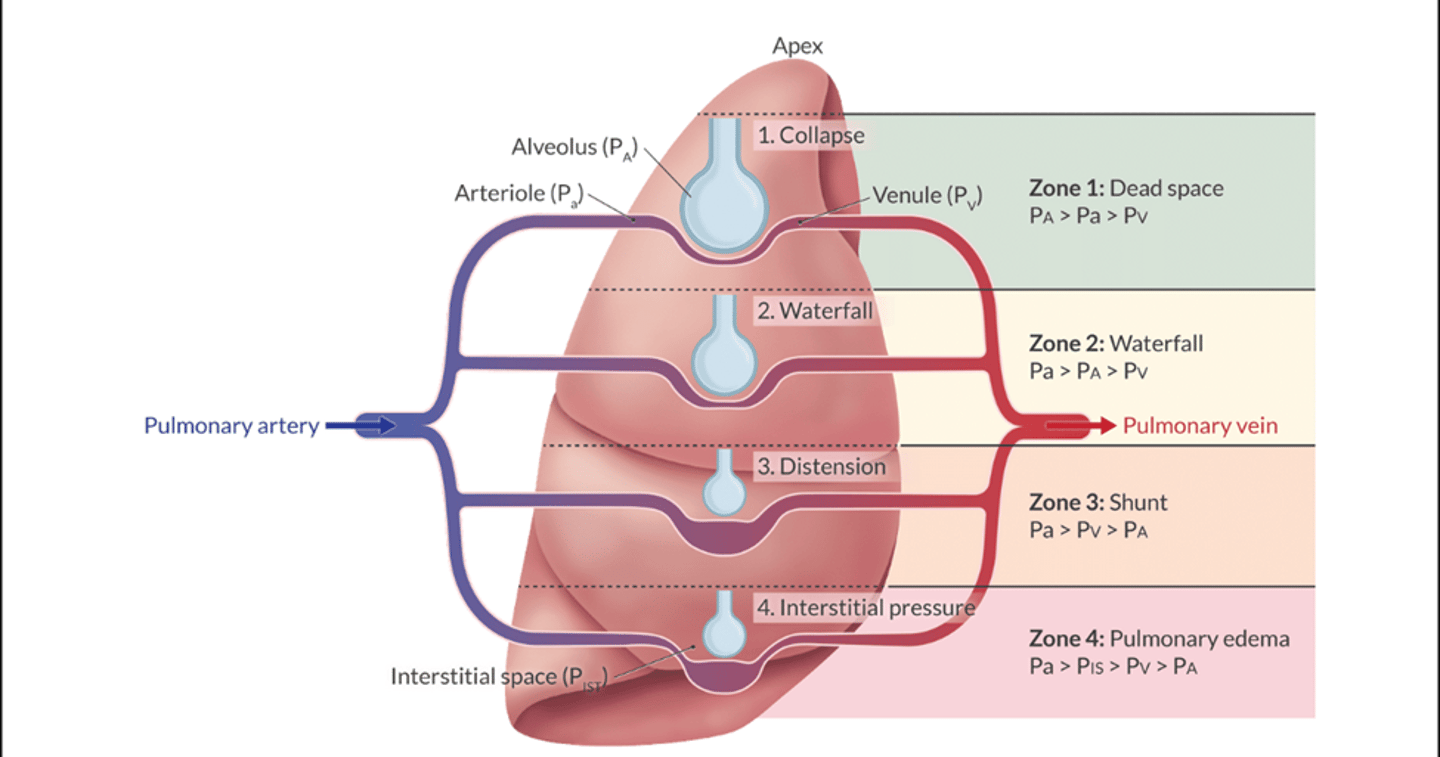
Zone 1
Deadspace
PA>Pa>Pv
V/Q= infinity
zone does not occur in normal lungs
increased by hTN, PE, or excessive airway pressure
To combat zone 1= bronchioles of under perfused alveoli constrict to reduce deadspace
Zone 2
Pa>PA>Pv
V/Q= 1
blood flow is directly proportional to the difference between Pa-PA. greater the difference= greater the blood flow
because pulmonary capillary pressure and alveolar pressure changes throughout the respiratory cycle, it is possible for zone 2 to transiently change to zone 1 or 3
zone 3
Shunt= Pa>Pv>PA
Blood flow is a function of pulmonary AV pressure difference (Pa-Pv)
V/Q=0
most zone 3 units are shunt like and are better perfused then ventilated
to combat this zone HPV constricts pulmonary BF to under ventilated units
since pressure in the capillary is always higher than the alveolus, the vessel is always open, and BF is moving
The tip of the PA catheter should be placed in zone 3
NOT ALL SHUNTS OCCUR IN THE LUNGS: anatomic shunts (venous blood emptying directly in the left side of the hear, blood bypasses lungs= no saturation of oxygen)
Ex: thebesian veins, bronchiolar veins (drain into bronchial circulation), pleural veins (drain bronchial circulation_
Zone 4
Pulmonary Edema
Pa>Pist>Pv>PA
the pressure in the interstitial space exceeds pressure of pulmonary capillaries and alveolus
pulmonary edema is a classic example of zone 4
rate of fluid accumulation exceeds the rate of removal by the lymphatic system
Result of 2 things
1. fluid is pushes across the capillary membrane by increase in capillary hydrostatic pressure (fluid overload, MS, severe pulmonary constriction)
2. fluid is pulled across the capillary membrane by profound reduction in pleural pressure (laryngospasm, NPPE)
Other Points about West zone
gravity is believed to be the most significant influence in distribution of perfusion
this hypothesis states the apex has less BF than the bases
BF follows more of a central to peripheral distribution
zone 3 at the lower core and zone 2 and zone 1 radiate out from that point to the periperhy
If a patient is breathing RA at sea level. The ABG reveals PaO2 of 60 mmHg and a PaCO2 of 70mmHg. Claculate patients alveolar oxygen concentration.
alveolar oxygen= fiO2 x (Pb- PH2O) - (PaCO2/RQ)
0.21 x (760-47) - (70/0.8)= 62.23 ~ 62
alveolar gas equation
this is used to estimate the partial pressure of O2 in the alveoli
must understand: hypoventilation can cause hypoxia and hypercarbia, supplemental O2 can easily reverse hypoxemia but not hypercarbia, hypercarbia can go undetected in pt that is breathing supplemental O2
FiO2= fraction of inspried oxygen
Pb= barometrc pressure
PH2O= humidity of inhaled gas (assume 47)
RQ= respiratory quotient (assume 0.8)
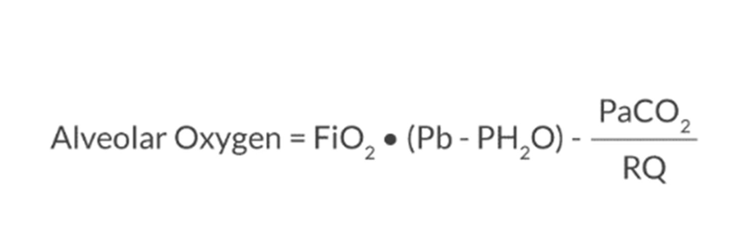
FiO2 in the alveolar gas equation
FiO2 is always higher than the partial pressure of O2 in the alveolus
This is because:
inspired O2 becomes 100% humidified as it moves to the alveoli, water takes up space and dilutes O2 concentration
inspired air (lots of O2 and no CO2) mixes the expired air and dilutes concentration of O2 going toward the alveoli
Hypoventilation increases PaCO2 and increased PaCO2 competes for space with O2 inside the alveoli and dilutes the O2 concentration
in this situation supplemental O2 can increase PAO2 and PaO2, this masks the cause and not treat it
R/Q
>1= lipogenesis= overfeeding
0.7= lipolysis= starvation
R/Q= CO2 production/ O2 consumption= 200/250= 0.8
Causes of increased A-a gradient include (select 2)
hypoventilation
V/Q mismatch
hypoxic mixture
diffusion limitation
V/Q mismatch
diffusion limitation
5 causes of hypoxemia
hypoxic mixture, hypoventilation, diffusion limitation, V/Q mismatch, and shunt
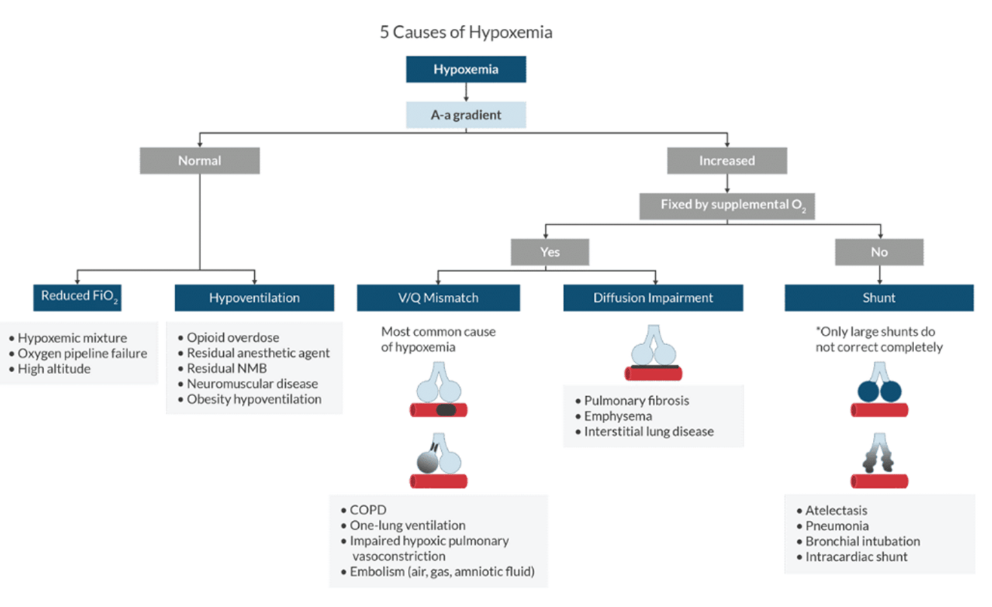
If A-a gradient is normal what is the cause
hypoxic mixture and hypoventilation
A-a gradient is increased, what is the cause
diffusion limitation
V/Q mismatch
shunt
When does hypoxemia occur
PaO2<80mmHg
hypoxia is insufficient O2 to tissues
Fixed by supplemental oxygen
V/Q mistmatch and diffusion impairment
NOT SHUNT
Examples of reducied fio2 causing hypoxemia
hypoexmia mixture
oxygen pipeline failure
high altitide
examples of hyperventialtion causing hypoxemia
Opioid OD
Residual anesthetic agent or NMB
Neuromuscular disease
obesity hypoventilion
V/Q mismatch examples causing hypoxia
Most common cause of hypoxemia*
COPD
OLV
Impaired HPV
embolism (air, gas, amniotic fluid)
Examples of diffusion impairement causing hypoxemia
pulmonary fibrosis
emphysema
interstitial lung disease
Examples of shunts causing hypoxemia
atelectasis
PNA
bronchial intubation
intracardiac shunt
A-a gradient calculation
PAO2 (from alveolar gas equation)- PaO2 (from ABG)
what is determined by the A-a difference
if there is a large difference the this implies the degree of shunt, V/Q mismatch, or diffusion limitation
Factors that ncrease A-a gradients
aging (increased CC compared to FRC)
vasodilators (decreased HPV)
Right to left shunt (atelectasis, bronchial intubation, intracardiac defect)
diffusion limitation (increased thickening of the alveolar capillary= decreased O2 diffusion)
Estimate the shunt
shunt increases 1% for every 20 mmHg of A-A gradient
Lung volume graph
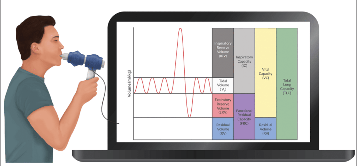
Lung volume chart
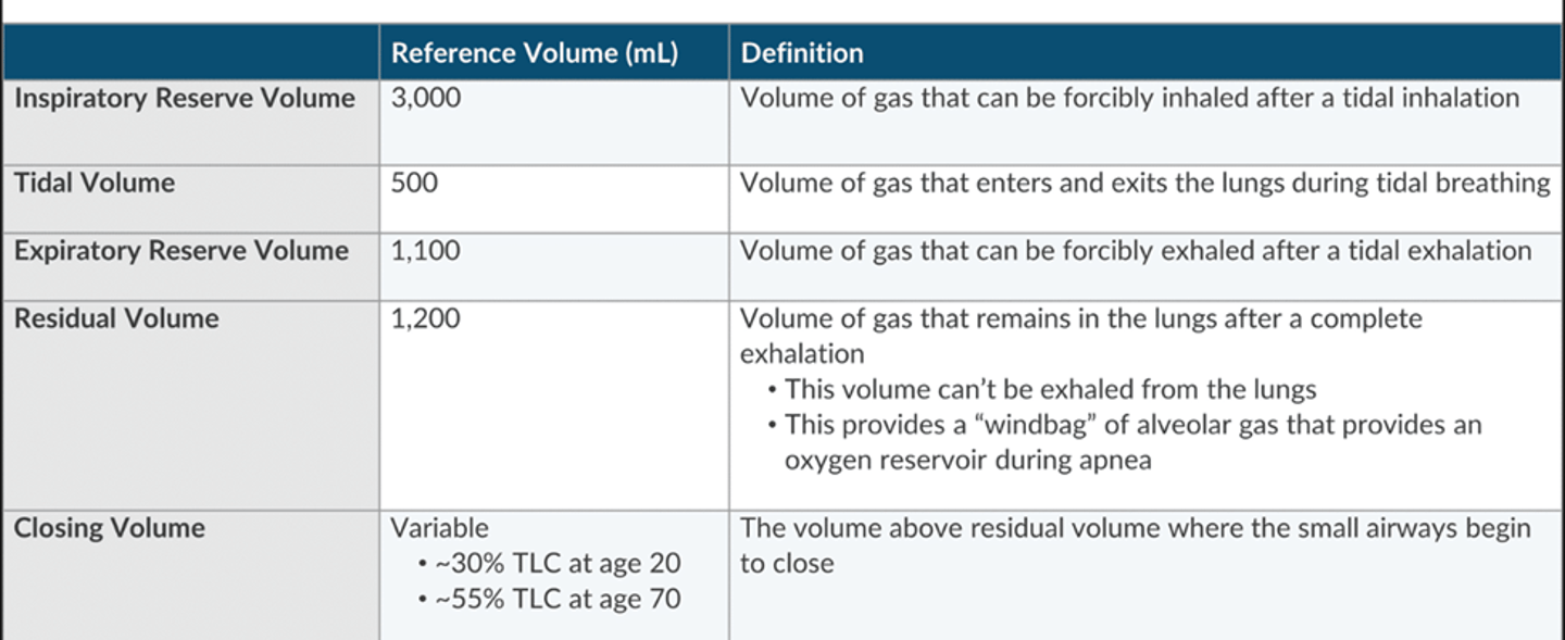
lung capacities chart
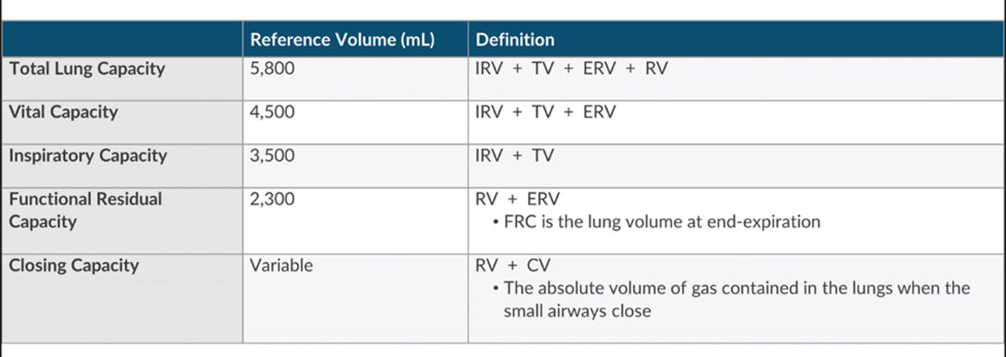
Key facts about the volumes and capacities
Vt= 6-8ml/kg
VC= 65-75ml/kg
FRC-35ml/kg
All of these are calculated from ideal body weight
lung volumes are 25% smaller in females
volumes change with position (larger when sitting and smaller when supine)
Obstructive disease= air trapping (increases RV, CC, TLC)
spirometry can measure everything except RV, therefore it cannot calculate FRC or TLC
CV and CC are dynamic measures and assess small airway closure (also cannot be measured with spirometry)
What conditions reduce FRC (select 2)
Advanced age
pulmonary edema
COPD
Obesity
pulmonary edema
obesity
How to measure FRC
FRC is the reservoir of O2 that prevents desaturation with apnea
FRC= inward elastic recoil of the lungs is balanced by outward elastic recoil of the chest wall (equilibrium)
diaphragmatic tone and position affect FRC
Normal= 35ml/kg
Measured indirectly by: nitrogen washout, helium wash in, body pelthysmography
NOT SPIROMETRY (cannot measure RV)
How long does FRC last during apnea
Time to desat= FRC/VO2 (oxygen consumption)
ensure the pt is preoxygenated
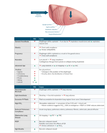
Conditions that alter FRC
these conditions reduce outward expansion or reduce lung compliance, FRC reduced= increased zone 3, alveolar recruitment act to restore FRC by reducing zone 2
CC is the sum of cv and:
RV
ERV
FRC
Vt
RV
what is closing volume
as a person exhales there is a point where pleural pressure exceeds airway pressure= small airway closure (d/t lack of cartilage)
since pleural pressure is higher in the dependent portion= these airways close first
CV is the point of dynamic compression of the airway begins (volume above RV where small airways collapse during expiration)
Factors that increase clsoing volume
(CLOSE-P)
COPD
LV failure
obesity
surgery
extremes of age
pregnancy
what is CC
CC is the absolute volume of gas contained in the lungs when the small airways begin to close
CC= CV+RV
What affects CC
important relationship of FRC and CC, will determine airway collapse in tidal breathing
normal= FRC>CC, airways dont collapse
CC>FRC= airway closure during tidal breathing= intrapulmonary shunting and hypoxemia
anything that decreases FRC to CC or increases CC to FRC will convert a normal V/Q to low V/Q units or shunt untis
PEEP can reverse this by increasing FRC
How does age affect CC
normal= CC just above RV
as we age pleural pressure progressively gets higher and the smaller airways will close sooner (this explains reduction in PaO2 with aging)
Aging=
increased FRC, CC, RV
and decreases VC
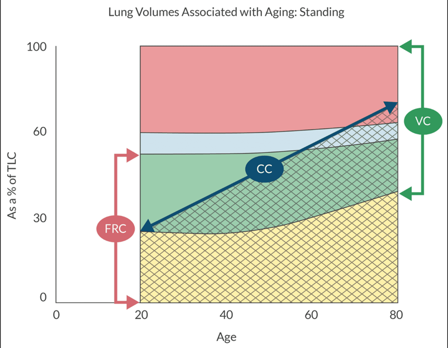
How does anesthesia affect CC
30= FRC= CC with GA
44= CC=FRC with supine
66= CC=FRC when standing
how to measure CC and CV
spirometry cannot measure
need nitrogen washout or xenon.
gas is inhales at RV and teh measurement is taken as the pt exhales from TLC
calculate patient’s arterial oxygen content from data set
Hgb 9
HR 100
SV 70
SaO2 90%
PaO2 60
11.034
CaO2= 1.34 x hgb x SaO2) + PaO2 × 0.003)
(1.34 × 9 × 0.9) + (60 × 0.003)
Oxygen content
how much O2 is present in 1 dL (100ml) of blood
O2 transported 2 ways
HGB (97%)
dissolved in plasma
O2 bound to HGB equation
1.34 X HGB X SaO2
men: 15 hgb 45 hct
female: 13 hgb 39%
Dissolved O2 equation
(PaO2 X 0.003)
dissolved O2 is measured by PaO2 (estimated gas exchange not a measure of O2 content in the blood)
dissolved according to HENRY's law (proportional to partial pressure of gas above to solution, solubility co-efficent 0.003, O2 is 20 times less soluble than CO2)
How to calculate O2 delivery
CaO2= O2 contained in the blood
DO2= delivery of the blood to tissues
CO is the driving mechanism of DO2
DO2- CaO2 X CO X 10
calculate VO2 aka O2 consumption
using FICK's principle to calculate
VO2 is the differnce of O2 that leaves the lungs and the amount of O2 that returns to the lungs
VO2= CO X (CaO2-CvO2) X 10
VO2= 3.5ml/kg
VO2= 250ml for 70 kg
P50 is reduced by select 3
HGB f
hyperthermia
hypocarbia
increased DPG
acidosis
carboxyhgb
HGBf
Hypocarbia
carbocyhgb
OxyHGB Curve
the curve tells us the tendency if HGB to bind to HGB
P50=26.5mmhg
max loading= 100mmHg (if increased FiO2 will increase dissolved fraction but not any bound to HGB past 100mmHg)
Tissues that have higher metabolic rate consume more O2 and produce more CO2, hydrogen, and heat
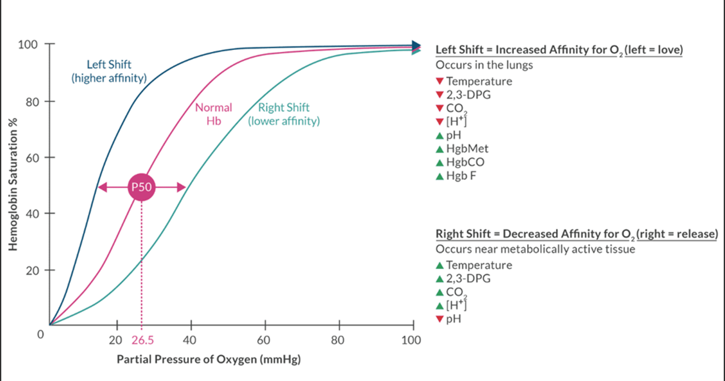
What causes left shift (higher affinity) in oxyhgb curve
(occurs in lungs)
Decreased temp
Decreased 2,3- DPG
Decreased CO2
Decreased H+
Increased pH
Increased Hgb MET (Methemoglobin)
Increased Hgb CO (carboxyhemoglobin)
Increased HgbF (fetal hgb)
What causes right shift (lower affinity) in oxyhgb curve
(Occurs near metabolically active tissue
Increased temperature
Increased 2,3 DPG
Increased CO2
Increased H+
Decreased pH
what is the Bohr effect
CO2 and hydrogen ions cause change in the HGB molecule, facilitates release of O2
increased partial pressure of CO2 and decreased pH= hgb will release O2
What is DPG and why is it important
produced by RBC glycolysis (rapoport-luebering path)
slightly shift to right at all times
hypoxia= increased DPG production
important compensation mechanism in anemia
in banked blood= less DPG= shift to the left and reduces O2 at tissue level
HGB f doesnt respond to DPG (explains why that shifts left and P50=19)
Identify the statement that best describes aerobic metabolism
NADH is the final electron acceptor during electron transport
1 molecule of glucose converts to 38 molecules of ATP
pyruvic acid is converted to lactate
electron transport occurs in the cytoplasm
1 molecule of glucose converts to 38 molecules of ATP