muscle anatomy 1
 Call Kai
 Learn
Learn Practice Test
 Spaced Repetition
 Match
Match Flashcards
 Knowt Play
1/33

There's no tags or description
Looks like no tags are added yet.
Name | Mastery | Learn | Test | Matching | Spaced | Call with Kai | Chat |
|---|
No analytics yet
Send a link to your students to track their progress
34 Terms
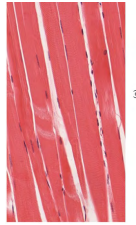
what is the cell shape, nuclei, control and location to skeletal muscle?
long cylindrical, striated
many nuclei
voluntary
attached to many bones
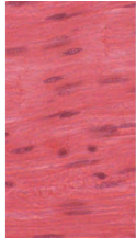
what is the cell shape, nuclei, control and location to smooth muscle?
long but tapered
1 nuclei, not straited
involuntary control, connected to organs
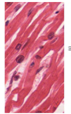
what is the cell shape, nuclei, control and location to cardiac muscle?
long, BRANCHED
striated
1 nuclei, involuntary found in heart!
what is the neuromuscular junction?
axons of motor neurons extend from spinal cord towards muscle!
divides into axon terminals that form NEUROMUSCULAR JUNCTIONS with muscle fibers across muscle. (1 per fiber)
action potential/electrical impulse travels down this axon into synaptic terminal, opeing voltage gated Calcium Channels
calcium enters neuron terminal.
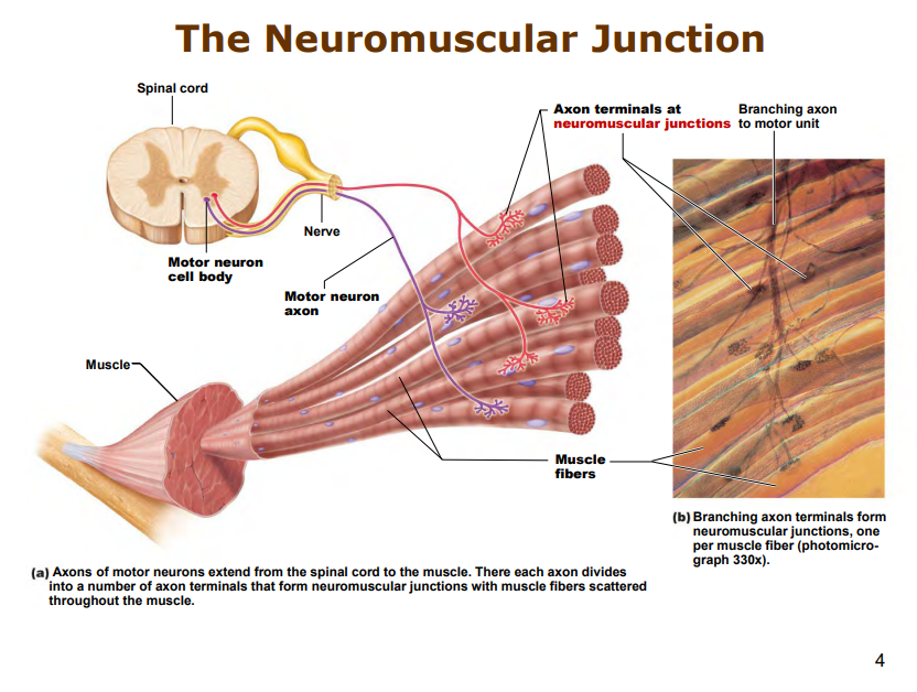
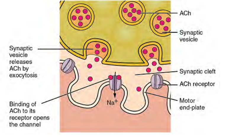
what is the anatomy of the neuromuscular junction?
calcium channels open, enters neuron terminal. triggers:
Synaptic vesicle contains acetylcholine (inside the axon terminal)
via exocytosis, releases Ach into synaptic cleft, connecting to the ACh receptor of motor end plate.
channel opens, Na+ floods in!
how does a muscle action potential generate?
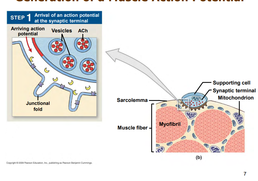
action potential will arrive at synaptic terminal
calcium channels open
enters synaptic terminal
acetylcholine released into synaptic cleft via exocytosis
binds to receptors, Na+ rushes into cell
action potential spreads across surface of sarcolemma
ACh broken down by AChE
returns to intial state, cycle beings again if another action potentia larrives at neuromuscular junction
Na+ channels outside of endplate open, causing more Na+ to rush in, K Out, etc eetc
What is excitation contraction coupling?
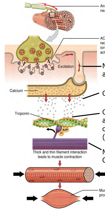
From the neuromuscular junction and after an action potential is generated in the sarcolemma, it travels along an down into the T Tubules
DHP receptors activated, opeing Ca release channels.
Ca2+ ions released from Sarcoplasmic reticulum
binds to troponin, removing the blocking action of tropmyosin
troponin shape changes, revealing binding sites for myosin on the thin filaments
binding occurs, cross bridges form, contraction begins.
what does the thin myofilament consist of? how does it contribute to contraction?
2 strands of actin sub units (twisted into helix)
two regulatory proteins
troponin
tropomyosin
calcium binds to troponin, reveals binding sites for myosin, binding to the actin and forming a cross bridge.
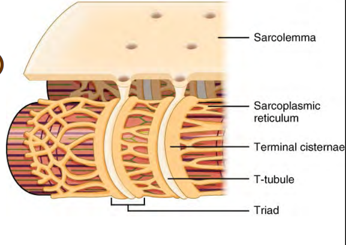
what is the anatomy of a muscle fiber/cell
surounded by sarcolemma (muscle membrane)
sarcoplasmic reticulum
terminal cisternae
T tubule
triad
What is the cross bridge formation?
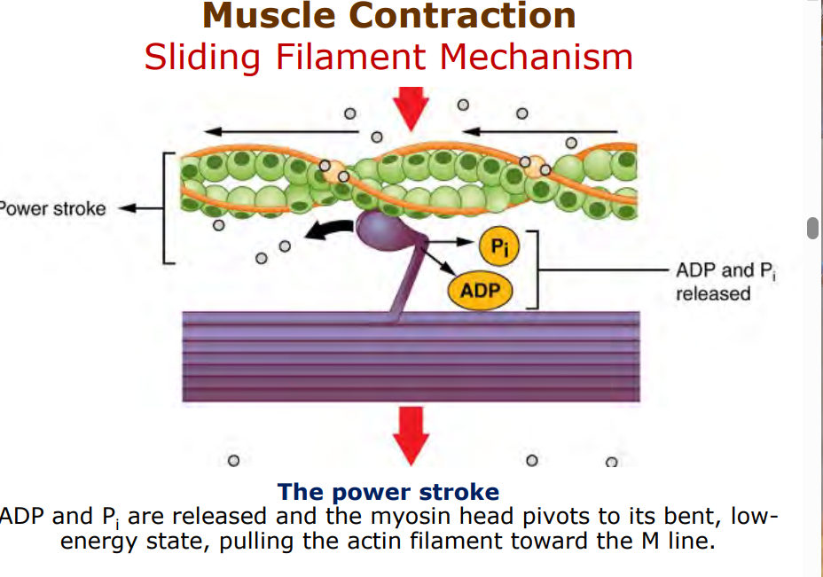
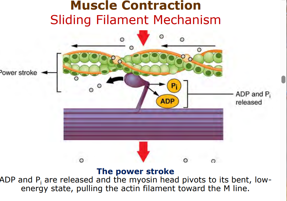
when mysoin (energized via holding ADP and P) attaches to newly exposed site on actin(via excitation contraction coupling).
myosin head pivots due to release of ADP P, pulling actin foilament towards center of sarcomere.
How does the myosin deattach from actin?
new molecule of ATP attches to myosin head, detaching the two
ATP is hydrolyzed into ADP P, “re cocking” myosin head into high energy position.
ready to pul again via ratchet mechanism if ATP is available
and if Ca is stil bound to troponin
how does the muscle relax?
Acetylcholine is broken down or diffuses away from neuromuscular junction
as a result no action potentials travel down
Ca2+ is returned to SR via protein pumps
Troponin stops binding to Ca2+, tropomyosin comes back and blocks binding site for myosin
what are 3 different sources for ATP for muscle contraction?
creatine phosphate - combines with ADP to create creatine and ATP
Anaerobic metabolism of glucose via glycolysis
aerobix metabolism of glucose and fatty acids.
what types of ATP are used in short duration exercise vs prolonged duration exercise?
6 seconds - ATP stored in muscles
10 seconds - ATP formed from creatine phosphate (phosphorylation)
30-40 - glycogen stored in muscles broken down to glucose, oxidized to pyruvate to generate ATP (anaerobically)
hours - full odixation of fats and carbs to CO2 and H2O
what is hypotonia and hypertonia
hypotonia - flaccid
hypertonia - spasticity
what influences muscle size
lack of use
aging
disease - dystrophy
Training
Hormones Anabolic - steroids
what is the microscopic anatomy of cardiac muscle
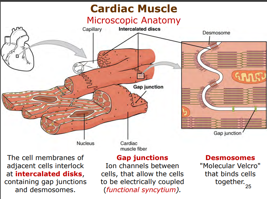
cells interlock at the cell membranes intercalated disks
connect gap junctions and desmosomes
gap junctions
ion channels between cells
allow cells to be coupled electrically
functional syncytium
desmosomes
molecular velcore
binds cells together
what is autorhythmicity? how does heart contract
craeted by pacemaker
clsuter of non contractile cardiac cells
unstable membrane potentials that depolarize sponatenously and rhythmically
triggers action potentials that spread throughout heart
uses ATP from aerobic metabolism only
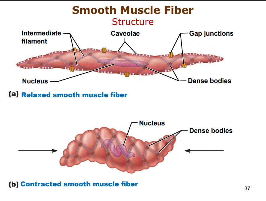
What is smooth muscle fiber anatomy?
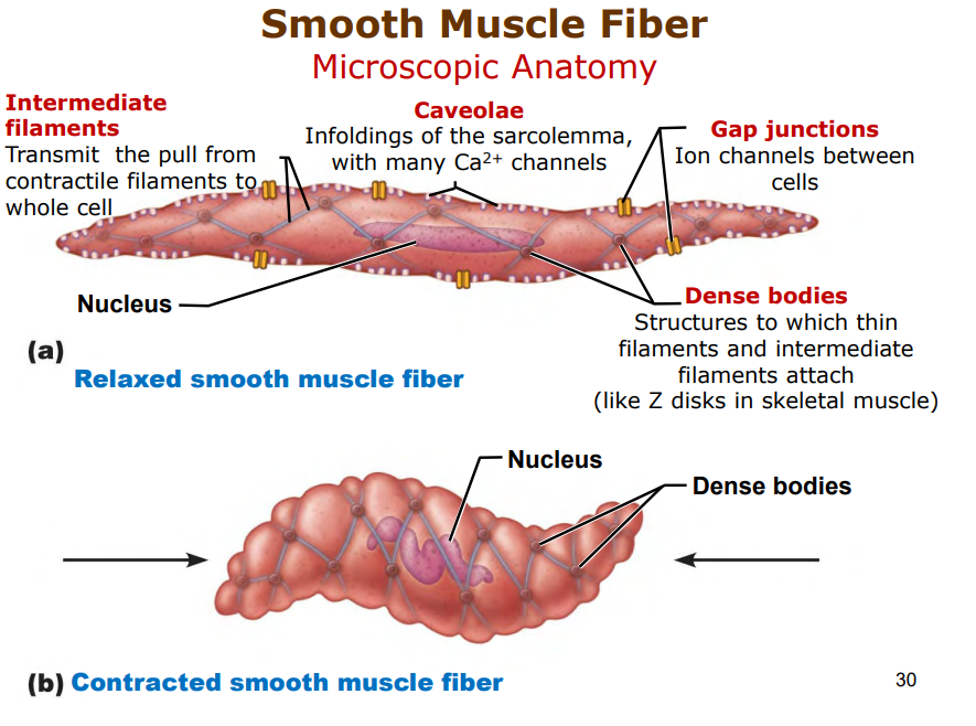
intermediate filaments- transmit pull from contractile filaments to whole cell
caveolae - infoldings of sarcolemma(many Ca2+ channels)
Gap Junctions - ion channels between cells
Dense bodies - attachment point for thin and intermdiate filamentes(similar to Z disks in skeletal muscle)
Where are neurotransmitters released from in smooth muscle fibers?
from swellings (varicosities) on the axons of autonomic neurons through diffusion
not localized to a neuromuscular junction!!
what is the excitation contraction coupling process like in smooth muscle?
calcium ions enter cytosol via Ca2+ channels or scant SR from ECF!
binds to and activates calmodulin
calmodulin activates myosin kinase
myosin kinase catalyzes transfer of phosphate to myosin
activated myosin forms cross bridges with actin of thin filaments, contraction!
how does smooth muscle relax
enzymes remove phosphate from mysoin, (doesnt activate)
no action potenials, Ca2+ channels wone open
moved out of the cell into SR by protein pumps
calmodulin wont activate, netiher will kinase, nor myosin
what is the affect of neurotransmitters o nsmooth muscle contraction? what about hormones?
acetylcholine - PNS stimulates contraction, bronchioles pupils constriction
norepinephrine - SNS - does both
dilation of bronchioles/pupils, but constriction of blood vessels depending on the area
gastrin, stimulates gastric motility when food in stomach
cholecystokinin relaxes hepatopancreatic sphincter when food is in intestine
oxytocin
stimulate contraction of uterus, ducts of mammary glands during breast feedings