CH. 7: MUSCLE SYSTEM
1/99
There's no tags or description
Looks like no tags are added yet.
Name | Mastery | Learn | Test | Matching | Spaced | Call with Kai |
|---|
No analytics yet
Send a link to your students to track their progress
100 Terms
What are the functions of skeletal muscle?
Produces voluntary movement
Stabilizes joints
Maintains posture
Generates body heat
Muscle tissue contracts in responce to
stimulation
What are the 3 types of muscle tissue?
Skeletal
Cardiac
Smooth
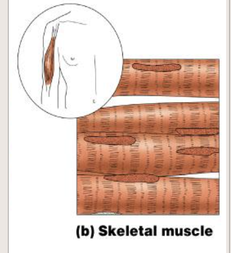
What are the characteristics of skeletal muscle?
Cylindrical cells
Striated
Multiple, peripheral nuclei
Voluntary
Attached to skeleton
What are other facts regarding the muscle tissue?
Makes up “flesh” of body (~40% by weight)
Most “meat” is skeletal muscle
Muscles are organs; give examples and what they do
Fibers (muscle cells)
Motor neurons (stimulate to contract)
Blood vessels (supply nutrients, oxygen)
Connective tissue (re-inforce)
Connective tissue coverings provide
strength, support, & insulation
What are the connective tissue coverings?
Endomysium
Perimysium
Epimysium
Fascia
Endomysium
Around each muscle fiber; insulates
Perimysium
Around fascicles (bundles of cells)
Epimysium
Around entire muscle (bundles of fascicles)
Fascia
collective term for C.T. around & between muscles
Connective tissue attachments join muscles to:
bones, cartilages, or to CT coverings of other muscles
Tendons
cordlike bundles of collagen fibers
Aponeuroses (sing. -sis)
sheetlike arrangements of collagen fibers
Muscle Fibers (skeletal muscle cells):
Long, cylindrical, multinucleate
Sarcolemma
cell membrane
T-tubules
Cell membrane extensions deep into the muscle cell
Sarcoplasm
cytoplasm
Muscle Fibers (cells) have numerous
mitochondria
Sarcoplasmic Reticulum
Smooth E.R., stores Ca2+
What are myofibrils?
Contractile organelles
Lie parallel to one another
Run entire length of cell
Composed of Myofilaments (Protein)
What are the 2 myofilaments in myofibrils?
Actin
myosin
Actin
Thin myofilament
Myosin
Thick myofilament
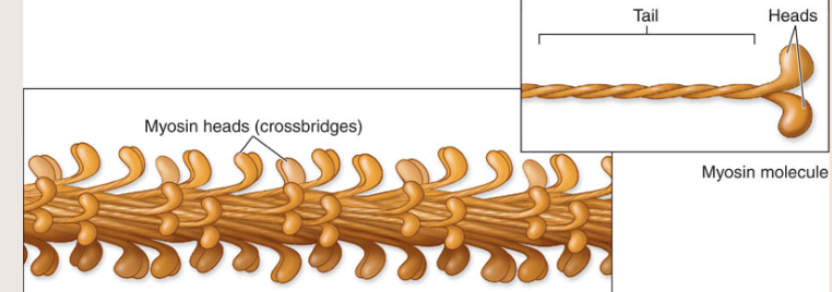
Describe thick myofilament
Myosin heads free, project out from ends
Myosin tails attached, central
Myosin heads can attach to actin, forming crossbridges
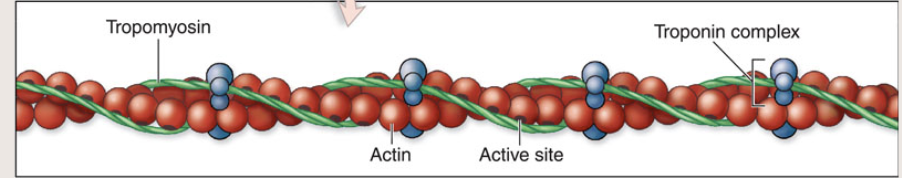
Describe Thin myofilament
Actin & regulatory proteins
Tropomyosin
Troponin
Tropomyosin
Covers sections of actin
Troponin
Attaches to actin & tropomyosin
Binding site for Ca2+
Describe Sarcomeres
Contractile units of myofibrils
Source of fiber’s striations
Banding caused by overlapping arrangement of myofilaments (Actin & Myosin)
What are the parts of a sarcomere?
A (dark) bands
I (light) bands
Z line
H zone
M line
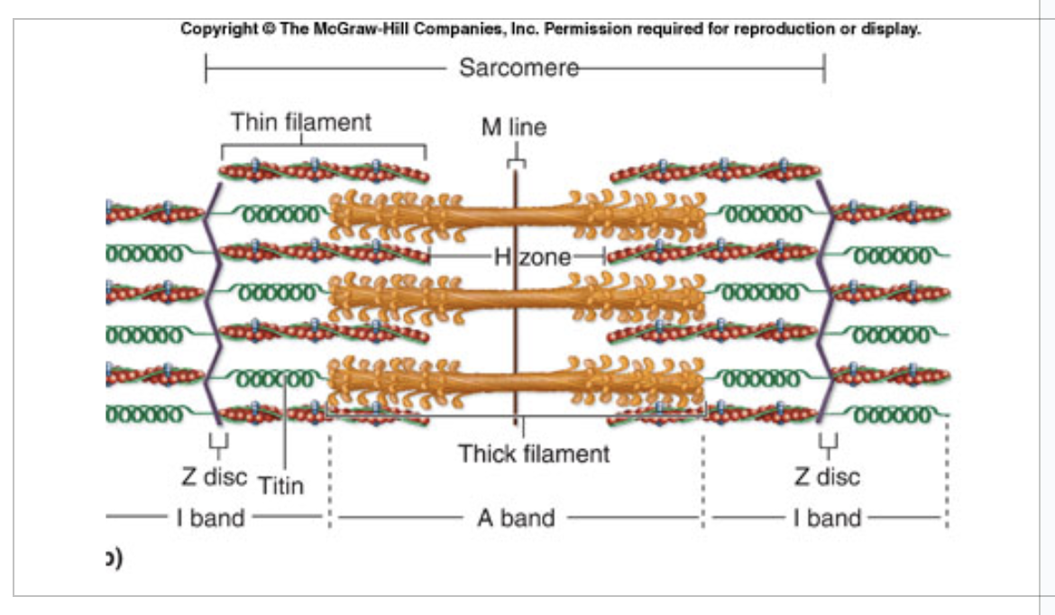
A (Dark) bands
correspond to length of myosin filaments
I (Light) bands
actin (no myosin)
Z line
anchor for myofilaments; separates sarcomeres
H zone
center of A band; no actin
M line
Narrow region at center of H zone; anchor for myosin
Fibers must be _________ to contract
stimulated
Motor neurons deliver the
stimulus
Neuromuscular Junction (NMJ)
point of communication between a motor neuron and a fiber
Fibers & Motors Neurons do not
touch
Neurotransmitter molecules bind to and stimulate
muscle fibers
Neuromuscular Junction (NMJ) Anatomy
Axon Terminal
Synaptic Cleft
Motor End Plate
Axon Terminal
end of motor neuron
Synaptic Cleft (gap)
space between axon terminal & motor end plate
Motor End Plate
Sarcolemma at NMJ
Invaginated (infolded)
High SA (surface area)
ACh (acetylcholine) receptors
“All-or-None” Law
At threshold, a fiber will contract to its maximum extent
Describe the “All-or-None” Law
No “partial” contractions of individual fibers
Increasing stimulus strength has no additional effect
In the “All-or-None”, a single nerve impulses
one contraction
A muscle is composed of
motor units
Motor Unit
a motor neuron and all the fibers it controls
Number of fibers varies
2-2000
Each motor unit responds
independently
All muscle cells in a motor unit respond
maximally, or they don’t respond at all
Strength of contraction is determined by
number of motor units stimulated
Recruitment
Process of increasing the number of motor units responding
Strength increases as number of
motor units recruited increases
Skeletal muscles are capable of
Graded Responses
Different degrees of shortening occur by
Changing the number of motor units activated
[Changing frequency of stimulation]
Red Slow is
Slow Oxidative
White Fast is
fast glycolytic
Intermediate
Fast Oxidative-Glycolytic
Whole muscles contain all 3 types of fibers, which are
Red Slow (Slow Oxidative)
White Fast (Fast Glycolytic)
Intermediate (Fast Oxidative-Glycolytic)
All fibers within same motor unit are of the
same type
Recruitment begins with
red-slow, followed by intermediate, then white-fast
Describe Red Slow:
Fewer myofibrils, weaker
Requires oxygen to make ATP
Lots of myoglobin, mitochondria, capillaries
Contracts slowly; fatigues slowly
Recruited first
Endurance, postural
Describe White Fast
Most myofibrils, strongest & widest in diam.
Makes ATP without oxygen
Low myoglobin, fewer mitochondria & capillaries
High glycogen stores
Contracts rapidly, fatigues rapidly
Short-term, powerful movements
Recruited last
May hypertrophy in response to training
Describe Intermediate
Intermediate diameter/number of myofibrils
Lots of myoglobin, mitochondria, capillaries
Can make some ATP without oxygen
Contracts rapidly, moderately resistant to fatigue
The second fiber type recruited
Postural & occasional rapid, powerful contractions (e.g. gastrocnemius)
Skeletal muscle cells do not undergo
mitosis
Exercise does not increase the number of
skeletal muscle cells
Hypertrophy
Enlargement of muscle cells due to exercise
Describe what happens during hypertrophy?
The number of actin and myosin myofilaments increases
Mitochondria increase
Blood supply increases
Atrophy
Decrease in the size of muscle cells due to lack of use
Describe what happens during Atrophy
The number of actin and myosin myofilaments decreases
Mitochondria decrease
Blood supply decreases
Body movements are produced by contraction of
skeletal muscle
Shortening of a skeletal muscle results in
movement of attachments
Movement depends on
joint, attachments
Skeletal muscles have at least two attachments
One attachment is relatively immobile
The other attachment is more mobile
Origin
Less movable attachment
Insertion
More movable attachment
Action
What the muscle “does”
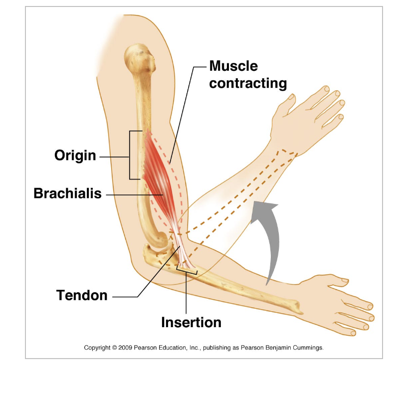
Describe Action
Moves insertion toward origin
The “movement” produced
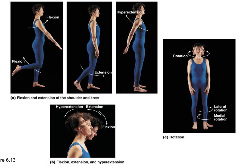
Flexion
decreases angle between bones
Extension
increases angle between bones
Rotation
movement around an axis
Abduction
moves appendage away from midline
Adduction
moves appendage toward midline
Circumduction
moves appendage in a circle around joint

BODY MOVEMENTS
DO THEM
WRITE THEM
ANYTHING TO KNOW THEM
Prime Mover (Agonist)
Muscle primarily responsible for an action
Antagonist
Muscle(s) that resist prime mover, or move opposite to it
Synergist
Muscle(s) that assist(s) prime mover
Fixator
Stabilize origin of prime mover or hold the bone still so all tension is used to move insertion
Muscle attachments
Origin and/or insertion (e.g. sternocleidomastoid)
Muscle action example
Adductor magnus
Direction of muscle fibers example
Rectus abdominis
Location of muscle example
Temporalis
Size of muscle example
Gluteus maximus
Number of origins/heads example
Biceps brachii
Shape of muscle example
Deltoid

MUSCLES
MEMORIZE THEM
ANATOMY
LOOK AT THEM
