The respiratory system
1/74
There's no tags or description
Looks like no tags are added yet.
Name | Mastery | Learn | Test | Matching | Spaced |
|---|
No study sessions yet.
75 Terms
What are the 2 portions of the respiratory tract
air conducting portion
respiratory portion
what is included in the upper respiratory tract
nasal cavities
nasopharynx
larynx
what is included in the lower respiratory tract
trachea
bronchi
bronchioles
respiratory bronchioles
alveolar ducts
alveolar sacs
alveoli
what kind of cells do we find in the air conducting portion?
epithelial lining, with supporting tissues: cartilage, smooth muscle, elastic fibres
What kind of cells do we find in the respiratory portion and why?
simple squamous epithelia + v. scarce loose connective tissue
why? - for optimal gas exchange and diffusion
Outline the structure of respiratory epithelium
brush cells
basal cells
ciliated cells
goblet cells
neuroendocrine cells
respiratory epithelium = ciliated, pseudostratified columnar epithelium
What 3 regions do we find in the nasal cavity
cutaneous region = nasal vestibule
respiratory region
olfactory region
what cells do we find in the cutaneous region
stratified squamous epithelium
what cells do we find in the respiratory region
ciliated, pseudostratified columnar epithelium
= respiratory epithelium
what cells do we find in the olfactory region (general)
olfactory epithelium
what is the structure of the nasal cavity
paired chambers separated by a bony and cartilaginous septum
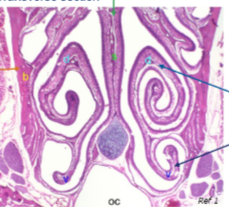
What is this a section of and what can we identify?
transverse section of the nasal cavity
orange arrow = maxillary bone
middle = midline septum
blue arrows = conchae (dorsal and ventral)
b = bone (maxilla)
d = dorsal conchae
oc = oral cavity
s = septum
v = ventral conchae
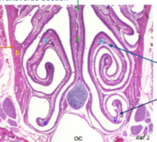
How can we refer to the conchae in chickens?
turbinates
what does the nasopharynx connect?
nasal cavities to the larynx
what makes up most of the structure of the nasopharynx
respiratory epithelium
stratified squamous epithelium caudodorsally along the soft palate
what does the submucosa contain in the nasopharynx and what does it form?
glands
lymph nodules
pharyngeal tonsil
what is the larynx
air passageway b/w oropharynx and trachea
what is the larynx formed from
cartilage plates
it’s a tubular region
what is the function of the larynx
producing sounds - vocal folds
what makes up most of the larynx
respiratory epithelium
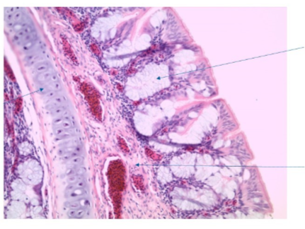
What can we identify in this section and what is it a section of?
cartilage (left arrow)
goblet cells
submucosa with blood vessels (RBCs in birds can be nucleated!)
chicken URT
what is the funciton of the trachea
air tube for air conduction and conditioning of inspired air
what are the 4 layers of the trachea?
mucosa (w. respiratory epithelium)
submucosa
cartilaginous layer
adventitia
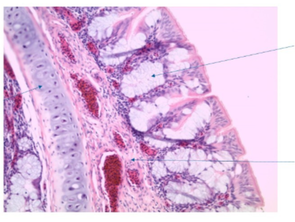
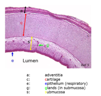
What is the function of the cartilaginous layer
keeps lumen open, cartilage and smooth muscle
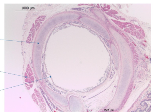
What structure is this and what has been identified
trachea
cartilage rings (top)
skeletal muscle running alongside the trachea
respiratory epithelium
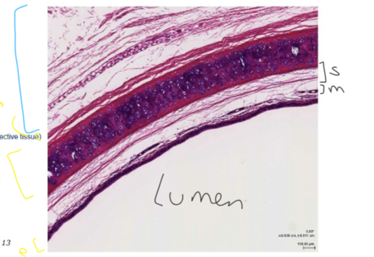
What are the layers we can identify here?
blue = adventitia
then cartilage
then submucosa
mucosa
epithelium
lumen
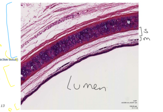
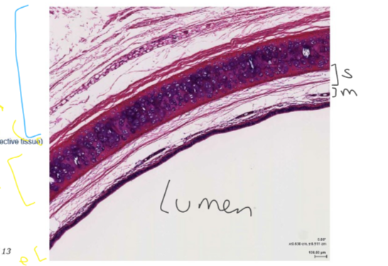
Why may the adventita appear much larger in a slide sample?
held together by weak connective links b/w cells
stretches
When do the bronchi form?
when the bifurcates into 2 main bronchi
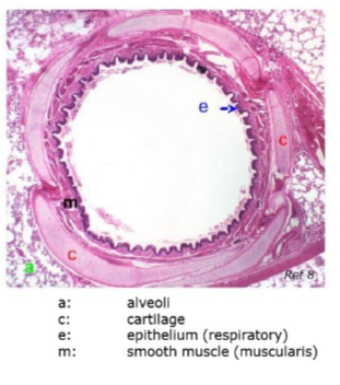
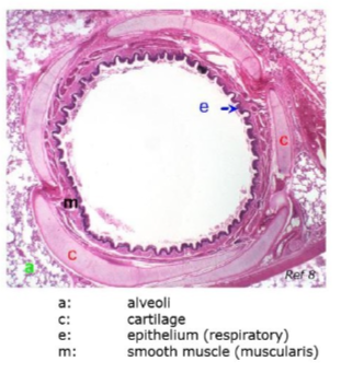
what do we see in the bronchi structure that’s not in the trachea
an additional layer of smooth muscle = muscularis
found b/w mucosa and submucosa
how do we get from the trachea to the alveoli?
trachea → bronchi → bronchioles → alveoli
what happens to the respiratory epithelium as we mov more distally down the bronchioles?
simple columnar
simple cuboidal
still ciliated!
How do the bronchi become bronchioles?
bronchi → intrapulmonary bronchi → terminal bronchioles
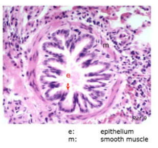
What cell type do we only see in the bronchioles?
epithelium with Clara/Club cells
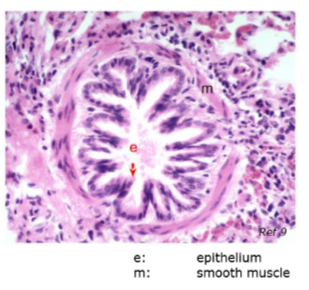
What kind of cell are Clara/club cells
bronchiolar exocrine cells
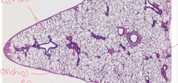
What can we find here?
respiratory pleura
bronchi
bronchioles
alveoli
what structures are ‘conducting’ in the airway?
trachea
extrapulmonary bronchi
intrapulmonary bronchi
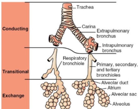
What are the transitional structures in the respiratory tract?
respiratory bronchiole
primary, secondary and tertiary bronchioles
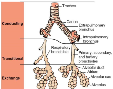
What are the sites of exchange in the airway tract?
alveolar duct
alveolar sac that contain alveoli
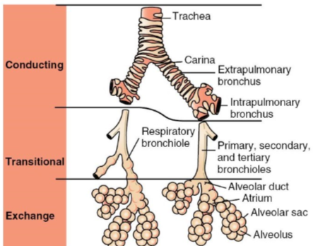
What are respiratory bronchioles
continuation of a terminal bronchiole, interrupted by thin-walled outpocketings = alveoli
what occurs in the respiratory bronchioles
air conduction and gas exchange
= transitional
what cells make up bronchioles
simple cuboidal
what cells make up alveoli?
simple squamous
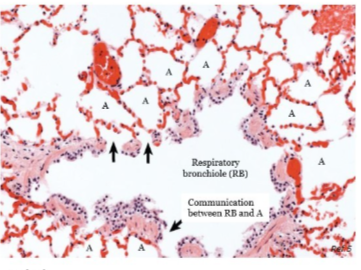
how does the amount of resp. bronchioles vary among species?
carnivores>>herbivores
absent in humans and small rodents
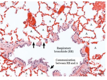
what is the function of alveoli
site of gas exchange b/w air and blood
what is the structure of alveolar ducts?
elongated airways that are lined by alveoli only
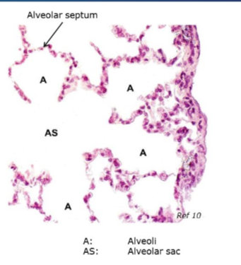
what is the structure of alveolar sacs
spaces surrounded by clusters of alveoli
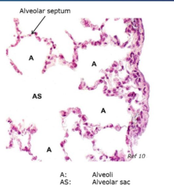
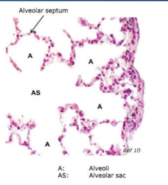
what is the alveolar septum
septal wall
tissue b/w adjacent alveolar air spaces
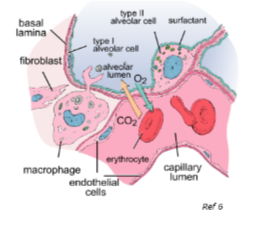
What are the 3 types of cells we find in alveolar epithelium
Type 1 cell
Type 2 cells
Brush cell (rare)
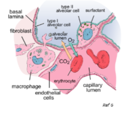
Outline type 1 alveolar cells
type 1 pneumocytes
v. thin, squamous cell, 95% of alveolar surface lining
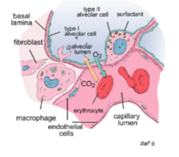
Outline type 2 alveolar cells
type 2 pneumocyte
cuboidal cell
secretes surfactant
covers ~5% alveolar surface
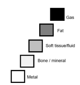
what are the 5 opacities in radiographs?
gas = anechoic = black
fat = dark grey
soft tissue/fluid = lighter grey
white = bone/mineral
bright white = metal
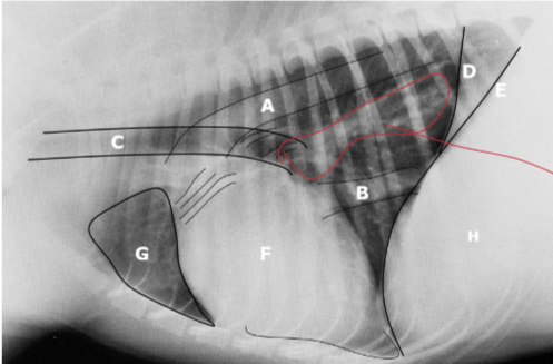
What are each of the letters
A = aorta
B = caudal vena cava
C = trachea
D = crura of diaphragm
E = crura of diphragm
F = cardiac silhouette
G = lung
H = liver
the red outline = normal bronchiole presence in a radiograph
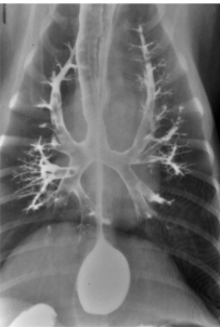
Where are the lobes in a DV radiograph
Working in a clock:
1-2 = left cranial
2-3 = left craniocaudal
3-4 = left caudal
4-5 = accessory
7-8 = right caudal
8-9 = right middle
9-11 = right cranial
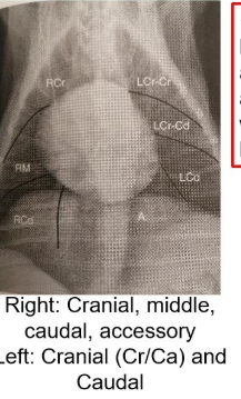
why is knowing about the accessory lung lobe useful?
can be surgically removed
what’s in the lung structures wise which we can see
bronchi
alveoli
interstitial tissue
blood vessels
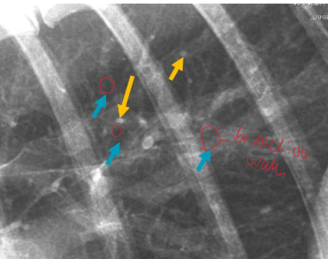
Are these normal? Should we refer for these?
yes, blue = bronchi - gas filled tube, soft tissue wall
yellow = blood vessel - soft tissue wall, fluid filled (same opacitites)
what is the white on abnormal radiographs coming from
usually something that’s already there
fluid
tumour etc
what are the 4 patterns of abnormal lungs on radiographs
bronchial
interstitial: diffuse or nodular
alveolar
vascular
what do normal bronchi look like on a radiograph?
we can see the walls or larger ones (usually towards the middle of the image)
they exist right tot he periphery of the lung lobes - we just can’t see them b/c they’re smaller and thinner walled
what do we see on a lung radiograph that indicates something wrong with bronchi, why and what may be wrong with them?
bronchi look like donuts if cut transversely or train tracks if cut longitudinally
we see bronchi more clearly in the periphery
why = wall becomes mineralised (opacity is more white) and therefore easier to see/wall becomes thicker and is easier to see
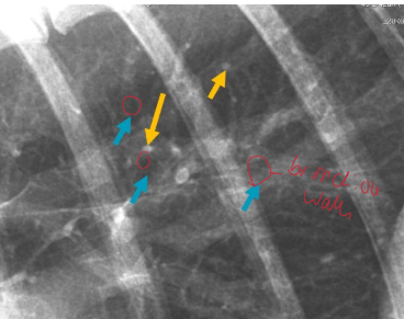
What do bronchial patterns look like on radiographs and what could a cause be
bronchi are more prominent due to thick/mineral opacity
bronchitis
Outline interstitial patterns:
what does it look like normally
what does it look like when abnormal
what are the 2 kinds and how do they differ in appearance?
don’t appreciate it
we see it - thickened e.g. haemorrhage or frank nodules
diffuse = unstructured nodular = soft tissue mass (be careful to not mistake for vessels)
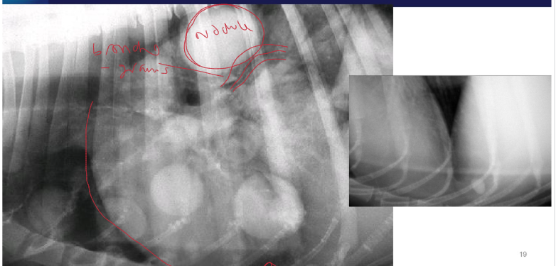
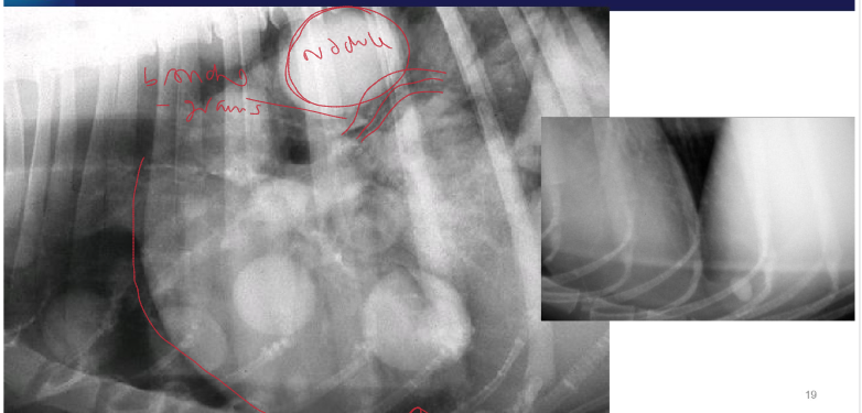
What are the 2 lung patterns present here
interstitial - nodular
alveolar (RHS)
what are key features of an unstructured/diffuse interstitial pattern
can still see soft tissue
air still where it should be (bronchi and alveoli)
can see cardiac silhouette
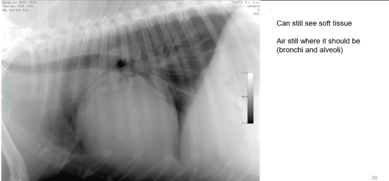
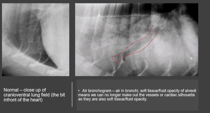
Outline alveolar lung pattern:
what is normal
what is abnormal
what do we see if abnormal
what would the fluid be?
what can’t we see compared to interstitial lung pattern
alveoli filled with air = anechoic = gas opacity
fluid filled or collapsed (soft tissue) but air still in bronchi
soft tissue/fluid opacity, air in bronchi b/c bronchi are currently fine. may be focal/diffuse depending on the case
could be anything e.g. blood/pus/oedema/other
can’t see soft tissue opacities - border obliteration of the heart
Outline vascular pattern:
what’s normal
what’s abnormal
where do we find veins
what should the diameter of the cranial lobar artery and vein be?
can see pulmonary vessels therefore soft tissue opacity
bigger/smaller
ventral and central
same diameter as proximal 1/3 of 4th rib

what should the diameter of the arteries and veins be the same as and at which rib?
9th rib - at the point when they cross this rib
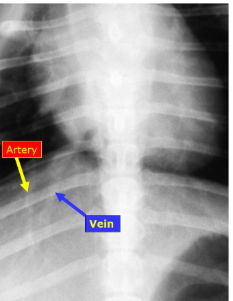
When we have a vascular lung pattern what does it mean when:
vessels are enlarged
if they’re smaller
too much circulation - fluid overload - left to right shunt
too little circulation e.g. hypovolaemia - R → L shunt
lungs look less white - seeing vessels is normal, if they’re small it will be less white, more black on the radiograph

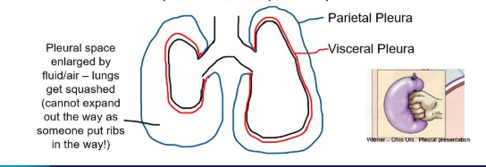
Outline the pleura:
what’s normal?
what’s abnormal?
pleura surrounds lungs but can’t be appreciated on the normal radiograph. Lungs occupy entire space - we can see vessels all the way to the periphery
lungs are pulled back from the edge due to contents (air/fluid/mass) in the pleura and heart may appear elevated
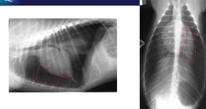
What’s wrong here?
gap beneath lung in right lateral recumbency due to fluid
collapsed lung on the DV view - tissue opacity = what’s left of the lung
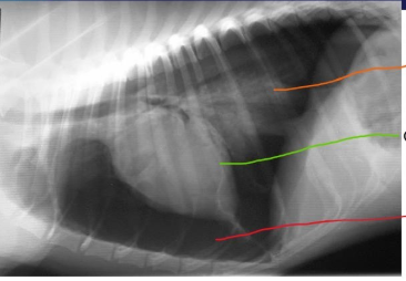
What’s wrong here?
increased opacity (orange)
cardiac silhouette elevated (green)
decreased opacity (red)
vessels aren’t visible in the area of decreased opacity, lungs aren’t there. Gas opacity in the pleural space - lungs unable to expand in with the gas therefore increased opacity
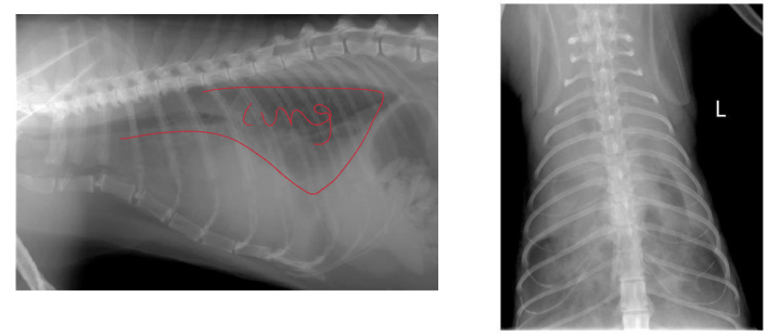
What do these radiographs show?
pleural effusion
can’t see cardiac silhouette, lungs are floating in something, fluid in the pleural space
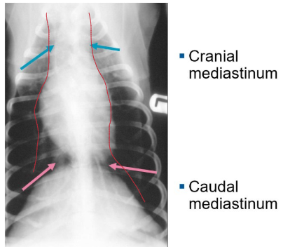
what is the mediastinum, what parts are there to it?
a ‘gap’ b/w the lung lobes in which we find the heart, oesophagus and trachea. It’s not a structure, just a space w/in the thorax.
has a cranial and caudal part
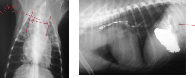
Is this a normal mediastinum?
no
it’s wider than normal, in the cranial mediastinum
soft tissue/fluid opacity that’s pushed the trachea upwards (in right radiograph with the radiopaque areas, that shows the trachea being pushed upwards)
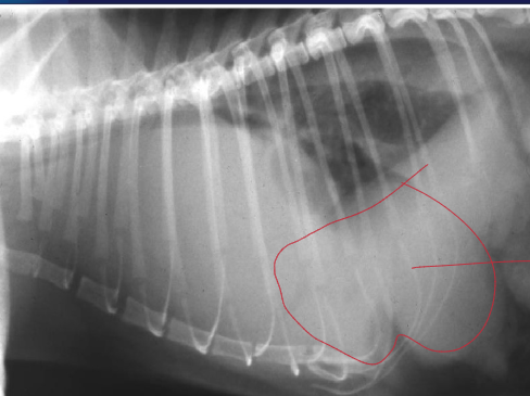
Is this a normal radiograph?
no
can’t see diaphragm, no cardiac outline, poor lung definition…why?
diaphragm has collapsed and the abdominal contents are in the thorax