rads week 8 - pediatric & breast
 Call Kai
 Learn
Learn Practice Test
 Spaced Repetition
 Match
Match Flashcards
 Knowt Play
1/161

There's no tags or description
Looks like no tags are added yet.
Name | Mastery | Learn | Test | Matching | Spaced | Call with Kai | Chat |
|---|
No analytics yet
Send a link to your students to track their progress
162 Terms
What 3 pediatric respiratory symptoms define reactive airway disease/bronchiolitis?
Wheezing, shortness of breath, and coughing.
How does reactive airway disease/bronchiolitis differ from asthma in chronicity?
It is usually transient/self-limited, unlike asthma, although it can progress to asthma.

What CXR sign is associated with peribronchial thickening in reactive airway disease/bronchiolitis?
The donut sign.
What additional linear CXR finding accompanies peribronchial thickening in reactive airway disease/bronchiolitis?
Tram-track densities.
What lung-volume change is common in reactive airway disease/bronchiolitis?
Hyperinflation (hyperaeration).
Why can atelectasis occur in reactive airway disease/bronchiolitis?
Mucus plugging can obstruct an airway and cause distal collapse. Causes mobile structures to go towards.
Classic CXR pattern for reactive airway disease/bronchiolitis?
Peribronchial thickening with donut/tram-track signs + hyperinflation; atelectasis may occur from mucus plugging.
How will asthma flares show on x-ray?
Look normal.
Is asthma primarily a radiologic diagnosis?
No. Asthma is a clinical diagnosis.

Why might a CXR be obtained during an asthma episode?
To evaluate another cause or detect complications.
What 3 CXR-detectable complications of asthma are emphasized?
Atelectasis from mucus plugging, pneumothorax, and pneumomediastinum.
On the asthma image, what does visibility of >10 ribs suggest?
Overinflation/hyperinflation.
On an asthma CXR, visible mediastinal borders due to air indicate what complication?
Pneumomediastinum.
Air in the soft tissues of the neck/chest on the asthma image represents what?
Subcutaneous emphysema.
Most common cause of pneumonia in neonates according to the lecture?
Group B Streptococcus.
How may neonatal pneumonia present?
Often only with fever.
Pneumonia in older infants is usually what type?
Viral.
Typical bacterial pneumonia symptoms in older infants/children?
Fever, chills, tachypnea, cough, chest pain, and shortness of breath.
Typical viral pneumonia symptoms listed in the lecture?
Cough, wheeze, and stridor.
Classic pediatric bacterial pneumonia imaging pattern?
Lobar consolidation.
What rounded opacity can bacterial pneumonia produce in children?
Round pneumonia.
What pleural complication may accompany bacterial pneumonia?
Pleural effusion.
Classic pediatric viral pneumonia imaging pattern?
Patchy/interstitial infiltrates or patchy areas of consolidation, usually on the bases.
A pediatric CXR with dense right middle lobe air-space opacity is an example of what?
Lobar consolidation from pneumonia.
A pediatric CXR with dense right middle lobe air-space opacity ~ how would the patient look like?
Sick, febrile, tachycardic.
A well-defined rounded pulmonary opacity in the bases in a child with pneumonia symptoms suggests what?
Round pneumonia.

Diffuse or patchy bilateral infiltrates in a child are more consistent with what type of pneumonia?
Viral pneumonia.
Most common age range for ingested foreign bodies in this lecture?
6 months to 6 years.
About what percentage of ingested foreign bodies pass spontaneously?
About 80%.
Examples of commonly ingested foreign bodies?
Toys, coins, fish bones, and chicken bones.
What complications can ingested foreign bodies cause if they do not pass normally?
Perforation, obstruction, and stricture formation.
Where can an ingested foreign body become stuck?
The esophagus.
Why is a button/disk battery ingestion an emergency?
It can cause severe burns and perforation.
How can you identify a button battery?
Black line within the outer edge.
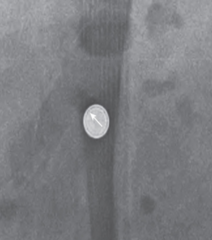
What should be done with an ingested button/disk battery?
It should be removed urgently.
Why are multiple ingested magnets especially dangerous?
They can attract across bowel loops and pull the loops together, causing injury.
Multiple ingested magnets ~ how would you get it out of the bowel?
Laxatives.
On frontal and lateral radiographs, how does an esophageal coin typically appear?
Round/en face on the frontal view (no black line)and edge-on on the lateral view.

What is the big radiology distinction for pediatric bacterial vs viral pneumonia?
Bacterial: lobar or round consolidation ± pleural effusion; viral: interstitial/patchy infiltrates.
What is mammography?
Low-dose x-ray imaging of the breast.
Why is the breast compressed during mammography?
To reduce tissue thickness, reduce scatter/radiation dose, improve image quality, and hold the breast still.
What is breast ultrasound most commonly used with?
Diagnostic mammography.
Main purpose of ultrasound when evaluating a breast mass?
Determine whether the mass is cystic (usually presents as plain black dot) or solid.
Why can ultrasound be especially useful in dense breasts?
It can improve cancer detection over mammography alone and is more sensitive than mammography in dense tissue.
What are the major breast MRI roles emphasized in the lecture?
Screen certain high-risk patients and you reach for it first when to evaluate patients with breast implants. Otherwise, it's not the first line.
Which breast imaging modality is the most sensitive?
MRI.
Which breast imaging modality is the most expensive?
MRI.
What 3 main tissue types make up the breast?
Fat, fibroglandular tissue, and connective tissue. The ratio of all these 3 things will determine how it looks like on imaging.
How does fatty breast tissue appear on mammography?
Darker/less dense.
How does fibroglandular/dense breast tissue appear on mammography?
Whiter/more dense.
Why does dense breast tissue make mammography harder to interpret?
Abnormalities can be obscured within white dense tissue.
What 4 major findings can mammography identify?
Masses, asymmetry, architectural distortion (ie dimpling), and calcifications.
What 4 breast conditions can imaging help diagnose according to the lecture?
Cancer, cysts, fibroadenomas, and abscesses.
What interventional procedure can be performed with breast ultrasound guidance?
Image-guided biopsy.
What 2 risks/downsides of breast imaging are listed? However, what is a caveat?
False positives and overdiagnosis. But it's not a high enough risk to not recommend it.
What factors should be considered for annual breast screening recommendations?
Age and breast cancer risk factors.
Who may recommend additional/special mammographic views?
The radiologist.
What modality is specifically emphasized for a palpable breast mass?
Ultrasound. or if Radiologist recommends.
What are practical limitations of breast MRI despite it being an excellent diagnostic tool?
It can be difficult to get approved, and patients may have difficulty lying still because of the positioning.
Who is a screening mammogram for?
An asymptomatic patient.
What information should accompany a screening mammogram order?
Pertinent history and the date of the last breast imaging.
Can an asymptomatic screening patient still need ultrasound?
Yes, especially with dense breasts or if ultrasound is indicated by the mammogram.
Who needs diagnostic breast imaging rather than routine screening?
A symptomatic patient, such as one with pain, a lump, or nipple discharge.
What histories can also make an exam diagnostic in this lecture?
History of breast cancer or breast implants.
What imaging combination is emphasized for diagnostic breast evaluation?
Mammography plus ultrasound.
What clinical detail should be included when ordering diagnostic imaging for a focal abnormality?
The location and description of the abnormality.
What scoring system is typically included with breast imaging results?
A BI-RADS score.
What does BI-RADS stand for?
Breast Imaging Reporting and Data System.
BI-RADS 0 means what?
Incomplete- additional imaging is needed.
BI-RADS 1 means what?
Negative; routine follow-up.
BI-RADS 2 means what?
They see something but think it's benign; routine follow-up.
BI-RADS 3 means what?
Probably benign; short-interval follow-up, typically in 6 months.
BI-RADS 4 means what?
Suspicious; breast biopsy is recommended/refer to a breast surgeon.
BI-RADS 5 means what?
Highly suggestive of breast cancer; but since you can't diagnose via imaging, refer to a breast surgeon for biopsy.
Why won't a breast imaging report say "no cancer"?
Imaging cannot definitively diagnose or rule out cancer; it only describes how suspicious the findings are. Biopsy is needed for a definitive diagnosis.
A mammogram report showing benign calcifications and BI-RADS 2 should get what follow-up?
Routine/annual follow-up.
How does increasing breast density affect mammography sensitivity?
Sensitivity decreases as density increases (whiter).
What phrase does the lecture use to describe finding cancer in dense breasts?
"Polar bear in a snowstorm." so ur prob ordering u/s and xray in this patient to see better.
What are the 2 standard mammographic views of each breast?
Craniocaudal (CC) [top to bottom] and mediolateral oblique (MLO) [side to side].
How are mammograms displayed relative to the patient?
Patient's right is on your left; patient's left is on your right.
What normal structures are highlighted on the benign mammogram slide?
Fat, fibroglandular tissue, and suspensory ligaments.

What does the MLO view commonly show superiorly along the chest wall?
The pectoralis muscle.
Are fat-containing breast masses usually benign or malignant?
Typically benign.
What 3 categories are used to describe a mammographic mass?
Shape, margins, and density.
What shapes are used to describe breast masses?
Oval, round, or irregular.
What margin descriptors are used for breast masses?
Circumscribed, obscured, microlobulated, indistinct, or spiculated.
What density descriptors are used for breast masses?
High density, equal density, low density, or fat-containing.
What mammographic mass pattern is more suspicious for malignancy?
Irregular + spiculated + high density.

What mammographic mass pattern is more suggestive of benign disease?
Oval + circumscribed + low density.
you can get fat suppression on mri if you're trying to see a fat filled are
Any fat-containing mass is typically:
Benign.
What mass margin is a principal mammographic finding of invasive ductal carcinoma?
Spiculated margins.
A dense spiculated breast mass should raise concern for what diagnosis?
Invasive ductal carcinoma.
How can a breast cyst appear on mammography?
A small usually white/gray oval mass.
Are breast cysts benign or malignant?
Benign.
Can breast cysts be single or multiple and change size over time?
Yes.
Coarse calcifications in a degenerating fibroadenoma are generally what type of finding?
Benign.

What is a fibroadenoma?
A common benign breast tumor.
Main breast ultrasound distinction?
Cystic vs solid mass.
In what patients is breast ultrasound especially useful?
Symptomatic patients and patients with dense breasts.