Psychology Unit 3 AOS 1
 Call Kai
 Learn
Learn Practice Test
 Spaced Repetition
 Match
Match Flashcards
 Knowt Play
1/138

There's no tags or description
Looks like no tags are added yet.
Name | Mastery | Learn | Test | Matching | Spaced | Call with Kai | Chat |
|---|
No analytics yet
Send a link to your students to track their progress
139 Terms
Neuron
Cell within the nervous system that transmits messages to and from the brain, with various functions.
Sensory neuron
Carry sensory information from the sensory organs to the CNS
Motor neuron
Carry motor information from the CNS to effector sites (muscles)
Interneuron
Found only in the CNS
Transmit neural signals/information from
sensory neurons to motor neurons
Structure of the motor neuron
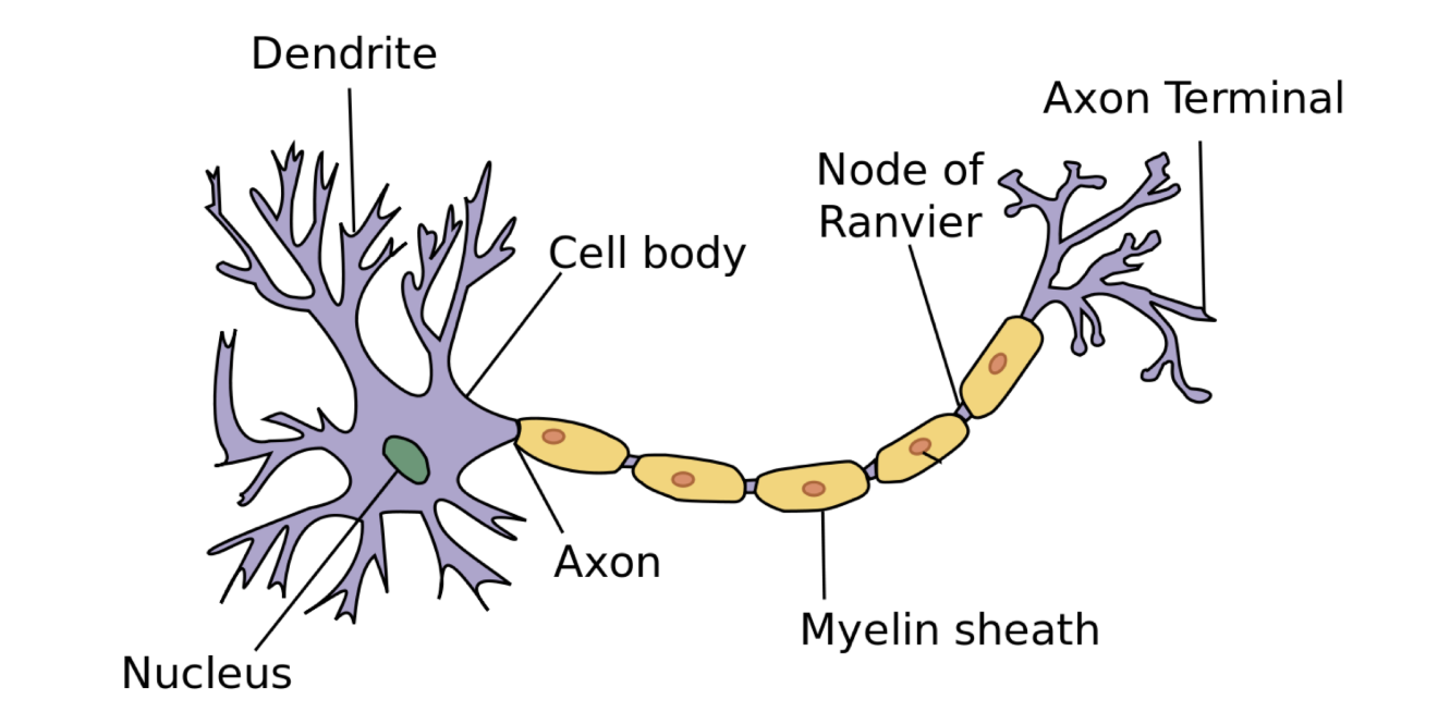
Dendrite
branched extensions of a neuron on which receptor sites are located.
receptor sites
receive neurotransmitters that have been released by another neuron and have travelled across the synapse
Axon
Long fiber of neuron
Transmits neural message (action potential) away from cell body, toward axon terminals
Axon terminals
Release neurotransmitters into the synapse (gap between neurons) so that neural message can be relayed to another neuron.
Presynaptic neuron
the neuron that releases neurochemicals into the neural synapse.
Postsynaptic neuron
the neuron that receives neurochemicals from the neural synapse.
Excitatory effect
Stimulate or activate post-synaptic neurons, increasing their likelihood of firing.
Inhibitory effect
Suppressing or slowing down post-synaptic neuron activity, decreasing their likelihood of firing.
Gamma-aminobutyric acid (GABA)
The main inhibitory neurotransmitter in the NS, that has a role in:
Regulating postsynaptic activation in neural pathways, preventing overexcitation of neurons:
Inhibits excitatory neural signals that contribute to anxiety.
Calms feelings of stress and fear.
Deficiency = low levels of neural inhibition = uncontrolled firing of postsynaptic neurons in neural pathways = anxiety and seizures.
Glutamate
The main excitatory neurotransmitter in the NS, which has a role in:
Learning and memory.
Excitatory effects form and strengthen synaptic connections between neurons that are repeatedly co-activated during learning (LTP).
Excess = overstimulation = neural damage/death, migraine, seizures.
Deficiency = inadequate stimulation of postsynaptic neurons = learning and concentration difficulties, and mental exhaustion.
Neural transmission
An action potential that releases neurochemicals stored the vesicles in the terminal buttons.
This energy goes from the presynaptic neuron into the synaptic gap (space between 2 neurons) for uptake by the receptor sites of the postsynaptic neuron.
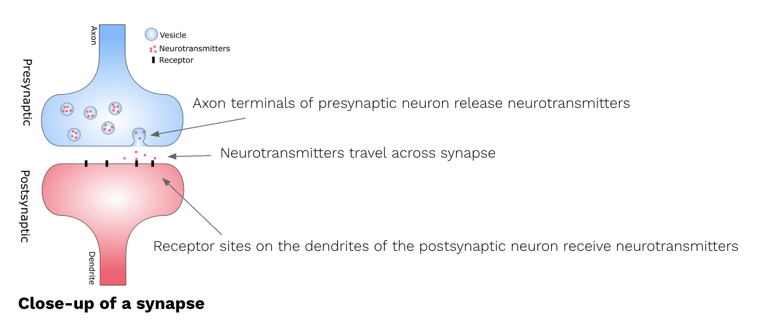
Neurotransmitter
Chemical molecules produced by a neuron that carries a message to, and has a specific effect on one postsynaptic neuron.
Enables rapid communication between 2 neurons across the synapse.
Each Neurotransmitter has a chemically distinct shape, and binds to s pecifiv receptor sites of postsynaptic neurons
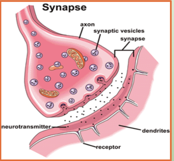
Neurochemical
a chemical substance that transmits neural information within the nervous system
(e.g., neurotransmitters and neuromodulators)
Receptor site
Specialized protein molecules located on the dendrites of a postsynaptic neuron (the receiving neuron)
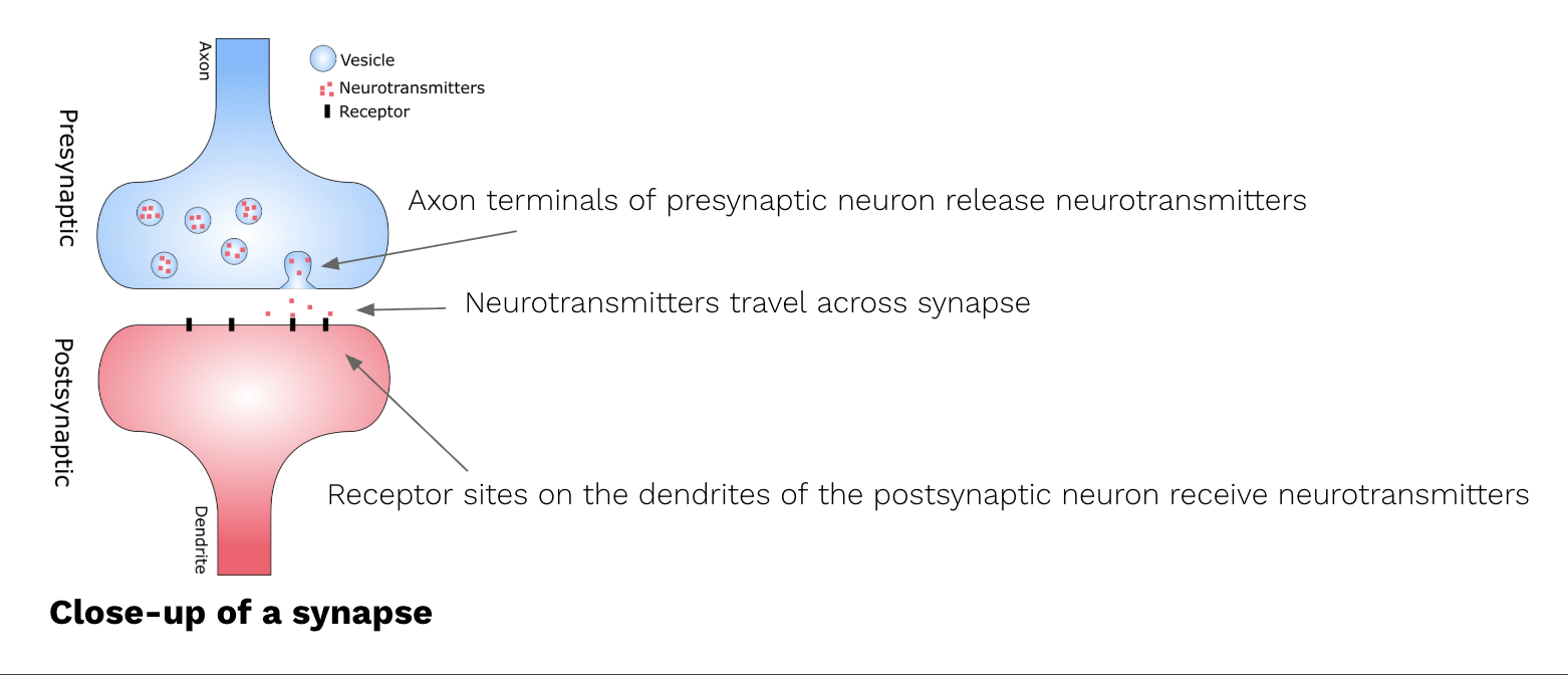
Synapse
The entire junctional area where communication occurs between neurons, comprising the presynaptic neuron, synaptic gap, and postsynaptic neuron
Synaptic gap
The microscopic space between the axon terminal of a sending (presynaptic) neuron and the dendrite of a receiving (postsynaptic) neuron
Synaptic vesicle
small, membrane-bound sacs located within the axon terminals (terminal buttons) of a presynaptic neuron.
They are essential for neural communication, specifically for the storage and release of neurotransmitters.
Synaptic Transmission
An action potential reaches the axon terminals of the presynaptic neuron.
Neurochemicals stored in synaptic vesicles are released into the synaptic gap.
Neurochemicals bind to specific receptor sites on the dendrites of the postsynaptic neuron.
This produces either:
an excitatory effect, making the postsynaptic neuron more likely to fire, or
an inhibitory effect, making the postsynaptic neuron less likely to fire.
Electrochemical Process
Neural communication is electrical within neurons and chemical between neurons.
Electrical component: Dendrites receive a neural signal and the neuron generates an action potential, which travels down the axon as an electrical impulse.
Chemical component: When the action potential reaches the axon terminal, it causes neurochemicals stored in synaptic vesicles to be released into the synapse.
Neuromodulator
A subclass of neurotransmitters.
A chemical molecule that is released into multiple neural synapses, having an effect on multiple postsynaptic neurons, therefore altering neural activity on a larger scale.
Neuromodulator effects on brain activity
Neuromodulators enhance or decrease the strength of communication between neurons
They do this in multiple brain areas
They are released diffusely, affecting multiple neurons (not only point to point like neurotransmitters)
Neuromodulator effects on neurotransmitters
‘Modulates’ the effect of other neurotransmitters by:
Increasing or decreasing the responsiveness of receptor sites to neurotransmitters, enhancing the excitatory or inhibitory effects.
Neuromodulator effects on postsynaptic neuron
Increase in dendritic receptors
Neuromodulator effects on presynaptic neuron
Increasing production and release of neurotransmitters
Neuromodulator effects in synapse
Neuromodulators can also team up and work together with another neurotransmitter in a synapse to make the other more or less potent.
Dopamine
A multifaceted neuromodulator with both excitatory and inhibitory effects, and primarily involved in many CNS functions:
enables smooth, coordinated, voluntary muscle movements.
Dopamine can motivate a person to engage in rewarding behaviours to experience pleasure, e.g. appetite
Associated with addictive behaviour
Attention
Mood
Cognition
Serotonin
A neuromodulator with inhibitory effects primarily responsible for the regulation of mood and sleep.
modulates excitatory effects of neurotransmitters.
Sufficient levels = calm, happy, stable mood.
Low levels = depression
Regulates sleep-wake cycle: influences quality and quantity of sleep (night), feelings of alertness (day).
Dopamine as a neuromodulator
When dopamine levels are higher, neurons in areas related to reward and motivation might react more strongly to incoming signals, enhancing focus, drive, and pleasure.
Serotonin as a neuromodulator
Adjusts the responsiveness of neurons to other signals.
When serotonin levels are higher, neurons might react more strongly to incoming signals, which can help improve mood, regulate sleep, and control appetite.
Neurotransmitters vs Neuromodulators
Neurotransmitters | Neuromodulators |
- Target: a single postsynaptic neuron - Speed of action: moderately fast - Site of release: into the synapse - Role: to transmit chemical signals to adjacent neuron | - Target: groups of neurons - Speed of action: moderately slow, long-lasting effects. - Site of release: outside the synapse into the neural tissue in brain regions - Role: to alter neural transmission between neurons |
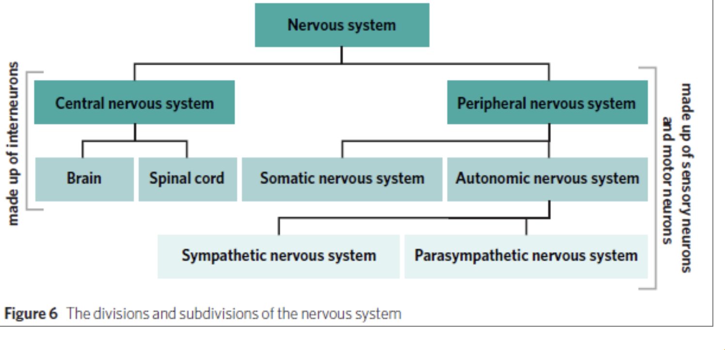
Subdivisions of the human nervous system
CNS—> Brain + Spinal cord
PNS—> Automatic nervous system + Somatic nervous system
ANS—> Sympathetic nervous system + parasympathetic nervous system + enteric NS
Explain the role of the central nervous system
The central nervous system (CNS) is made up of the brain and the spinal cord.
Responsible for transmitting neural messages to and receiving neural messages from the peripheral nervous system.
Explain the role of the central nervous system- Brain
The body’s information center, responsible for coordinating and processing actions, thoughts and behaviour.
Spinal cord
A long cable of nerve tissue (neurons) that extends from the brain, connecting it to the peripheral nervous system.
Explain the role of the central nervous system- Spinal cord
Carry sensory information such as pain, heat, or an itch on your skin, from the various areas of the body such as the arms, legs, and external organs, to the brain for processing
Carry motor (movement) information from the brain to the relevant parts of the body such as the muscles, glands, and organs so that action can be taken
Explain the role of the peripheral nervous system
It has two functions:
1. Carrying information to the spinal cord: communicates sensory information from the body’s organs, glands and muscles to the CNS. This includes information from the outside world to the CNS.
2. Carrying information from the spinal cord: Sends motor information from the CNS to the muscles, organs and glands of the body.
Explain the role of the somatic nervous system
network of nerves- carries sensory info @ receptor sites to the central NS via sensory neurons.
carries motor info from the central NS to the voluntary muscles of the body via motor neurons.
Explain the role of the autonomic nervous system
The autonomic nervous system (ANS) is responsible for regulating visceral muscles, organs and glands
e.g heart, stomach and liver,
It transmits neural messages to the central nervous system about their activity and
initiates the responses of the body without conscious control.
Explain the role of the sympathetic nervous system
Immediately increases the activity of visceral muscles, organs and glands at times of vigorous activity, stress or threat.
Ready to deal with stressful or threatening situations
(fight/flight response)
Explain the role of the parasympathetic nervous system
Gradually decreases the activity of visceral muscles, organs and glands once the threat has been eliminated.
It aims to maintain a balanced state of the body called homeostasis (normal levels of functioning most of the day).
Conscious response
Deliberate and voluntary actions that are intentionally initiated by the brain and performed by the body.
sensory receptor
nerve ending that detects internal sensations in the body and external sensations from the environment
Process of a Conscious Response
Sensory stimulus comes into contact with sensory receptors on sensory organs.
This sensory neural message is transmitted via afferent pathways in the somatic NS.
Interneurons in the brain receive and processes this sensory information, coordinating and initiating a conscious motor response to perform the voluntary action.
This motor neural message is transmitted via efferent pathways in the spinal cord, and then the somatic NS, to skeletal muscles.
Skeletal muscles carry out the conscious motor response to the sensory stimulus.
Unconscious response
Automatic and involuntary actions that are performed by the body independently of the brain (however, these actions still rely on feedback from the brain)
They occur without conscious awareness in response to internal and external sensory stimuli
Unconscious physiological responses of the sympathetic nervous system
Heart rate increases
Breathing rate increases
Pupils dialate
Unconscious physiological responses of the parasympathetic nervous system
Steady and regular heart rate
Steady and regular breathing rate
Pupils constrict according to external light levels
Spinal reflex
An unconscious, automatic, involuntary withdrawal response to intense sensory stimuli that is initiated by interneurons in the spinal cord independently of the brain.
Requires less processing than conscious responses and neural signals involved travel shorter distances to the spinal cord than conscious responses do to the brain.
Adaptive: evolved to protect humans from harm
How the spinal reflex enhances survival
allows for responses to danger before consciously registering it.
Faster reaction time = increased chance of survival.
process of spinal reflex
Sensory receptors in (part of the body) detect the extreme sensation of pain caused by the stimulus.
This sensory information is sent via afferent tracts in the somatic nervous system to the spinal cord.
Interneurons intercept and relay a motor message to withdraw/move (body part) via efferent tracts out to the relevant skeletal muscles.
The individual will immediately withdraw/move away from the harmful stimulus.
While the spinal reflex is occurring, the sensory information continues to the brain for processing and coordination of further conscious responses.
SAME (acronym)
Sensory Afferent Motor Efferent
Afferent= sensory messages toward the brain (through afferent tracts)
Efferent= motor messages away from the brain(through efferent tracts)
Neuroplasticity
The ability of the brain to physically change in response to activity and experience.
Formation of new neurons, increase in brain size/mass, changes to where particular functions are performed in the brain.
Synaptic plasticity
Type of neuroplasticity – the ability of synaptic connections to change over time in response to activity or experience
3 mechanisms of synaptic plasticity
formation, strengthening, or weakening of synaptic connections
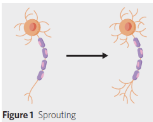
Sprouting
Dendrites or axons develop new extensions
Increases the reach of the neuron
Enables the strengthening of synaptic connections.
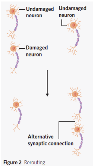
Rerouting
A neuron connected to a damaged neuron creates an alternative synaptic connection with an undamaged neuron
Reestablishes a synaptic connection
restores brain functioning.
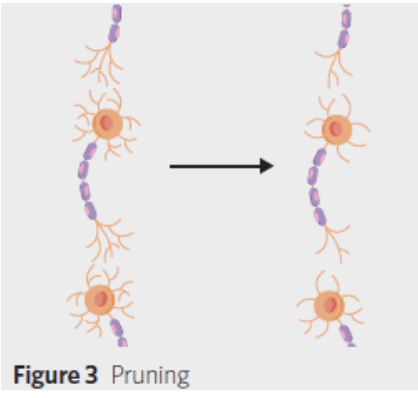
Pruning
Synaptic connections inadequately activated are eliminated
Accommodates for stronger and more essential synaptic connections
Enhances the efficiency of brain functioning
Learning
Acquiring knowledge, skills, or behaviours through experience.
Memory
Encoding, storing, and retrieving information that has been previously encountered.
Co-activation
the simultaneous firing of the pre-synaptic and post-synaptic neurons.
Memory Trace definition + example
Neural pathways that form during learning as a result of synaptic plasticity.
Each memory trace represents a different memory.
Learning how to ride a bike.
Neurons involved in bike riding are regularly coactivated. As you continue to practise, these neurons are repeatedly coactivated (pre and postsynaptic neurons activated at the same time).
E.g. neural pathway associated with balance and neural pathway associated with motor movement of pedalling activated at the same time = synaptic connection strengthened.
Synaptic connections between these neurons physically change (synaptic plasticity: strengthened).
A memory trace forms that represents the learnt info of how to ride a bike.
Long-Term Potentiation – Synaptic Plasticity
Long-lasting and experience-dependent strengthening of synaptic connections between neurons in neural pathways that are regularly coactivated.
Strengthening at synapse
= neurons more likely to fire an action potential together again more efficiently in the future, and so, form a pathway
= neural info can travel quickly.
“Neurons that fire together, wire together”
LTP during Memory and Learning
Glutamate is repeatedly released by presynaptic neuron and received by postsynaptic neuron (repeated coactivation)
= high-intensity stimulation of postsynaptic neuron
= strengthens synaptic connection between neurons
LTP- Change in Structure to Neural Synapse
- Growth of axon extensions on axon terminals of presynaptic neuron = more terminal buttons.
- Increase in dendritic spines on postsynaptic neuron due to sprouting = bushier dendrites = more receptor sites.
LTP- Change in Function to Neural Synapse
Increase in the amount of the neurotransmitters (glutamate) produced and released by the presynaptic neuron.
Postsynaptic neurons are more receptive to neural signals from presynaptic neurons and more readily activated → increases the efficiency of synaptic transmission.
LTP- Memory and learning example
Learning to play the guitar – repeatedly practises the guitar.
Synaptic connections involved in playing the guitar are repeatedly coactivated, and thus strengthened (LTP).
Memory trace that represents the process of playing the guitar is formed and strengthened so playing the guitar becomes easier.
Long-Term Depression – Synaptic Plasticity
The long-lasting and experience-dependent weakening of synaptic connections between neurons in neural pathways that are infrequently coactivated.
LTD during Memory and Learning
Previously established neural pathway is infrequently activated
lack of high-intensity postsynaptic neuron stimulation
LTD weakens synaptic connections of neural pathway no longer necessary
accommodates for more necessary memory traces.
LTD- Change in Structure to Neural Synapse
- Decreased no. of dendrites on postsynaptic neurons due to pruning = decreased number of receptor sites.
- Decreased no. of synaptic connections between neurons due to pruning.
- Less glutamate is produced and released by presynaptic neuron.
LTD- Change in Function to Neural Synapse
Post synaptic neuron less receptive to neural signals from presynaptic neuron
= less rapidly activated
LTD- Memory and learning example
Stops playing guitar – stops repeatedly practising the guitar.
Synaptic connections involved in playing the guitar are no longer repeatedly coactivated and thus, weakened (LTD).
Memory trace that represents the process of playing the guitar is weakened so playing the guitar becomes more difficult.
(Overview) Synaptic Plasticity in Learning and Memory
Repeated experience strengthens synapses (LTP), forming durable memory traces
unused pathways weaken (LTD), allowing efficient learning.
Dopamine and addiction
Process of addiction:
Unhealthy behaviours, such as computer gaming or gambling
Increased dopamine in the reward pathway, producing feelings of pleasure
Over time, less dopamine is produced, diminishing the brain’s supply and therefore the feelings of pleasure.
This increases the urge to continue playing and seeking out the same feelings of pleasure
reuptake
Neurochemicals that do not bind to receptors in the postsynaptic neuron are absorbed back into the terminal buttons of the presynaptic neuron
Stress
A psychological and physiological experience
Occurs when an individual encounters something of significance that demands their attention and/or efforts to cope.
Stressor
An internal or external stimulus that prompts the stress response
E.g. Living in a country experiencing war, moving to a new city, having two exams in a day
psychological stress response
How we think or feel about a stressor
influences an experience of stress
differs between people
The same stressor can cause someone to experience a positive psychological state (e.g., excitement)
At the same time, it can cause someone else to experience a negative psychological state (e.g., worry).
Physiological stress response
How the body reacts to a stressor. An immediate threat requires a quick response.
E.g. The flight-or-fight-or- freeze response
Internal stressor
a stimulus from within a person’s body that prompts the stress response
E.g. Attitude, Rumination, Low self-esteem
External stressor
A stimulus from outside of a person’s body that prompts the stress response.
E.g. vce, deadlines, car accident, ill family member
Acute stress
A form of stress characterised by intense psychological and physiological symptoms that are brief in duration, such as the fight-flight-freeze response.
E.g. slamming on brakes, taking an exam, public speaking.
The flight-or-fight-or-freeze response
An involuntary and automatic response to a threat that takes the form of either escaping it, confronting it, or freezing in the face of it.
Fight-Flight response physiological features
Both are initiated by the activation of the Sympathetic Nervous System
Increases heart rate & respiration
Diverts blood to muscles
Dilation of pupils
Energises body overall
Freeze response physiological features
Body movements and vocalisations stop (immobility and shock)
Racing heart slows significantly
Blood pressure drops very quickly due to activating and dominating parasympathetic NS
How is the fight-flight response activated
Threat is detected by the amygdala
Activates the hypothalamus
activates the sympathetic NS
Stimulates the adrenal medulla
Adrenal glands secrete adrenaline and noradrenaline into the bloodstream
Circulation activates fight-flight reactions
How the freeze response is activated
Sympathetic NS activates first.
Parasympathetic NS then activates at the same time and dominates whilst both are active.
This immobilises the individual.
Allows rapid switch to full sympathetic fight-or-flight if needed.
Chronic stress defintion + examples
A state of prolonged and persistent arousal involving a stressor that exists for an extended period and the release of cortisol.
E.g. prolonged financial hardship, abusive or unhappy relationships, high-pressure job
Cortisol
A hormone that is released in times of stress to aid the body in initiating and maintaining heightened arousal.
Positive effects of the short term increase of cortisol
Adaptive process because it
energises the body by increasing energy supplies such as blood sugar and enhancing metabolism.
has anti-inflammatory effects - blocking the activity of white blood cells that contribute to inflammation.
Negative effects of the increase of cortisol
Maladaptive because it suppresses the immune system
Prolonged cortisol (cortisol starts to become dysfunctional)
Slows down tissue repair - widespread inflammation
Impairs the immune system
Vulnerability to disease
The three stages of GAS
Alarm reaction: - Shock + countershock
Resistance
Exhaustion
The body’s level of resistance to stress in each stage of the GAS model
Alarm reaction- shock: ability to deal with stress drops
Alarm reaction- countershock: ability to deal with stress rises back up to ‘normal’ levels
Resistance- ready to fight the challenge → resistance rises above normal
Exhaustion- ability to deal with stress starts to decline
In which stage of the GAS model is cortisol released?
Alarm reaction: counter shock
Resistance
Cortisol’s role in the alarm reaction- countershock stage
energise the body and facilitate the fight-flight-freeze response
ensures the body has sustained energy to continue dealing with the stressor
Cortisol’s role in the resistance stage
Cortisol allows for unnecessary physiological processes to shut down: digestion, growth, menstruation stops etc..
Cortisol boosts our energy system; this is what increases your resistance to stress
Latter half/Prolonged release of cortisol= immune system vulnerable.
‘Flu like’ symptoms develop, ability to resist stress starts to decline
Cortisol’s role in the exhaustion stage
Cortisol levels decrease after a sustained period of increased levels during resistance
Cortisol depletes your body’s physiological resources
Makes you vulnerable to serious illness and or mental health disorders
E.g. high blood pressure, heart conditions
Alarm reaction- shock: role of parasympathetic NS
blood pressure and body temperature drop