Lecture 5: Ultrasonography, Computed Tomography, and MRI
 Call Kai
 Learn
Learn Practice Test
 Spaced Repetition
 Match
Match Flashcards
 Knowt Play
1/39

There's no tags or description
Looks like no tags are added yet.
Name | Mastery | Learn | Test | Matching | Spaced | Call with Kai | Chat |
|---|
No analytics yet
Send a link to your students to track their progress
40 Terms
How is an ultrasound image generated?
high-frequency sound waves penetrate tissue (or don’t) and bounce back to transducer → converted by crystals to electric current → computer converts electric current to image
When a substance appears black on an ultrasound, what does that mean is happening to the sound waves?
waves are passing through (fluid)
When a substance appears white on an ultrasound, what does that mean is happening to the sound waves?
waves reflect fully (bone/air)
When a substance appears gray on an ultrasound, what does that mean is happening to the sound waves?
partial reflection (soft tissue)
Why is lung ultrasound imaging difficult?
ultrasounds hate “air” and all waves bounce back
What is the ultrasonography terminology?
anechoic: black
hypoechoic: darker
isoechoic: same
hyperechoic: lighter
reflective (air)
What is an acoustic shadow?
an ultrasound artifact: cannot see below a structure that reflects back all waves
What is acoustic enhancement?
an ultrasound artifact: brightening deep to an anechoic structure
What is reverberation?
an ultrasound artifact: repeated reflections between strong surfaces (common in lung)
What is an ultrasound mirror image?
an ultrasound artifact: duplicate structures across a reflector (most common from thorax/abdomen interface
What is slice-thickness in an ultrasound?
occurs when imaging a 3D structure with anechoic fluid (most common in gall bladder = artificial sludge)
What is edge-shadowing artifact in an ultrasound?
sound waves bend as they a curved surface tangentially
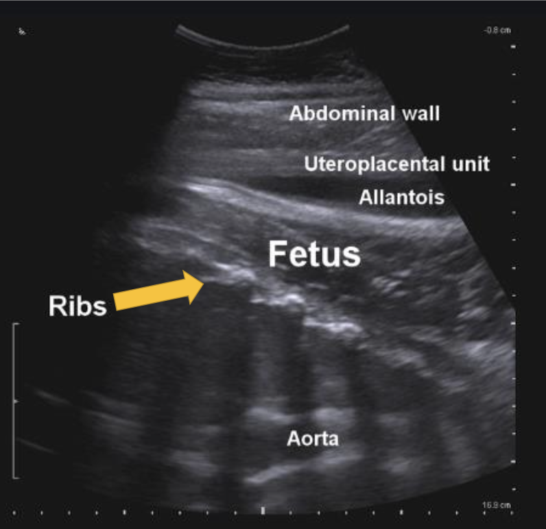
What ultrasound artifact is being demonstrated here?
acoustic shadow
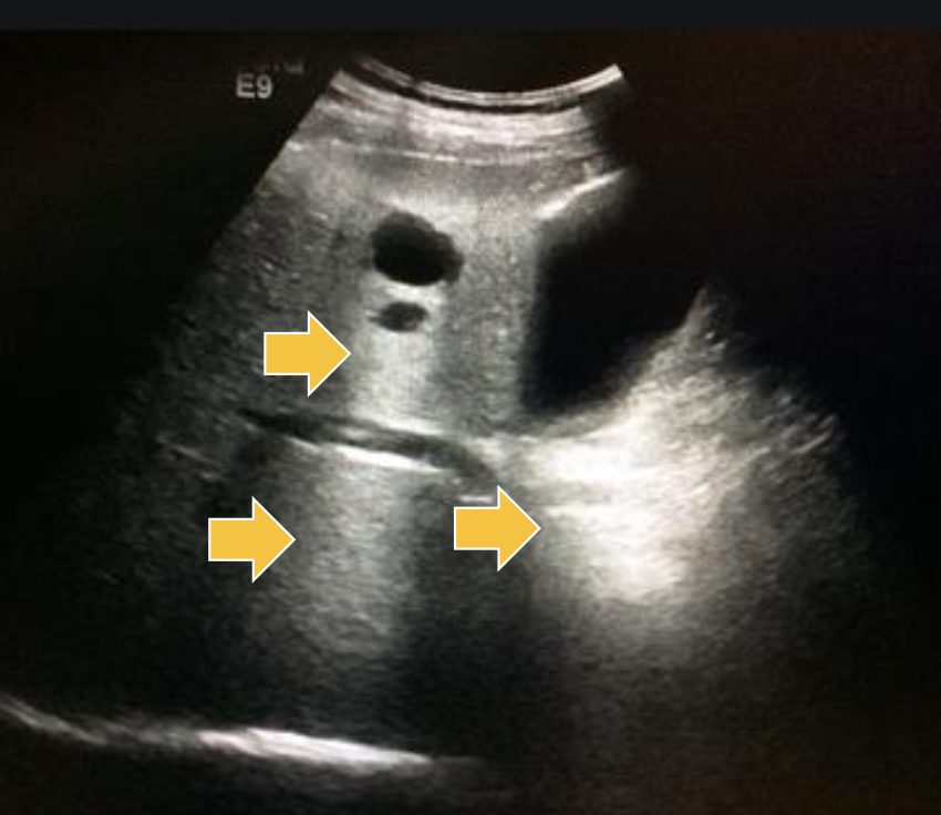
What ultrasound artifact is being demonstrated here?
acoustic enhancement
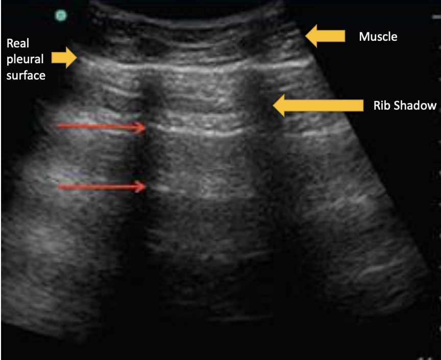
What ultrasound artifact is being demonstrated here?
reverberation effect
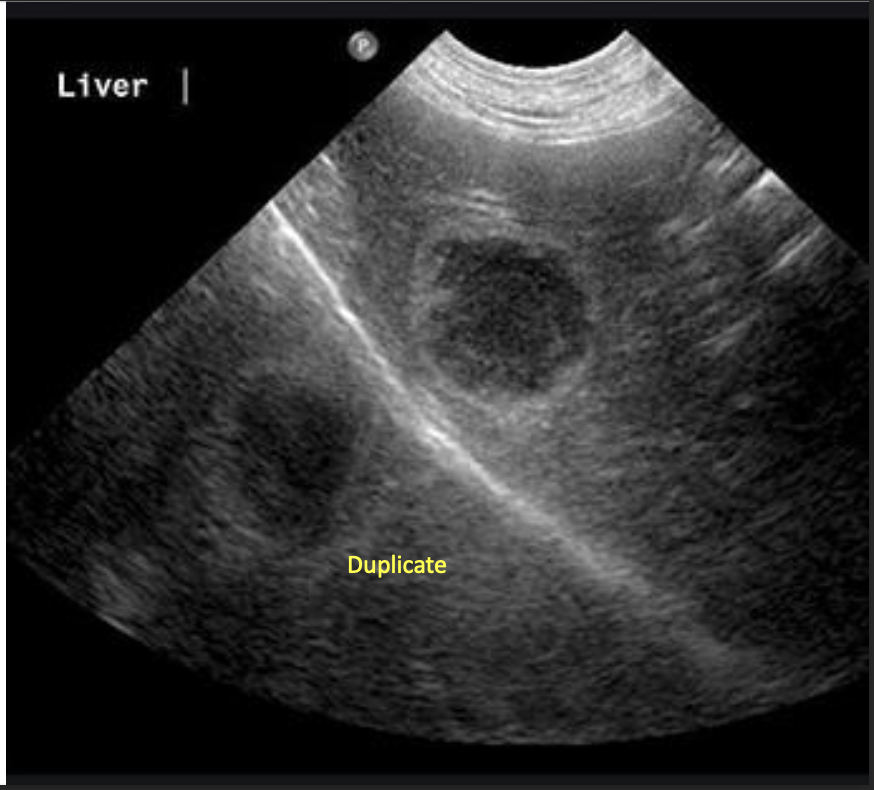
What ultrasound artifact is being demonstrated here?
mirror image artifact
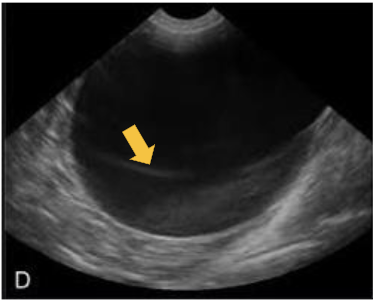
What ultrasound artifact is being demonstrated here?
slice thickness artifact
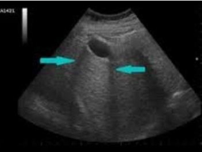
What ultrasound artifact is being demonstrated here?
edge-shadowing artifact
What are the two types of probes typically used in ultrasound?
linear (high frequency) = excellent resolution, shallow depth (equine tendons, SA abdomen or thorax)
curvilinear/sector = deeper penetration, lower resolution (LA thorax and abdomen)
What does frequency determine in ultrasound probes?
increased frequency: greater resolution but less depth
decreased frequency: greater depth but less resolution
What is the function of real-time ultrasound imaging (doppler mode)?
can measure movement (heart beat)
can assess direction of flow (ex. regurgitation of blood through a leaky valve)
How is a computed tomography (CT) scan generated?
x-ray tube rotates → detectors collect intensity data → computer reconstructs slices → software makes 3D images
What practical application do CT scans have in veterinary medicine?
excellent for bone detail and fracture planning
What is needed for soft tissue CT evaluation?
contrast injection
What are the advantages of CT scans?
fast
detailed bone images
3D reconstructions
What are the disadvantages of CT scans?
usually require anesthesia
radiation exposure
contrast risk
How is an MRI image generated?
tissues have hydrogen protons (water) → strong magnetic field aligns protons → radiofrequency pulse excites them → relaxation emits signal → different tissues relax at different rates → superior soft tissue contrast
What must be removed on a horse before an MRI?
horseshoes (not metal)
What are the advantages to MRI?
superior soft tissue contrast
evaluates cartilage and ligaments better than CT
What are the disadvantages of MRI?
expensive
requires removal of ferromagnetic materials (horseshoes)
anesthesia
specialized equipment
What are the MRI indications in small animals?
neuroimaging
musculoskeletal
tumor staging
What are the indications for ultrasound?
Tendons, ligaments, joints, abdomen, reproductive tract, heart, thoracic fluid, superficial bone irregularity.
What are the MRI indications for horses?
Foot lesions, musculoskeletal injuries not accessible to ultrasound, soft tissue imaging
How is an image generated using nuclear scintigraphy?
inject radiopharmaceutical → binds active bone formation sites → gamma camera detects decay signals → increased uptake = increased bone turnover
What are the equine indications for nuclear scintigraphy?
Localize lameness when radiographs/blocks fail, multiple limb lameness, axial skeleton issues, occult fractures
What are the small animal indications for nuclear scintigraphy?
Renal function, thyroid evaluation, musculoskeletal studies
If I want a 3D image of bone, what is my choice of imaging?
CT
If I want a 3D image of soft tissue, what is my choice of imaging?
MRI
If I want a 2D image of soft tissue, what is my choice of imaging?
ultrasound
If I want a 2D image of bone, what is my choice of imaging?
x-ray