hhp 210L: unit 1 pictures??? decide how to divide it up later

0.0(0)
Studied by 0 people
call kaiCall Kai
Locked
learnLearn
examPractice Test
spaced repetitionSpaced Repetition
heart puzzleMatch
flashcardsFlashcards
GameKnowt Play
Card Sorting

1/9

encourage image

There's no tags or description

Looks like no tags are added yet.

Last updated 3:33 AM on 9/1/26
Name
Mastery
Learn
Test
Matching
Spaced
Call with Kai
Chat

No analytics yet

Send a link to your students to track their progress

10 Terms
1
New cards
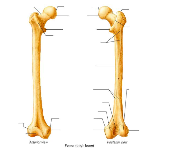
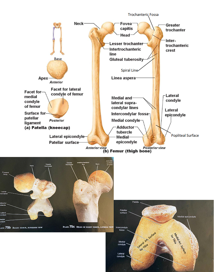
<p>Label these parts of a femur</p><ul><li><p>head</p></li><li><p>fovea capitis</p></li><li><p>neck</p></li><li><p>greater trochanter</p></li><li><p>lesser trochanter</p></li><li><p>trochanteric fossa</p></li><li><p>intertrochanteric crest </p></li><li><p>intertrochanteric line</p></li><li><p>gluteal tuberosity</p></li><li><p>spiral line</p></li><li><p>linea aspera: medial and lateral lips??</p></li><li><p>condyles: medial and lateral</p></li><li><p>epicondyles: medial and lateral</p></li><li><p>supracondylar ridges: medial and lateral</p></li><li><p>adductor tubercle</p></li><li><p>intercondylar fossa</p></li><li><p>popliteal surface</p></li><li><p>patellar surface</p></li><li><p>articular surface for the tibia: medial and lateral</p></li></ul><p></p>

Label these parts of a femur

  • head

  • fovea capitis

  • neck

  • greater trochanter

  • lesser trochanter

  • trochanteric fossa

  • intertrochanteric crest

  • intertrochanteric line

  • gluteal tuberosity

  • spiral line

  • linea aspera: medial and lateral lips??

  • condyles: medial and lateral

  • epicondyles: medial and lateral

  • supracondylar ridges: medial and lateral

  • adductor tubercle

  • intercondylar fossa

  • popliteal surface

  • patellar surface

  • articular surface for the tibia: medial and lateral

knowt flashcard image
2
New cards
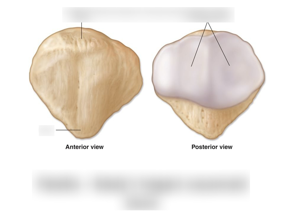
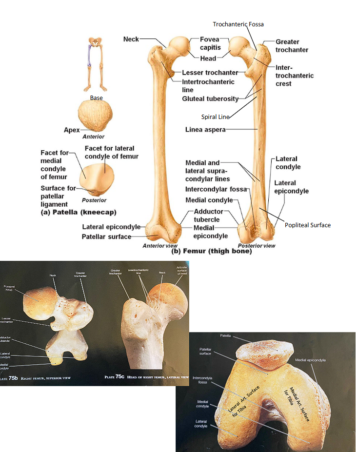
<p>label these parts of a patella</p><ul><li><p>base</p></li><li><p>apex</p></li><li><p>facets for the femur: medial and lateral</p></li></ul><p></p>

label these parts of a patella

  • base

  • apex

  • facets for the femur: medial and lateral

knowt flashcard image
3
New cards
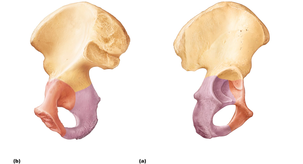
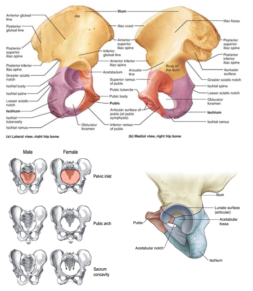
<p>label these parts of an innominate bone</p><ul><li><p>obturator foramen</p></li><li><p>iliac crest</p></li><li><p>iliac fossa</p></li><li><p>anterior superior iliac spine</p></li><li><p>anterior inferior iliac spine</p></li><li><p>posterior inferior iliac spine</p></li><li><p>greater sciatic notch</p></li><li><p>gluteal lines: anterior, posterior and inferior</p></li><li><p>auricular surface</p></li></ul><ul><li><p>ischial tuberosity</p></li><li><p>ischial spine</p></li><li><p>lesser sciatic notch</p></li><li><p>ischial ramus</p></li></ul><ul><li><p>pubis body</p></li><li><p>surface for pubic symphysis</p></li><li><p>superior ramus</p></li><li><p>inferior ramus</p></li></ul><ul><li><p>lunate articular surface</p></li><li><p>acetabular notch</p></li><li><p>acetabular fossa</p></li></ul><p></p>

label these parts of an innominate bone

  • obturator foramen

  • iliac crest

  • iliac fossa

  • anterior superior iliac spine

  • anterior inferior iliac spine

  • posterior inferior iliac spine

  • greater sciatic notch

  • gluteal lines: anterior, posterior and inferior

  • auricular surface

  • ischial tuberosity

  • ischial spine

  • lesser sciatic notch

  • ischial ramus

  • pubis body

  • surface for pubic symphysis

  • superior ramus

  • inferior ramus

  • lunate articular surface

  • acetabular notch

  • acetabular fossa


ilium

  • obturator foramen

  • iliac crest

  • iliac fossa

  • anterior superior iliac spine

  • anterior inferior iliac spine

  • posterior inferior iliac spine

  • greater sciatic notch

  • gluteal lines: anterior, posterior and inferior

  • auricular surface

ischium

  • ischial tuberosity

  • ischial spine

  • lesser sciatic notch

  • ischial ramus

pubis

  • body

  • surface for pubic symphysis

  • superior ramus

  • inferior ramus

acetabulum

  • lunate articular surface

  • acetabular notch

  • acetabular fossa


<p>ilium</p><ul><li><p>obturator foramen</p></li><li><p>iliac crest</p></li><li><p>iliac fossa</p></li><li><p>anterior superior iliac spine</p></li><li><p>anterior inferior iliac spine</p></li><li><p>posterior inferior iliac spine</p></li><li><p>greater sciatic notch</p></li><li><p>gluteal lines: anterior, posterior and inferior</p></li><li><p>auricular surface</p></li></ul><p>ischium</p><ul><li><p>ischial tuberosity</p></li><li><p>ischial spine</p></li><li><p>lesser sciatic notch</p></li><li><p>ischial ramus</p></li></ul><p>pubis</p><ul><li><p>body</p></li><li><p>surface for pubic symphysis</p></li><li><p>superior ramus</p></li><li><p>inferior ramus</p></li></ul><p>acetabulum</p><ul><li><p>lunate articular surface </p></li><li><p>acetabular notch</p></li><li><p>acetabular fossa</p></li></ul><p></p>
4
New cards
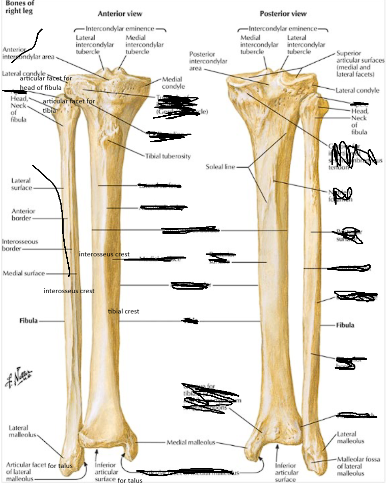
<p>label these parts of the tibia and fibula</p><ul><li><p>medial and lateral condyles</p></li><li><p>superior articulate surfaces: medial and lateral</p></li><li><p>intercondylar eminence: medial and lateral tubercles</p></li><li><p>intercondylar fossa: anterior and posterior</p></li><li><p>tibial tuberosity</p></li><li><p>articular facet for head of fibula</p></li><li><p>soleal line</p></li><li><p>tibial crest</p></li><li><p>interosseus crest</p></li><li><p>medial malleolus</p></li><li><p>fibular notch</p></li><li><p>articular surface for talus</p></li></ul><p></p><ul><li><p>head</p></li><li><p>articular facet for tibia</p></li><li><p>neck</p></li><li><p>interosseus crest</p></li><li><p>lateral malleolus</p></li><li><p>articular facet for talus</p></li><li><p>malleolar fossa</p></li></ul><p></p>

label these parts of the tibia and fibula

  • medial and lateral condyles

  • superior articulate surfaces: medial and lateral

  • intercondylar eminence: medial and lateral tubercles

  • intercondylar fossa: anterior and posterior

  • tibial tuberosity

  • articular facet for head of fibula

  • soleal line

  • tibial crest

  • interosseus crest

  • medial malleolus

  • fibular notch

  • articular surface for talus


  • head

  • articular facet for tibia

  • neck

  • interosseus crest

  • lateral malleolus

  • articular facet for talus

  • malleolar fossa


tibia

  • medial and lateral condyles

  • superior articulate surfaces: medial and lateral

  • intercondylar eminence: medial and lateral tubercles

  • intercondylar fossa: anterior and posterior

  • tibial tuberosity

  • articular facet for head of fibula

  • soleal line

  • tibial crest

  • interosseus crest

  • medial malleolus

  • fibular notch

  • articular surface for talus


fibula

  • head

  • articular facet for tibia

  • neck

  • interosseus crest

  • lateral malleolus

  • articular facet for talus

  • malleolar fossa


<p>tibia</p><ul><li><p>medial and lateral condyles</p></li><li><p>superior articulate surfaces: medial and lateral</p></li><li><p>intercondylar eminence: medial and lateral tubercles</p></li><li><p>intercondylar fossa: anterior and posterior</p></li><li><p>tibial tuberosity</p></li><li><p>articular facet for head of fibula</p></li><li><p>soleal line</p></li><li><p>tibial crest</p></li><li><p>interosseus crest</p></li><li><p>medial malleolus</p></li><li><p>fibular notch</p></li><li><p>articular surface for talus</p></li></ul><p></p><p>fibula</p><ul><li><p>head</p></li><li><p>articular facet for tibia</p></li><li><p>neck</p></li><li><p>interosseus crest</p></li><li><p>lateral malleolus</p></li><li><p>articular facet for talus</p></li><li><p>malleolar fossa</p></li></ul><p></p>
5
New cards
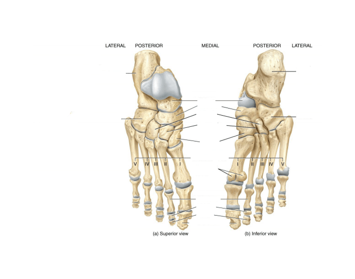
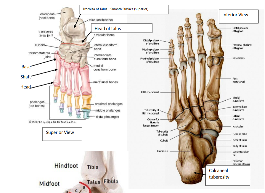
<p>label the parts of the foot</p><ul><li><p>talus: trochlea and head</p></li><li><p>navicular</p></li><li><p>calcaneus: calcaneal tuberosity and sustentaculum tali</p></li><li><p>cuboid</p></li><li><p>cuneiforms: medial, intermediate, and lateral</p></li><li><p>metatarsals (1-5): base shaft and head</p></li><li><p>phalanges (1-5): proximal, intermediate, and distal; base shaft and head</p></li></ul><p></p>

label the parts of the foot

  • talus: trochlea and head

  • navicular

  • calcaneus: calcaneal tuberosity and sustentaculum tali

  • cuboid

  • cuneiforms: medial, intermediate, and lateral

  • metatarsals (1-5): base shaft and head

  • phalanges (1-5): proximal, intermediate, and distal; base shaft and head


#1 phalange only has a proximal and distal part
<p>#1 phalange only has a proximal and distal part</p>
6
New cards
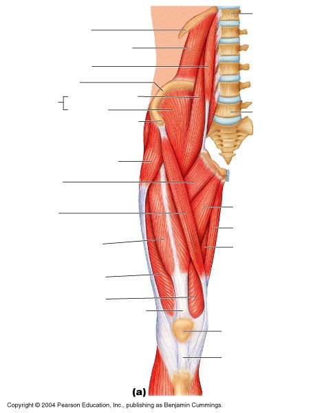
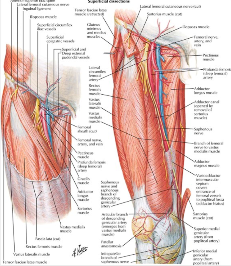
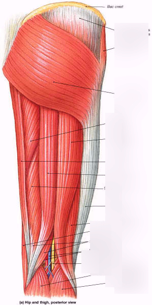
<p>label the anterior thigh muscles, arteries, veins, and nerves</p><ul><li><p>vastus lateralis</p></li><li><p>vastus medialis</p></li><li><p>vastus intermedius</p></li><li><p>rectis femoris</p></li><li><p>sartorius</p></li><li><p>iliacus</p></li><li><p>tensor fascia lataae</p></li><li><p>adductor longus</p></li><li><p>adductor brevis</p></li><li><p>adductor magnus</p></li><li><p>pectineus</p></li><li><p>gracilis</p></li><li><p>femoral nerve, artery, and vein</p></li><li><p>profunda femoral artery</p></li><li><p>lateral femoral circumflex artery</p></li><li><p>great saphenous vein</p></li><li><p>adductor canal</p></li></ul><p></p>

label the anterior thigh muscles, arteries, veins, and nerves

  • vastus lateralis

  • vastus medialis

  • vastus intermedius

  • rectis femoris

  • sartorius

  • iliacus

  • tensor fascia lataae

  • adductor longus

  • adductor brevis

  • adductor magnus

  • pectineus

  • gracilis

  • femoral nerve, artery, and vein

  • profunda femoral artery

  • lateral femoral circumflex artery

  • great saphenous vein

  • adductor canal


  • vastus intermedius is deep to rectus femoris

  • adductor brevis and magnus is deep to adductor longus

  • great saphenous vein and gracilis run together

<ul><li><p>vastus intermedius is deep to rectus femoris</p></li><li><p>adductor brevis and magnus is deep to adductor longus</p></li><li><p>great saphenous vein and gracilis run together</p></li></ul><p></p>
7
New cards
<p>other image of parts of the anterior thigh</p>

other image of parts of the anterior thigh
8
New cards
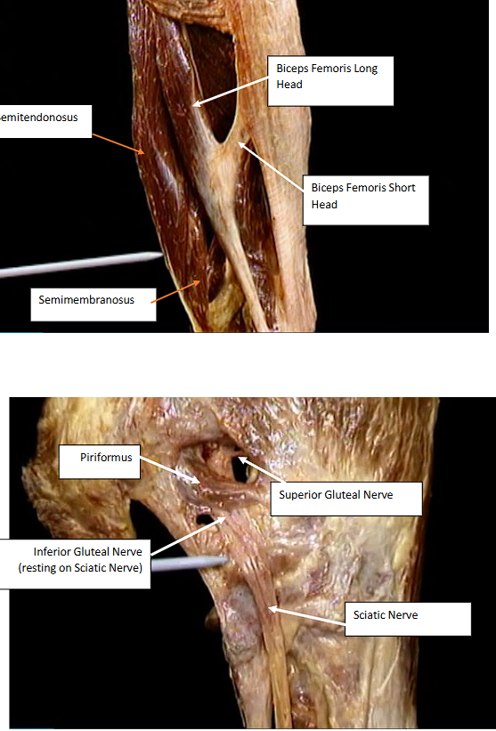
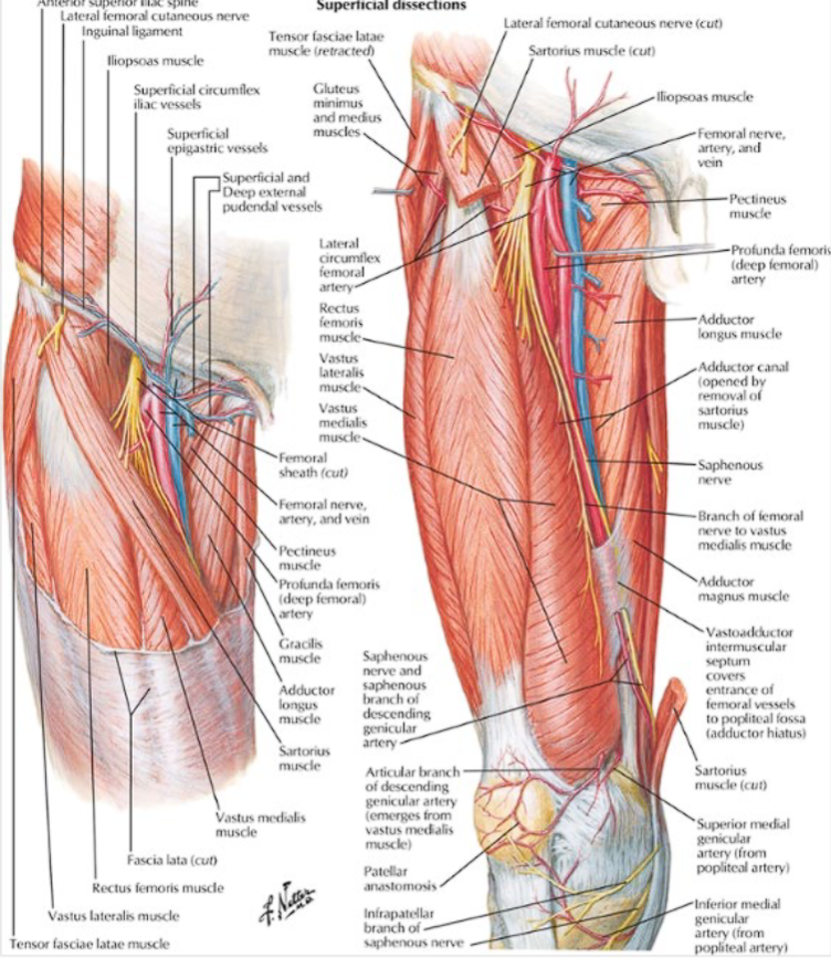
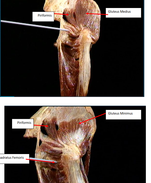
<p>label these parts of the posterior thigh and gluteal region</p><ul><li><p>semitendinosus</p></li><li><p>semimembranosus</p></li><li><p>biceps femoris (long and short)</p></li><li><p>gluteus maximus</p></li><li><p>gluteus minimus</p></li><li><p>piriformis</p></li><li><p>quadratus femoris</p></li><li><p>superior gluteal nerve</p></li><li><p>inferior gluteal nerve</p></li><li><p>sciatic nerve</p></li><li><p>adductor hiatus</p></li><li><p>popliteal vein</p></li><li><p>popliteal artery</p></li><li><p>tibial nerve</p></li><li><p>common peroneal nerve</p></li></ul><p></p>

label these parts of the posterior thigh and gluteal region

  • semitendinosus

  • semimembranosus

  • biceps femoris (long and short)

  • gluteus maximus

  • gluteus minimus

  • piriformis

  • quadratus femoris

  • superior gluteal nerve

  • inferior gluteal nerve

  • sciatic nerve

  • adductor hiatus

  • popliteal vein

  • popliteal artery

  • tibial nerve

  • common peroneal nerve


  • gluteus maximus is superficial to gluteus medius which is superficial to gluteus minimus

  • popliteal vein is flat while popliteal artery is round

  • sciatic nerve splits into the tibial nerve and common peroneal nerve

<ul><li><p>gluteus maximus is superficial to gluteus medius which is superficial to gluteus minimus</p></li><li><p>popliteal vein is flat while popliteal artery is round</p></li><li><p>sciatic nerve splits into the tibial nerve and common peroneal nerve</p></li></ul><p></p>
9
New cards
<p>more images for posterior thigh and gluteal region (flip)</p>

more images for posterior thigh and gluteal region (flip)
knowt flashcard image
10
New cards
<p>more images for posterior thigh and gluteal region </p>

more images for posterior thigh and gluteal region