comd 474 L3 Organization of the Central Nervous System Pt. II
 Call Kai
 Learn
Learn Practice Test
 Spaced Repetition
 Match
Match Flashcards
 Knowt Play
1/50

There's no tags or description
Looks like no tags are added yet.
Name | Mastery | Learn | Test | Matching | Spaced | Call with Kai | Chat |
|---|
No analytics yet
Send a link to your students to track their progress
51 Terms
Peripheral Nervous System
Comprised of cranial nerves, spinal/peripheral nerves, and peripheral parts of the autonomic nervous system.

Spinal Cord - Transverse Section
The central gray area is unmyelinated nerve fibers, cell bodies, and the white surrounding area are closely packed myelinated fibers.

Spinal Cord Definitions - Gray Commissure
A thin, horizontal band of gray matter that connects the left and right halves of the spinal cord.

Spinal Cord Definitions - Dorsal/Posterior Horn
Acts as the main receiving station for incoming signals from sensory nerves outside the central nervous system.

Spinal Cord Definitions - Ventral/Anterior Horn
The front section of the butterfly shaped gray matter inside the spinal cord. It controls motor neurons by sending signals through the ventral roots of the spinal nerve to tell the skeletal muscles to contract.

Spinal Cord Definitions - Intervertebral Foramen
An oval-shaped opening between adjacent vertebrae that allows for the spinal nerves and blood vessels to pass from the spinal canal to the rest of the body

Spinal Cord Definitions - Spinal Ganglion
A cluster of sensory neurons cell bodies located along the dorsal root of a spinal nerve just outside the spinal cord, near or in each of the foramen, and are used to relay sensory information from the PNS to the CNS

Spinal Cord Definitions - Rami or Ramus
Small branchlike structures extending from a longer one into two or more parts. Each type is a mixed nerve carrying both a sensory and motor nerve fiber.

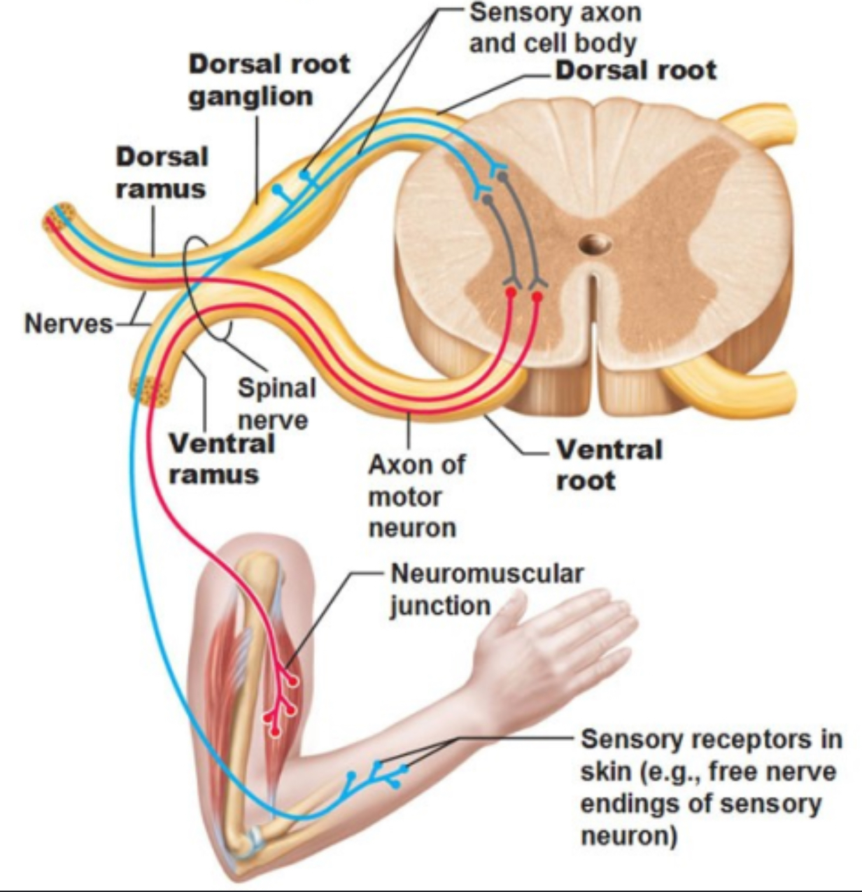
Anterior Root
Made of bundles of nerve fibers that transmit impulses away (efferent) from the CNS to the PNS.
Originates from the ventral horn cells
Anterior/Ventral Horn Cells
Groups of cells or motor nuclei located in the front gray matter of the anterior horn of the spinal cord that sends vital movement information signals to the skeletal muscles
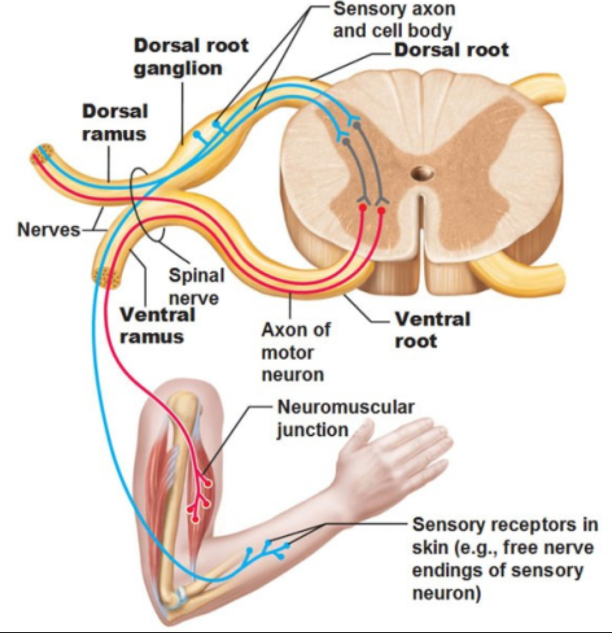
“Final Nerve Pathway” - Charles Sherrington
The lower motor nerves of the spinal cords that have left the neuraxis and act as the terminal route for all the impulses acting on the muscles.
Posterior Root
Made of bundles of sensory nerve fibers that transmit impulses away (efferent) from the CNS and are located on the posterior root of the spinal cord facing your back as poster root ganglions.

Anterior and Posterior Roots
These nerve bundles leave the intervertebral foramen and unite to form a spinal nerve that carries both sensory and motor impulses.

The Vertebrae and Corresponding Nerves
7 Cervical Vertebrae - 8 pairs Cervical Spinal Nerves
12 Thoracic Vertebrae - 12 pairs Thoracic Nerves
5 Lumbar Vertebrae - 5 pairs Lumbar Nerves
1 Sacrum - 5 pairs Sacrum Nerves
1 Coccyx - 1 pair Coccygeal Nerves
Spinal Nerve Distribution
Almost immediately after leaving the intervertebral foramen, each spinal nerve divides into a dorsal and ventral ramus.

Spinal Nerve Distribution - Dorsal Ramus
Branches of motor and sensory nerves that supply the muscles and skin of the back

Spinal Nerve Distribution - Ventral Ramus
C1-C4 cervical sensory and motor nerves supply the muscles and skin of the front of the neck.
C5-C8 and parts of T1 from the Thoracic nerve form the brachial plexus by a series of communicating branches.
Plexus
A branching network if intersecting nerves that organize and distribute signals from the spinal cord to different parts of the body.
Cervical Plexus
Comprised of the ventral rami of C1-C4 nerves and the upper part of C5.

Cervical Plexus - C1
Controls the carotid plexus which determines blood pressure, blood supply to the head, and causes headaches.

Cervical Plexus - C2
Controls the eyes, ears, and sinuses.

Cervical Plexus - C3
Controls the face and teeth

Cervical Plexus - C4
Controls the pharynx and the larynx.

Cervical Plexus - Upper part of C5
Controls the neck and throat.

Brachial Plexus
Comprised of the ventral rami of the lower ½ of the C4, C5, -C8, T1, and the upper ½ of T2

Brachial Plexus - T1
Controls the trachea, esophagus, and esophageal sphincter.
Segmental Distribution
The human body has 31 spinal nerves that are distributed across 5 distinct anatomical regions that connects the spinal cord to specific body areas. This process is called __.

Autonomic Nervous System
Comprised of
The Enteric Division
Sympathetic Division
Parasympathetic Division
The Endocrine System
Autonomic Nervous System - The Enteric Division
Formed by a neuronal plexus in the GI tract
Autonomic Nervous System - sympathetic Division
The body’s “alerting system” during stressful or dangerous situations such as flight or fight.
Autonomic Nervous System - Parasympathetic Division
Calms the body down and controls the rest and digest functions, slows the heart rate, conserves energy, and manages waste.
Autonomic Nervous System - The Endocrine System
Comprised of a network of glands and organs that make and release hormones directly into the blood to control important body functions such as:
Growth and development
Regulating metabolism
Managing sleep, mood, and stress responses
Guides sexual function and reproduction
Meninges
Three productive layers of membrane that cover and support the brain and spinal cord.
Dura Mater
A two layer protective sheath lining the cranial vault that extends out from the cranial and spinal nerves to form an epineurium.

Dura Mater - Epineurium
The outer connective tissue layer of a peripheral nerve that is a direct structural continuation of the central nervous system’s outer protective layer.
Dura Mater - Periostal Layer
The outer layer that is closely attached to the inner surface of the cranium
Dura Mater - Meningeal Layer
The inner layer with four major folds that separates the large parts of the brain.
Dura Mater - Meningeal Layer - Falx Cerebri
The longitudinal fissure between the two hemispheres
Dura Mater - Meningeal Layer - Tentorium Cerebelli
Separates the cerebellum from the occipital lobes
Dura Mater - Meningeal Layer - Falx Cerebelli
Separates the two cerebellar hemispheres
Dura Mater - Meningeal Layer - Diapragma Sella
Covers the stella turcica “Turkish Chair” of the sphenoid bone which holds the pituitary gland
Arachnoid Membrane
Made of a delicate web of reticular (connecting) fibers lined with mesothelium and projects into the venous sinuses in some points to form an arachnoid villi and joins together as arachnoid granulations.

Subarachnoid Space
Space between the arachnoid membrane and the pia mater filled with cerebrospinal fluid.
Pia Mater
This is the deepest layer of the meninges and extends into the ventricles where it forms the choroid plexus, which is the major site that forms cerebrospinal fluid.
This layer is very vascular and is responsible for the blink color of the brain.

Ventricular System - 1 and 2 Lateral Ventricles
These ventricles are C-shaped cavities paired with one on each hemisphere, located in the parietal lobe and extending into the frontal, occipital, and temporal lobes. They connect to the 3rd ventricle by the intraventricular foramen, or the foramen of Munro.

Ventricular System - 3rd Ventricle
The thalamus is a paired structure of the central nervous system and sits on the brainstem. It functions as a switchboard for information, relaying between the sub cortical structures and the cerebral cortex. The small slit between the thalami and to the 4th ventricle through the cerebral aqueduct, or the aqueduct of Sylvius, is the ___.

Ventricular System - 4th Ventricle
This sits anterior to the cerebellum, posterior to the pons and the superior half of the medulla.

Cerebrospinal Fluid
This is the cushion between the CNS and the bones of the body, regulates intracranial pressure, protects against trauma, nourishes the CNS, and removes waste products from the brain.
Cerebralspinal Fluid Pathway
Flows into the lateral ventricles, 3rd ventricle, 4th ventricle, and into the subarachnoid space. This travels into the inferior surface of the cerebrum, superiority over the lateral aspect of each hemisphere and some into the subarachnoid space of the spinal cord.

Cerebralspinal Fluid - Diagnostics
Measures the the pressure
High pressure is indicative of:
Intracranial tumor
Hemorrhage
Hydrocephalus
Meningitis
Encephalitis
Chemical and cell studies
Lumbar puncture
Cerebralspinal Fluid - Neuodegenerative Diseases
In 1977, Oxford did research on cord distortion and vascular lesions leading to Multiple Sclerosis due to placating on the brain
Dr. Raymond v Damaidan, MD and Dr. Scott Rosa DC pioneered the Cine MRI which showed that aberrant cerebralspinal fluid flow can erode the brain leading to neurodegenerative diseases. The multiple sclerosis study showed an average of 11 years from the time of trauma to the start of the multiple sclerosis.