A Level Biology, 3.6 - Organisms Respond to Changes in their Internal & External Environments
1/126
Earn XP
Description and Tags
A stimulus is a change in the internal or external environment. A receptor detects a stimulus. A coordinator formulates a suitable response to a stimulus. An effector produces a response. Receptors are specific to one type of stimulus. Nerve cells pass electrical impulses along their length. A nerve impulse is specific to a target cell only because it releases a chemical messenger directly onto it, producing a response that is usually rapid, short-lived and localised. In contrast, mammalian hormones stimulate their target cells via the blood system. They are specific to the tertiary structure of receptors on their target cells and produce responses that are usually slow, long-lasting and widespread. Plants control their response using hormone-like growth substances. (AQA) Sources: Seneca Learning & CGP A-Level Biology Student Book WORK IN PROGRESS
Name | Mastery | Learn | Test | Matching | Spaced |
|---|
No study sessions yet.
127 Terms
What is a tactic response?
directional movement in response to stimulus
What affects the tactic response?
stimulus direction
What is a kinetic response?
non directional (random) movement in response to stimulus
What affects the kinetic response?
stimulus intensity
Investigating Simple Animal Responses to a Stimulus using a _______________
To investigate the effect of light intensity on woodlouse movement, cover one half of the lid (including the sides) with black paper. This will make one side of the chamber dark. Put ________________ in both sides of the base to make the humidity constant throughout the chamber.
Place 10 woodlice on the ________ in the centre of the chamber and position the lid on the mesh so it’s lined up with the base below.
After 10 minutes, take off the lid and record the number of woodlice on each side of the chamber. Try to minimise the amount of time the lid is off, so that the environmental conditions created aren’t disturbed.
Repeat the experiment after gently moving the woodlice back to the centre. You can use a small, soft paintbrush to help with moving the woodlice if necessary. You should find that most woodlice end up on the dark side of the choice chamber (____________________).
To investigate humidity, place some damp filter paper in one side of the base and a desiccating (drying) agent (such as anhydrous calcium chloride) in the other side. Don’t cover the lid with paper. Put the lid on and leave the chamber for 10 minutes for the environmental conditions to stabilise before carrying out steps 3-5 above.
choice chamber / damp filter paper / mesh / phototaxis
Which element of the plant in positively phototropic? Which element of the plant in negatively phototropic?
shoot / root
Which element of the plant in positively gravitropic? Which element of the plant in negatively gravitropic?
root / shoot
How is IAA transported around the plant over short distances? How about for longer distances? What does it stand for?
diffusion & active transport / via phloem / indoleacetic acid
A* - How does IAA cause Cell Elongation?
Auxin helps to promote _____________________ via activating a membrane _________________ or triggering their gene expression.
The protein activity is known to break the ________________ bonds of the cell wall compositions.
The cell wall compositions help to withstand the _______________________ of the cytoplasm.
But according to the enzyme-linked cell wall loosening, auxin tends to trigger ____________________ of a few ___________________, catalysing hydrolysis of the cell wall polysaccharides.
protein extrusion / H+-ATPase / acid-labile / osmotic pressure / gene expression / glycanases
A* - What hypotheses are the the theory of auxin causing cell elongation based on?
acid growth theory / enzyme linked cell wall loosening
Responses in Plants
Shoots are _________________________.
If a shoot is exposed to an uneven light source, IAA is transported to the ___________________.
A now higher concentration of IAA in these parts cause ________________.
This causes the shoot to ______________________.
positively phototropic / more shaded part / cell elongation / bend towards light
Responses in Plants
Roots are _________________________.
If a root is exposed to an uneven light source, IAA is transported to the ___________________.
A now higher concentration of IAA in these parts cause ________________.
This causes the root to ______________________.
negatively phototropic / more shaded part / cell elongation inhibition / bend away from light
Responses in Plants
Roots are _________________________.
If a root is exposed to an uneven gravitational pull, IAA is transported to the __________.
A now higher concentration of IAA in these parts cause ________________.
This causes the shoot to ______________________.
positively gravitropic / underside / cell elongation inhibition / bend towards gravitational pull
Responses in Plants
Shoots are _________________________.
If a shoot is exposed to an uneven gravitational pull, IAA is transported to the __________.
A now higher concentration of IAA in these parts cause ________________.
This causes the shoot to ______________________.
negatively gravitropic / underside / cell elongation / bend away from gravitational pull
What happens when sensory receptors are stimulated?
sensory neuron generator potential creation
Receptors - Pacinian Corpuscles
Pacinian corpuscles are ________________________ - they detect mechanical stimuli, e.g. pressure and vibrations. Found in skin.
Pacinian corpuscles contain the end of a sensory neuron.
This _____________________ is wrapped in loads of layers of _____________________ called lamellae.
When a Pacinian corpuscle is stimulated, e.g. by a tap on the arm, the lamellae are deformed and press on the sensory nerve ending.
The sensory neurone’s cell membrane stretches, deforming the _______________________________.
The channels open and sodium ions diffuse into the cell, creating a generator potential.
If the generator potential reaches the threshold, it triggers an __________________.
mechanoreceptors / sensory nerve ending / connective tissue / stretch-mediated Na+ channels / action potential
What happens when a pacinian corpuscle is stimulated?
lamellae deform & press on sensory nerve endings
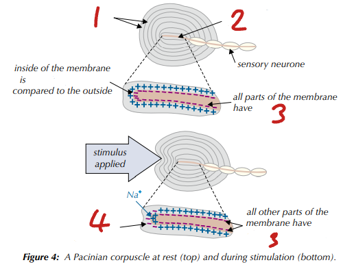
Label the Pacinian Corpuscle (1→4)
lamellae / sensory nerve ending / Na+ channels closed / Na+ channels open so Na+ diffuses into cell
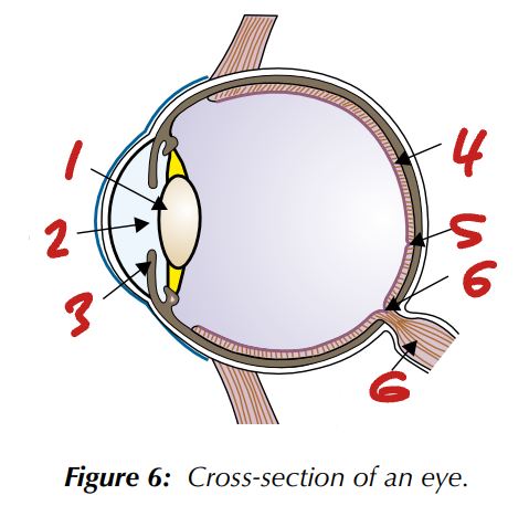
Label the Eye Cross-Section (1→6)
lens / pupil / iris / retina / fovea / blind spot / optic nerve
What is visual acuity?
ability to distinguish between close objects or two points
Photoreceptors - Rods vs Cones
Mainly located in the ___________________; Mainly located in the _________.
Give information in _____; Give information in ________.
_________ join one bipolar neurone; _________ joins one bipolar neurone.
_________ sensitivity to light; _________ sensitivity to light.
Give _________ visual acuity; Give _________ visual acuity
retina periphery / fovea / b&w / colour / many rods / one cone / high / low / low / high
What is the frequency of rod cells? How are they distributed?
high / evenly on retina periphery
What is the frequency of cone cells? How are they distributed?
fewer than rod cells / fovea
How sensitive are rod cells to light? How sensitive are cone cells to light?
highly / less
What is the visual acuity (ability to distinguish between close objects or two points) of rod cells? What is the visual acuity of cone cells?
low / high
Why do cones have _______ visual acuity?
high / multiple action potentials (one from each cone) go to brain
Why do rods have low visual acuity?
many rods join same bipolar neuron
Photoreceptors - Pigment
Rod cells
Use a pigment called _____________ which detects ___________.
Rod cells are _________________ - only detect one wavelength of light.
Cone cells
Use pigment called ______________ which detects ___________.
Cone cells are _______________ - divided into three types and each responds to a different wavelength of light, either red, blue or green.
rhodopsin / light & dark / monochromatic / lodopsin / colour / trichromatic
Photoreceptors - Detecting Light
Light is absorbed by the pigments in photoreceptor cells:
Rhodopsin is the pigment in rod cells
Iodopsin is the pigment in cone cells.
Absorption of light induces a change in the _______________________________. This causes Na+ ions to flood into the cell and a ______________________ is established.
If the generator potential reaches the threshold, an ______________________ flows along a _________________.
pigment membrane permeability / generator potential / action potential / bipolar neuron
Photoreceptors - Neurons
What is the name for the relay neuron which the photoreceptor synapses with?
What is the name for the sensory neuron which the bipolar neuron synapses with?
How do these send a signal to the brain?
bipolar neuron / ganglion cell / axons leave eye via optic nerve
Photoreceptors - Sensitivity to Light
Differences in sensitivity to light are due to differences in how rod and cone cells connect to bipolar neurons.
_____________________________________________________.
Sufficient light must stimulate the cone cell to generate an action potential in the bipolar neuron.
Several rod cells synapse with the same bipolar neuron.
Light stimulating a single rod cell may not be sufficient to generate an action potential in the bipolar neurone.
_____________________ - cumulative stimulation of more than one rod cell can create an _____________________ in the bipolar neuron.
Results in ______________________ - several rod cells generate a signal in a single sensory neuron (ganglion cell).
each cone cell synapses with single bipolar neuron / spatial summation / action potential / retinal convergence
Control of Heart Rate - Sinoatrial Node
The SAN is located in the wall of the ______________.
The SAN acts as a ___________________ by transmitting __________________________ along the walls of the atria at regular intervals.
The electrical waves from the SAN cause the right and left atria to contract together.
This forces blood in the right atrium into the right ventricle and blood in the left atrium into the left ventricle.
right atrium / pacemaker / electrical activity waves
Control of Heart Rate - Atrioventricular Node
The waves of electrical activity cannot pass from the atria to the ventricles due to a collection of ________________________________.
This creates a _________ to ensure the atria are empty before the ventricles begin to contract.
The electrical activity passes through the AVN to the _____________________.
This is a collection of conducting tissue that transmits the electrical activity to the _________ (bottom) of the heart and around the ventricle walls along the __________________.
As the waves of electrical activity pass along these, the ventricles contract together.
Blood is forced out of the ventricles and out of the heart.
non-conducting collagen tissue / delay / bundle of His / apex / Purkyne fibres
What is meant by cardiac muscle being myogenic?
self stimulating / contracts & relaxes without nervous stimulation
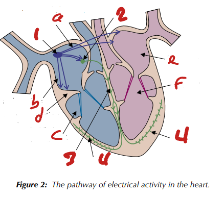
Label the Heart’s Pathway of Electrical Activity (in order, 1→4)
SAN / AVN / bundle of His / Purkyne tissue
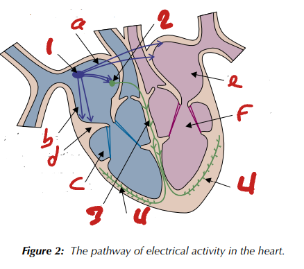
Label the Heart’s Pathway of Electrical Activity (a→f)
electrical activity wave / right atrium / right ventricle / non-conducting collagen tissue / left atrium / left ventricle
What are the two main receptors involved in controlling heart rate? What do these do when stimulated?
chemoreceptors & baroreceptors / send signal to medulla oblongata
What is the region in the medulla which modifies heart rate called? What are its two regions?
cardiovascular centre / cardioinhibitory & cardioacceleratory centre
What are chemoreceptors sensitive to changes in? Where are they found?
CO2 conc / aortic body (aorta wall) & carotid body (carotid artery wall in neck)
What are baroreceptors sensitive to changes in? Where are they found?
blood pressure / carotid sinus (carotid artery wall) & various artery walls
Control of Heart Rate - High Blood Pressure
Detected by _________________.
Impulses are sent from the medulla along ________________________ neurons to the ________.
_____________________ is released.
Heart rate _______________ and blood pressure decreases.
baroreceptors / parasympathetic / SAN / acetylcholine / decreases
Control of Heart Rate - Low Blood Pressure
Detected by _________________.
Impulses are sent from the medulla along _______________________ neurons to the ________.
_____________________ is released.
Heart rate _______________ and blood pressure increases.
baroreceptors / sympathetic / SAN / noradrenaline / increases
Control of Heart Rate - Low CO2 / High O2
Detected by ____________________.
Impulses are sent from the medulla along _____________________ neurons to the ________.
________________________ is released.
Heart rate ____________________ and CO2 levels increase / O2 levels decrease.
chemoreceptors / parasympathetic / SAN / acetylcholine / decreases
Control of Heart Rate - High CO2 / Low O2
Detected by ____________________.
Impulses are sent from the medulla along _____________________ neurons to the ________.
________________________ is released.
Heart rate ____________________ and CO2 levels decrease / O2 levels increase.
chemoreceptors / sympathetic / SAN / noradrenaline / increases
In basic neuronal structure, what do axons do?
carry action potentials away from cell body
In basic neuronal structure, what do dendrites do?
carry action potentials toward cell body
Which type of animal usually have myelinated motor neurons?
vertebrates
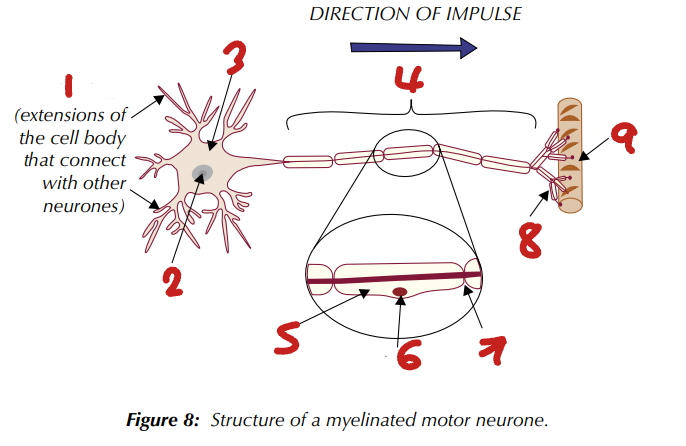
Label the Structure of a Myelinated Motor Neuron (1→9)
dendrites / nucleus / cell body / axon / myelin sheath / Schwann cell nucleus / node of ranvier / axon terminal / effector
Structure of Neurons - Myelinated Motor Neurons
What is usually wrapped around the axon of a neuron?
What do these form?
What are gaps between adjecent ones of these called?
Schwann cells / myelin sheath / nodes of Ranvier
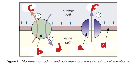
Label the Resting Neuron Cell Membrane (a→f)
neuron cell membrane / Na+/K+ pump / 3Na+ / 2K+ / K+ channel / K+
Resting Potentials
In a neuron’s resting state (when it’s not being stimulated), the outside of the membrane is more ______________ than the inside.
This is because there are _____________________________.
The membrane is _________________ - there’s a potential difference across it.
The voltage across the membrane when it’s at rest is called the resting potential, approx. ___________.
negative / more cations outside cell / polarised / -70 mV
Resting Potentials
The sodium-potassium pumps move Na+ _______ the neuron, but the membrane isn’t __________________ to Na+, so they can’t diffuse back in. This creates a Na+ ________________________ because there is more Na+ outside the cell than inside.
The sodium-potassium pumps also moves K+ _____ the neuron.
When the cell’s at rest, most K+ channels are open. This means that the membrane is permeable to K+, so some diffuse back out through K+ channels.
out of / permeable / electrochemical gradient / into
What are the stages in an action potential?
stimulation / depolarisation / repolarisation / hyperpolarisation / resting potential
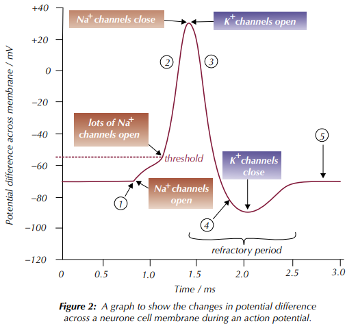
Label the Changes in Potential Difference across a Neuron Cell Membrane during an Action Potential (1→5)
stimulation / depolarisation / repolarisation / hyperpolarisation / resting potential
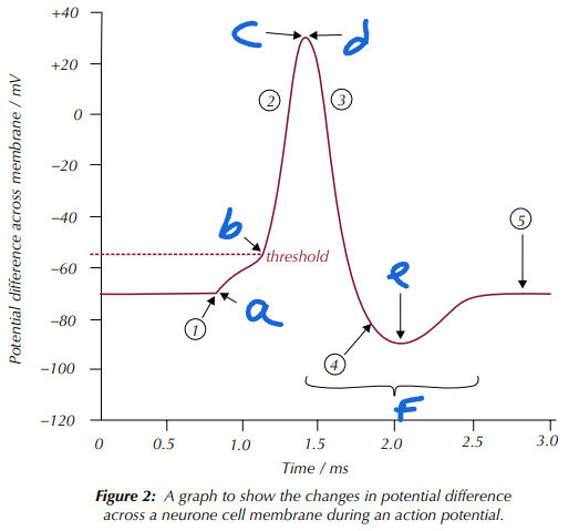
Label the Changes in Potential Difference across a Neuron Cell Membrane during an Action Potential (a→f)
Na+ channels open / lots more Na+ channels open / Na+ channels close / K+ channels open / K+ channels close / refractory period
Action Potentials - Stage 1: Stimulation
_______________________ in the cell membrane _________ when a neuron is stimulated.
Na+ ions flood into the neuron.
The potential difference across the membrane changes to become ______________________ inside the neurone.
Na+ channels / open / more positive
Action Potentials - Stage 2: Depolarisation
If the potential difference increases above the ________________________ (about −55mV) then the membrane will become depolarised.
More _______________________ and there is a sharp increase in potential difference to about +30mV.
Depolarisation is an ___________________________.
If the potential difference reaches the threshold, depolarisation will always take place and the change in potential difference will always be the same.
If the stimulus is stronger, action potentials will be produced ________________________, but their size will not increase.
threshold value / Na+ channels open / all-or-nothing response / more frequently
Action Potentials - Stage 3: Repolarisation
After the neuron membrane has depolarised to +30mV, the _____________________________________.
K+ ions are transported back out of the neuron and the potential difference becomes ____________________.
Na+ channels close & K+ channels open / more negative
Action Potentials - Stage 4: Hyperpolarisation
There is a short period after repolarisation of a neuron where the potential difference becomes ________________________________________.
______________________: hyperpolarisation prevents the neuron from being restimulated instantly.
slightly more negative than resting potential / refractory period
Action Potentials - Wave of Depolarisation
When an action potential happens, some Na+ that enter the neuron ______________________ along the neuron axon.
Na+ presence creates a change in potential difference further along neuron membrane.
If this reaches the _____________________, Na+ channels open so Na+ diffuses into that part.
This causes a wave of depolarisation to travel along the neuron.
The wave moves away from the parts of the membrane in the ______________________ because these parts can’t fire an action potential.
diffuse sideways / threshold value / refractory period
In the refractory period (hyperpolarisation)…
What are the ion channels doing?
What does this mean for action potentials?
What does this mean for the wave of depolarisation?
recovering / can’t be instantly restimulated / unidirectional
What are 3 factors which affect the speed of action potential conduction?
myelination / temperature / axon diameter
Speed of Action Potential Conduction - Temperature
An increase in temperature increases _________________.
Ions move across the membrane more rapidly when they have more kinetic energy.
Channel proteins begin to ______________ (after approx. 40°C) so rate of movemenr decreases.
kinetic energy / denature
Speed of Action Potential Conduction - Axon Diameter
Action potentials are conducted quicker along axons with bigger diameters because there’s ____________________________ to the flow of ions than in the cytoplasm of a smaller axon.
____________________ reaches other parts of the ____________________ quicker.
Greater axon diameter means there is a greater ____________________ for the movement of ions across the cell membrane.
less resistance / depolarisation / neuron cell membrane / surface area
Speed of Action Potential Conduction - Myelination
In a myelinated neuron, depolarisation only happens at the ___________________ (where Na+ can get through the membrane).
_____________________: the neuron’s __________________ conducts enough electrical charge to _____________ the next node, so the impulse ‘jumps’ from node to node - very fast.
In a non-myelinated neuron, the impulse travels as a wave along the ___________________________________.
Depolarisation along the whole length of the membrane - slower.
nodes of Ranvier / salatory conduction / cytoplasm / depolarise / axon membrane whole length
How can the myelin sheath act as an electrical insulator?
impermeable to ions
What is concentrated at the nodes of Ranvier?
Na+ channels
Synapse Structure - _________________
End of presynaptic neuron’s axon - a swelling which contains synaptic vesicles.
Synapstic vesicles contain neurotransmitters, which bind to specific receptors on the postsynaptic membrane.
Location where the ___________________ is transmitted across the synpatic cleft.
Lots of ___________________ - great amount of energy needed to synthesise neurotransmitters.
synaptic knob / action potential / mitochondria
Cholinergic Synapses (part 1/2) - Arrival of an Action Potential & Fusion of Vesicles
An action potential arrives at the synaptic knob of the presynaptic neuron.
Stimulation of __________________________ in the presynaptic neuron to open so Ca2+ diffuses into the synaptic knob (they’re pumped out afterwards by active transport).
The ______________________________ causes the synaptic vesicles to _________ with the presynaptic membrane. The vesicles release the neurotransmitter acetylcholine (ACh) into the synaptic cleft by _________________.
voltage-gated Ca2+ channels / Ca2+ influx into synaptic knob / fuse / exocytosis
Cholinergic Synapses (part 2/2) - Diffusion of Acetylcholine
ACh diffuses across the synaptic cleft and binds to specific ___________________________ on the postsynaptic membrane.
This causes Na+ channels in the postsynaptic neuron to open.
The influx of Na+ into the postsynaptic membrane causes __________________.
An _______________________ on the postsynaptic membrane is generated if the threshold is reached.
ACh is removed from the synaptic cleft so the response doesn’t keep happening.
Breakdown by the enzyme _______________________ and the products are reabsorbed by the presynaptic neuron - used to make more ACh.
nicotinic cholinergic receptors / depolarisation / action potential / acetylcholinesterase
What do inhibitory neurotransmitters do? What does this lead to?
hyperpolarise postsynaptic membrane / prevent action potential firing
What do excitatory neurotransmitters do? What does this lead to?
depolarise postsynaptic membrane / action potential firing promotion

What is shown by the image?
temporal summation
Synaptic Transmission - Temporal Summation
Takes place when multiple action potentials arrive at the same synaptic knob within ______________________.
More neurotransmitter is released into the synaptic cleft, so more neurotransmitter is available to bind to __________________ on the postsynaptic membrane.
Together the neurotransmitters can establish a _________________________ that reaches the threshold value and an action potential is generated.
quick succession / receptors / generator potential

What is shown by the image?
spatial summation
Synaptic Transmission - Spatial Summation
Takes place when multiple presynaptic neurons form a junction with a _____________________.
Release of many neurotransmitters that bind to the receptors on one postsynaptic membrane.
Together the neurotransmitters can establish a generator potential that reaches the _____________________ and an action potential is generated.
If some neurons release an ____________________________ then the total effect of all the neurotransmitters might be no action potential.
single neuron / threshold value / inhibitory neurotransmitter
Synaptic Transmission - Cholinergic Synapses in Neuromuscular Junctions
Types of postsynaptic cell
Cholinergic synapses are between ________________.
Neuromuscular junctions are between a ____________________________.
Number of receptors - ________ receptors in the postsynaptic membrane at a cholinergic synapse than at a neuromuscular junction.
Type of response
A cholinergic synapse can trigger an inhibitory or excitatory response in the postsynaptic membrane.
An action potential at a neuromuscular junction always triggers an _______________ response in the muscle cell.
two neurons / motor neuron & muscle cell / less / excitatory
Synaptic Transmission - Cholinergic Synapses in Neuromuscular Junctions
Result of depolarisation
In a cholinergic synapse, depolarisation of the postsynaptic membrane results in an __________________.
At a neuromuscular junction, depolarisation of the postsynaptic membrane results in ______________________.
Acetylcholinesterase
In cholinergic synapses, the enzyme is located in the __________________.
At a neuromuscular junction, the enzyme is stored in __________________________.
action potential / muscle contraction / synaptic cleft / postsynaptic membrane clefts
Drugs at Synapses - Examples of Effects
_________ - same shape as neurotransmitters so mimic their action at receptors - more receptors activated.
_______________ - block receptors so they can’t be activated by neurotransmitters - fewer receptors (if any) can be activated.
Inhibition - prevents function of enzyme that breaks down neurotransmitters - more neurotransmitters in the _________________ to bind to receptors and available for longer time.
Receptor activation
Stimulate the release of neurotransmitter from the presynaptic neuron - more receptors activated.
Inhibit the release of neurotransmitters from the presynaptic neuron - fewer receptors activated.
agonists / antagonists / synaptic cleft
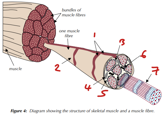
Label the Skeletal Muscle Structure (1→9)
T tubule / sarcolemma / SR / nucleus / sarcoplasm / mitochondrion / myofibril
Skeletal Muscle Fibre Structure - Sarcolemma
The ____________ of muscle fibres.
________________: folds inwards to the sarcoplasm at certain points.
Very important in ___________________________.
membrane / T tubules / initiating muscle contraction
Skeletal Muscle Fibre Structure - Sarcoplasmic Reticulum
Organelle in the _________________.
Store for ___________.
Important in muscle contraction.
sarcoplasm / Ca2+
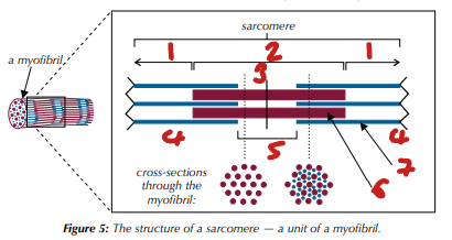
Label the Sarcomere Structure (1→7)
I band / A band / M line / Z line / H zone / myosin / actin
Myofibril Structure
_____________________: units that run end-to-end along the myofibril.
______________: the end of this structure.
Made from thick (myosin) & thin (actin) ______________________ arranged in an alternating pattern.
Thick myosin filaments overlap with the thin actin filaments at each end.
_______________: overlapping region of myofilaments at each end.
______________: thin actin filaments only overlap with myosin filaments in the middle of the sarcomere.
________________: region with only actin filament.
________________: region with only myosin filament.
sarcomeres / Z line / myofilaments / A band / M line / I band / H zone
When Muscle Fibre Contracts, what Happens to the…
A band?
I band?
H zone?
no change / shortens / shortens
What are thick myofilaments made of? What are thin myofilaments made of?
myosin / actin
Skeletal Muscle
What attaches skeletal muscle to bones?
What are ligaments?
What is the pair of muscles that work together to move the bone called?
What is the muscle that is relaxing called?
What is the muscle that is contracting called?
tendons / strong connective tissue bands that connect bones / antagonistic pair / antagonist / agonist
According to the sliding filament theory, how is muscle contraction coordinated by myofibrils?
sarcolemma depolarisation / sarcomere contraction / muscle contraction / muscle relaxation
How does muscle contraction happen?
action potential arrival / actin filament movement / cross bridge breaking / return to resting state
Muscle Contraction - Step 1: Action Potential Arrival
When an action potential from a motor neuron stimulates a muscle cell, it ________________ the ____________________.
This spreads down the _________________ to the ______.
Influx of Ca2+ into the sarcoplasm triggers muscle contraction.
Ca2+ binds to a protein attached to tropomyosin, causing the protein to change shape.
Pulling of the attached tropomyosin out of the actin-myosin binding site on the actin filament.
Exposure of the binding site, which allows the __________________ to bind.
__________________________: the bond formed when a myosin head binds to an actin filament.
depolarises / sarcolemma / T-tubules / SR / myosin head / actinomyosin cross bridge
Muscle Contraction - Step 2: Actin Filament Movement
Ca2+ also activates the enzyme _________________, which hydrolyses ATP to provide the energy needed for muscle contraction.
The energy released from ATP causes the ____________________, which pulls the actin filament along in a kind of rowing action.
ATP hydrolase / myosin head to bend
Muscle Contraction - Step 3: Breaking of the Cross Bridge
Another ATP molecule provides the energy to break the actinomyosin cross bridge, so the ______________ detaches from the actin filament after it’s moved.
The myosin head then returns to its starting position, and reattaches to a different binding site further along the actin filament.
A new actinomyosin cross bridge is formed and the cycle is repeated (attach, move, detach, reattach to new binding site...).
Many actinomyosin cross bridges form and break very rapidly, pulling the __________________ along - _________________________, causing the muscle to contract. The cycle will continue as long as Ca2+ is present.
myosin head / actin filament / shortening the sarcomere
Muscle Contraction - Step 4: Return to Resting State
When the muscle stops being stimulated, Ca2+ leaves its binding site and is moved by _____________________ back into the _______.
This causes the ____________________ molecules to move back, so they block the actin-myosin binding sites again.
Muscles aren’t contracted because no myosin heads are attached to actin filaments (so there are no ________________________). The actin filaments slide back to their relaxed position, so the ________________________.
active transport / SR / tropomyosin / actinomyosin cross bridges / sarcomere lengthens
Sliding Filament Theory of Muscle Contraction - Myosin Filaments
Have _____________________ that are _________, allowing ______________________________.
Each myosin head has a binding site for _________ and another for ______.
globular heads / hinged / back-and-forth movement / actin / ATP
Sliding Filament Theory of Muscle Contraction - Actin Filaments
Have ____________________________ (binding sites for ____________________).
Another protein called ____________________ is found between actin filaments.
Blocks the binding site when muscle fibres at rest.
Upon stimulation, protein is moved so binding can occur and myofilaments move past each other.
actin-myosin binding sites / myosin heads / tropomyosin
Energy for Muscle Contraction - Respiration
Aerobic
Most ATP is generated via ___________________________ in the cell’s mitochondria.
Aerobic respiration only works when there’s oxygen so it’s good for long periods of low-intensity exercise,
Anaerobic
ATP is made rapidly by _______________. The end product is _____________, which is converted to ___________ by ________________________.
Ccan quickly build up in the muscles and cause muscle fatigue.
Anaerobic respiration is good for short periods of hard exercise.
oxidative phosphorylation / glycolysis / pyruvate / lactate / lactate fermentation
Energy for Muscle Contraction - ATP-Phosphocreatine (PCr) System
____________________ is a molecule that can supply ATP for muscle contraction.
During intense muscular effort, donation of phosphate to ADP to produce ATP. The ATP produced is used to sustain muscle contraction.
During low periods of muscle activity, ATP can be used to ______________________ creatine back to phosphocreatine.
This process is _______________________ but phosphocreatine is in short supply.
phosphocreatine / phosphorylate / anaerobic & alactic
Skeletal Muscle Fibres - Slow Twitch
Found in muscles used for _____________ such as the back and neck.
Adapted for _______________ and slow movement over long periods of time.
Muscle fibres are _______________.
The muscles fatigue slowly and contract slowly.
Rely on energy released through ____________ respiration.
posture / endurance / long & thin / aerobic
Skeletal Muscle Fibre Cell Structure - Slow Twitch
Lots of _______________ to maintain aerobic respiration.
Lots of _______________ to supply muscle fibres with oxygen.
_______ levels of glycogen.
_______ levels of phosphocreatine.
_________ stores of _____________ (pigment that stores oxygen), so appear ____________.
Less ______ (containing _______).
mitochondria / capillaries / low / low / large / myoglobin / reddish / SR / Ca2+
Skeletal Muscle Fibres - Fast Twitch
Found mainly in muscles such as the ________________.
Adapted for fast or strong movement over short periods of time.
Muscle fibres are ________________.
The muscles fatigue quickly and contract quickly.
Rely on energy released through _________________ respiration.
arms & legs / short & wide / anaerobic