Seasons LG LO's wk3 pt.3
 Call Kai
 Learn
Learn Practice Test
 Spaced Repetition
 Match
Match Flashcards
 Knowt Play
1/34

Earn XP
Description and Tags
Cellular (Reversible/Irreversible) Injury
Name | Mastery | Learn | Test | Matching | Spaced | Call with Kai | Chat |
|---|
No analytics yet
Send a link to your students to track their progress
35 Terms
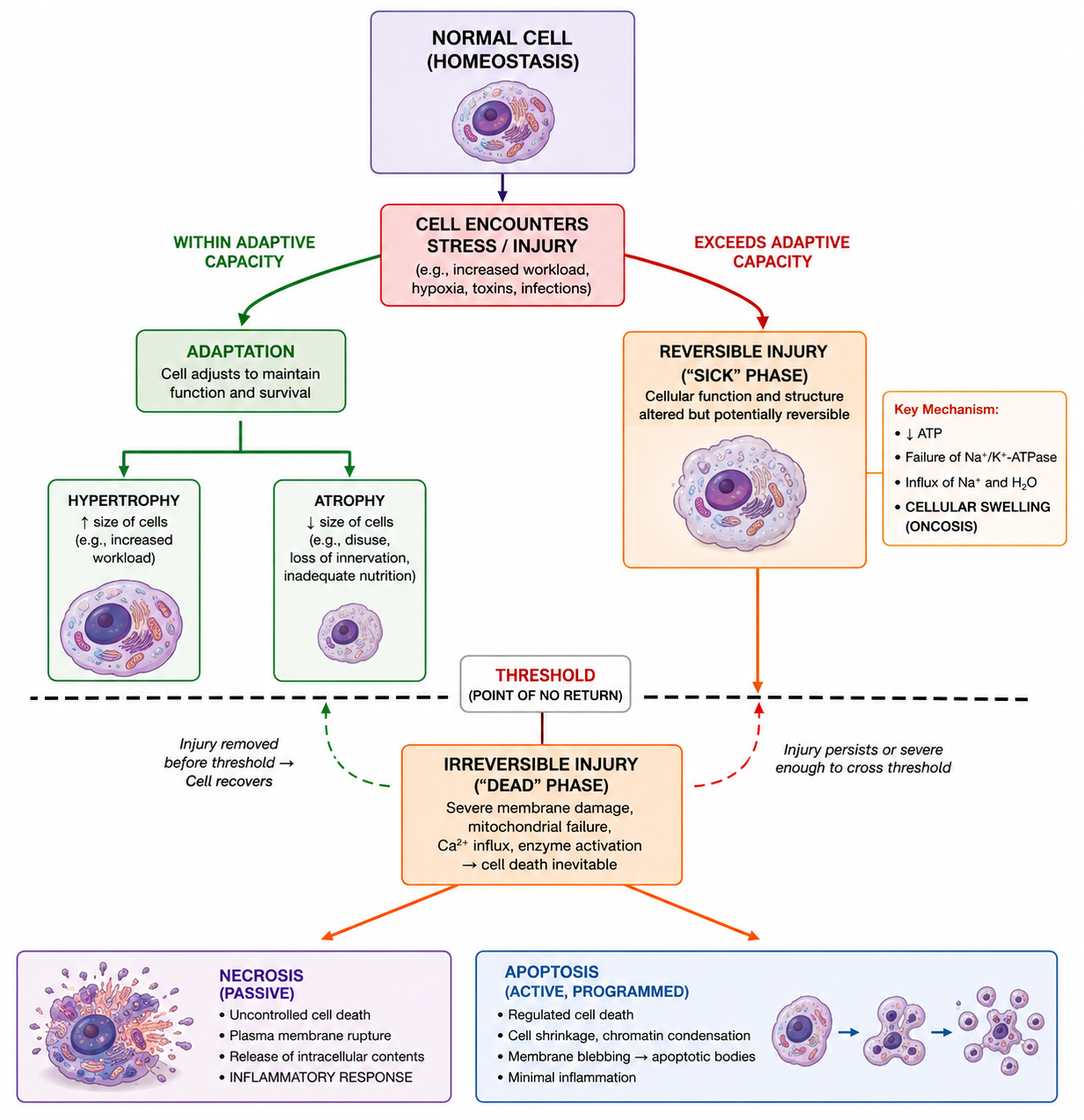
LO 1: cells are usually in homeostasis but when stressed or injured how do they respond
they first try to adapt to survive
If the stress is too severe or the adaptation limits are exceeded, cell injury occurs.
This injury is reversible if the stimulus is removed, but it crosses a "point of no return" into irreversible injury and cell death if the damage is persistent or profound.

LO 1: what are 4 components involved in cell response to injury
reversible injury
ultrastructural changes
threshold
irreversible injury
LO 1: cell response to injury (reversible injury)
what occurs in early stages
what is a major morphological hallmark: what its called and what it is caused by
Reversible Injury (The "Sick" Phase):
In the early stages, the cell's function and structure are altered but correctable.
The earliest morphological hallmark is cellular swelling (hydropic change).
This is caused by the failure of the energy-dependent Na⁺-K⁺-ATPase plasma membrane pump. Without ATP, sodium accumulates intracellularly, creating an osmotic gradient that drives water influx
LO 1: cell response to injury (ultrastructural changes)
what do reversibly injures cells exhibit
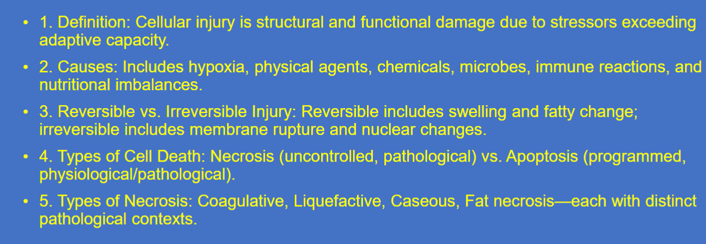
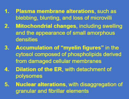
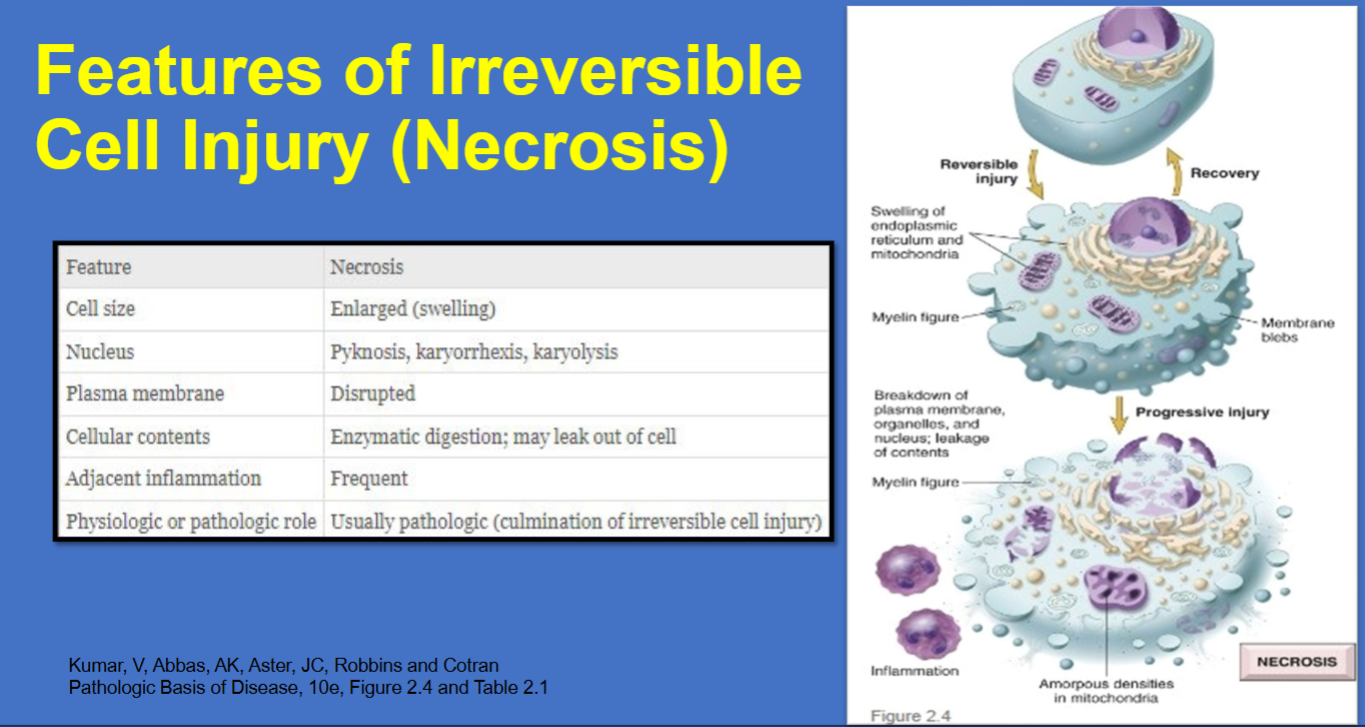
Ultrastructural Changes: Reversibly injured cells exhibit plasma membrane blebbing (bubble like buldges), loss of microvilli, mitochondrial swelling, and detachment of ribosomes from the rough endoplasmic reticulum (ER), which impairs protein synthesis
LO 1: cell response to injury (threshold)
what 2 things is irreversibility characterized by
Threshold (The "Point of No Return"): Irreversibility is consistently characterized by two phenomena: the inability to reverse mitochondrial dysfunction (lack of oxidative phosphorylation and ATP generation) and profound disturbances in membrane function
LO 1: cell response to injury (irreversible injury)
can the cell recover
how does cell death occur
morphologic hallmarks
Irreversible Injury (The "Dead" Phase):
Once the threshold is crossed, the cell cannot recover even if the original injury is resolved.
This leads to cell death, principally through necrosis or apoptosis.
Morphologic hallmarks include severe mitochondrial densities, lysosomal membrane rupture (leaking acid hydrolases), and nuclear degradation: pyknosis (shrinkage/basophilia), karyorrhexis (fragmentation), and karyolysis (dissolution )

LO 1: mitochondria reversive vs irreversible injury (necrosis)
what occurs

LO 1: membranes reversive vs irreversible injury (necrosis)
what occurs

LO 1: nucleus reversive vs irreversible injury (necrosis)
what occurs

LO 1: cell contents reversive vs irreversible injury (necrosis)
what occurs

LO 1:
Oncosis is ______________________________
The threshold for cell death is marked by _______ _________ _____________ and______________ _____________.
Biomarkers of necrosis include the detection of _________ _________ (e.g., transaminases) leaking through damaged membranes into the circulation
Oncosis (cellular swelling) is the first sign of reversible injury.
The threshold for cell death is marked by irreversible mitochondrial damage and membrane rupture.
Biomarkers of necrosis include the detection of intracellular proteins (e.g., transaminases) leaking through damaged membranes into the circulation
LO 2: what does hypoxia disrupt
Hypoxia (oxygen deficiency) disrupts aerobic oxidative respiration, the primary source of cellular ATP. This energy failure triggers a metabolic crisis that initially allows for survival through backup mechanisms but eventually leads to enzymatic self-digestion of the cell
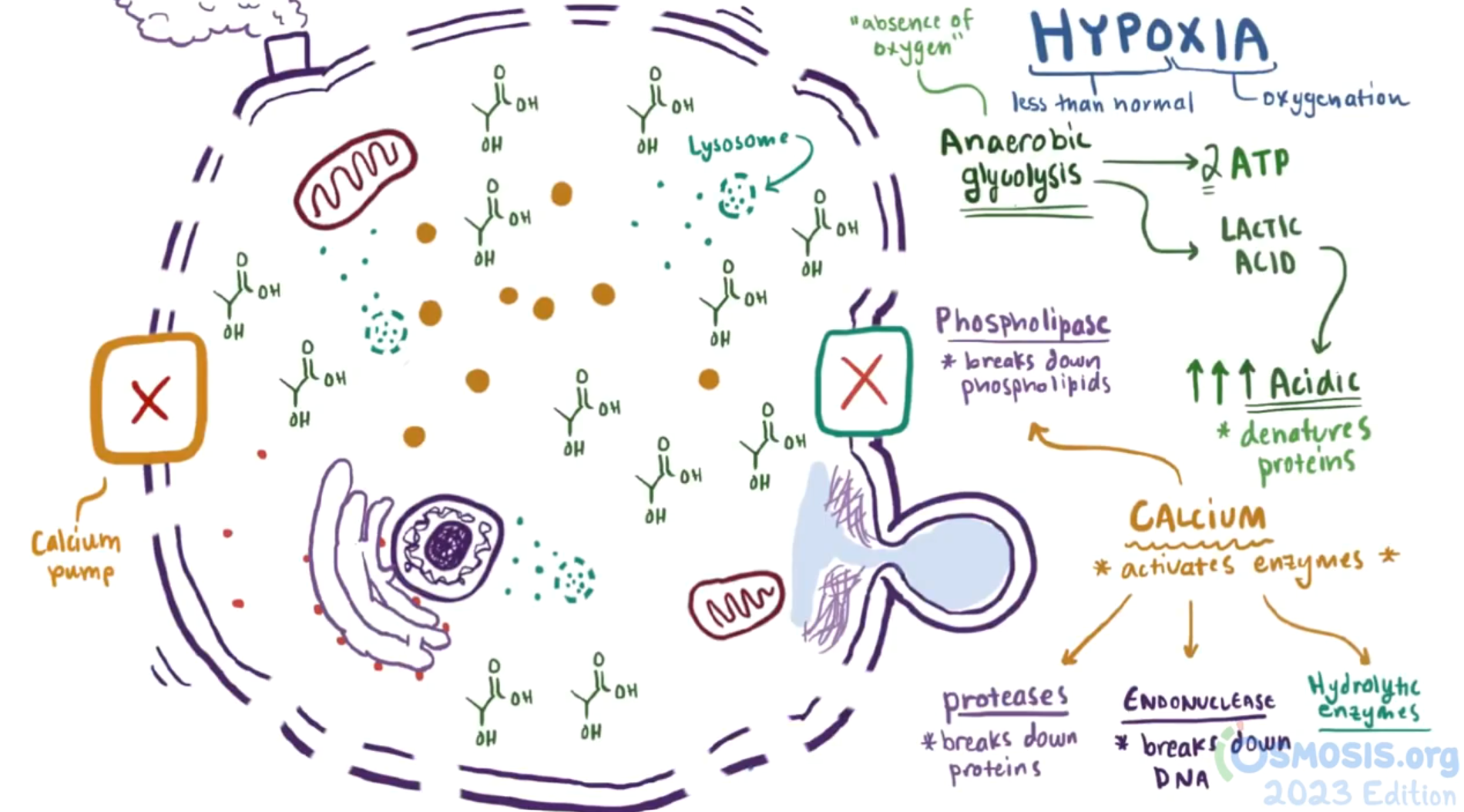
LO 2: what are 6 cellular responses to hypoxia & reversible vs irreversible
ATP depletion (reversible)
intracellular acidosis
pump failure (reversible)
calcium influx (pivot toward irreversible)
membrane/mitochondrial rupture (irreversible)
LO 2: cellular responses to hypoxia (ATP depletion)
what occurs when O2 levels drop
type of metabolism that is utilized & what it produces
ATP Depletion (Reversible):
As oxygen levels drop, oxidative phosphorylation fails.
The cell switches to anaerobic glycolysis, which is inefficient and produces lactic acid, lowering the intracellular pH and potentially denaturing proteins.
LO 2: cellular responses to hypoxia (intracellular acidosis)
what causes this & what occurs
Intracellular Acidosis: The byproduct lactic acid lowers the intracellular pH, causing clumping of nuclear chromatin and potentially denaturing cytosolic enzyme

LO 2: cellular responses to hypoxia (pump failure)
what pumps fail & what causes them to fail
what occurs as a result
ATP depletion causes the Na⁺-K⁺ pump and Ca²⁺ pump to fail
Pump Failure (Reversible):
The Na⁺-K⁺ pump stops working due to lack of ATP. Sodium (and then water) rushes into the cell, causing swelling, blebbing, and detachment of ribosomes from the rough ER (decreasing protein synthesis).

LO 2: cellular responses to hypoxia (calcium influx)
what occurs if hypoxia persists
what is activated & what they do
what does calcium induce
Calcium Influx (The Pivot toward Irreversible):
If hypoxia persists, the calcium pump also fails.
Excess cytosolic calcium activates destructive enzymes: proteases, endonucleases, and phospholipases.
Calcium also induces the mitochondrial permeability transition pore to open, allowing cytochrome c to leak into the cytosol, which triggers apoptosis

LO 2: cellular responses to hypoxia (membrane/mitochondrial rupture)
what occurs
what does this lead to
Membrane/Mitochondrial Rupture (Irreversible):
Severe damage to lysosomal membranes leaks digestive enzymes into the cytosol, and cytochrome c leaks from damaged mitochondria, signaling the cell to undergo apoptosis or necrotic dissolution.
LO 2:
________ ________leads to decreased pH via lactic acid.
Ribosome detachment from the rough ER occurs during the_________ phase of hypoxia.
Rupture of the plasma membrane is the most critical sign that hypoxic injury has become _______
Anaerobic glycolysis leads to decreased pH via lactic acid.
Ribosome detachment from the rough ER occurs during the reversible phase of hypoxia.
Rupture of the plasma membrane is the most critical sign that hypoxic injury has become irreversible
LO 2: mneumonics reversible vs irreversible response to hypoxia
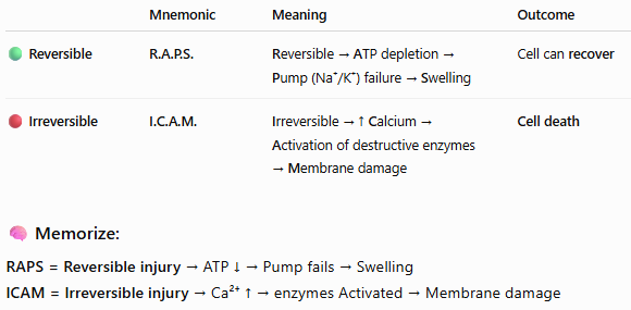
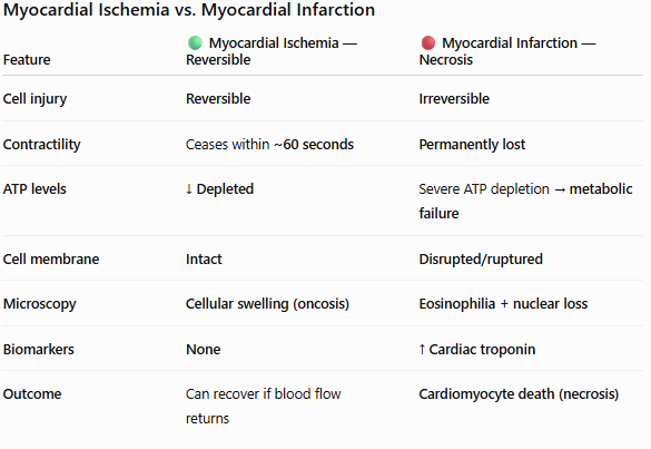
LO 3: what is myocardial ischemia vs infarction
Ischemia is a reduced blood and oxygen supply to tissue, while infarction is the actual death (necrosis) of tissue caused by a complete or severe lack of blood flow.
Ischemia can be reversible if blood flow returns, but infarction causes permanent tissue damage
LO 3: biochemical findings in MI
what occurs within seconds of ischemia
what significant protein leaks out & how and when can it be detected
Biochemical:
Within seconds of ischemia, myocardial contraction ceases.
Within 1-2 hours, ATP depletion leads to irreversible mitochondrial densities.
Troponin, a cardiac-specific contractile protein, leaks through the ruptured plasma membrane into the blood and can be detected within 2 hours

LO 3: morphologic findings in MI (microscopic) → increased eosinophilia
what do dead cells look like & why
Increased Eosinophilia:
Dead cells look "pinker" on hematoxylin & eosin stains because they lose RNA which binds the blue hematoxylin (which is blue) and accumulate denatured proteins (which are red). Eosinophilia = increased pink/red staining with eosin.
RNA is lost/degraded | ↓ binding of hematoxylin (blue/purple) → less blue |
Proteins become denatured | ↑ binding of eosin (pink/red) → more pink |
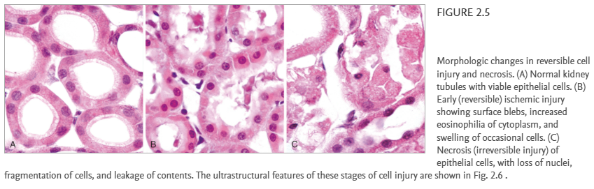
LO 3: morphologic findings in MI (microscopic) → nuclear changes
what are 3 things that can happen to the nucleus
Nuclear Changes: The nucleus may shrink (pyknosis) followed by fragment into pieces (karyorrhexis), or simply fade away (karyolysis).
LO 3: morphologic findings in MI (microscopic) → preserved architecture
what is coagulative necrosis
what occurs in coagulative necrosis
Preserved Architecture:
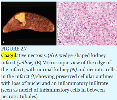
Coagulative necrosis is a type of cell death caused by a sudden lack of blood flow and oxygen (ischemia).
In coagulative necrosis, you see "ghost-like" outlines of the myocytes (with no nucleus) preserved for days because the injury denatures the very enzymes required for proteolysis stopping the cells from breaking down completely.

LO 3: morphologic findings in MI (gross morphology)
what does an MI typically look like
Gross Morphology: A wedge-shaped pale area (infarct) appears, with the apex pointing toward the site of vascular obstruction
LO 3:
______ _______ is the hallmark of ischemic death in all organs except the brain.
_________ is a biomarker of lost membrane integrity.
________ and _____ ______ are the classic microscopic signs of necrosis.
Coagulative necrosis is the hallmark of ischemic death in all organs except the brain.
Troponin is a biomarker of lost membrane integrity.
Eosinophilia and nuclear loss are the classic microscopic signs of necrosis.
LO 4: what typically causes compromise in coronary blood flow
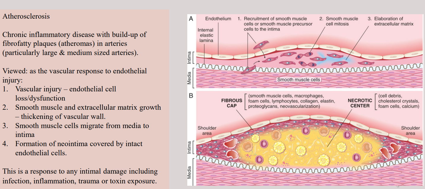
A compromise in coronary blood flow (often due to atherosclerosis or a thrombus) causes ischemia, which is more damaging than simple hypoxia because it also cuts off the delivery of glucose and the removal of toxic metabolites.
LO 4: what are 4 components of cellular progression of coronary compromise
vascular obstruction
ischemic penumbra
mitochondrial failure
ischemia-reperfusion injury
LO 4: cellular progression of coronary compromise (vascular obstruction)
what occurs
timing
Vascular Obstruction: Plaque buildup or a clot restricts flow, reducing oxygen and nutrient delivery while allowing metabolite accumulation.
LO 4: cellular progression of coronary compromise (ischemic penumbra)
what is ischemic penumbra
what occurs
timing
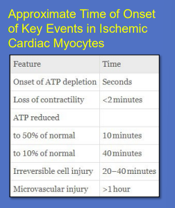
ischemic penumbra: It is the area of ischemic tissue surrounding a region of more severely injured/necrotic tissue that has reduced blood flow but is still viable.
Ischemic Penumbra: This is the viable tissue surrounding a necrotic core that can still be salvaged if reperfusion occurs quickly.

LO 4: cellular progression of coronary compromise (mitochondrial failure)
what occurs
timing
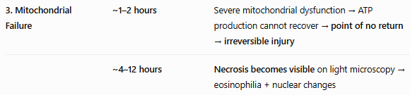
Mitochondrial Failure: Without flow, the mitochondrial permeability transition pore opens, collapsing the membrane potential and ending ATP production.
Normally, the inner mitochondrial membrane keeps H⁺ (protons) separated, creating a membrane potential/proton gradient.
When the MPTP opens:
MPTP opens → inner membrane becomes abnormally permeable → H⁺ gradient collapses → ATP synthase cannot make ATP → ATP production stops
LO 4: cellular progression of coronary compromise (ischemia-reperfusion injury)
what occurs
what components cause this to occur (3)
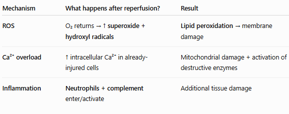
Ischemia-Reperfusion Injury: Restoring blood flow to damaged tissue can paradoxically cause more harm. This occurs via:
Reactive Oxygen Species (ROS): Reintroduction of oxygen leads to the generation of superoxide anions and hydroxyl radicals, which cause lipid peroxidation of membranes.
Calcium Overload: Reperfusion brings a "flush" of calcium that overloads already damaged mitochondria.
Inflammation: Reperfusion recruits neutrophils and activates the complement system (inflammatory response), which further digests the tissue.
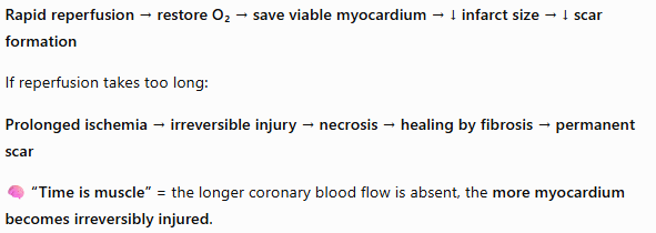
LO 4: why is minimizing the time to perfusion important ex.) patient with infarct
In a patient with a "red infarct," blood has re-entered an area of necrosis (often after a clot dissolves), giving the tissue a dark red appearance.
The goal of emergency medicine is to minimize the time to reperfusion to save as much of the penumbra (viable if perfusion is quick) as possible before it turns into a permanent scar.
LO 4:
Ischemia is worse than hypoxia because it also prevents _______washout.
Myocardial contraction stops within _______, long before the cells actually die.
Reperfusion injury is largely driven by ________ and ______ influx.
Ischemia is worse than hypoxia because it also prevents metabolite washout.
Myocardial contraction stops within minutes, long before the cells actually die.
Reperfusion injury is largely driven by ROS and calcium influx.