Cardiac Contractability
 Call Kai
 Learn
Learn Practice Test
 Spaced Repetition
 Match
Match Flashcards
 Knowt Play
1/24

There's no tags or description
Looks like no tags are added yet.
Name | Mastery | Learn | Test | Matching | Spaced | Call with Kai | Chat |
|---|
No analytics yet
Send a link to your students to track their progress
25 Terms
What determines cardiac output?
Cardiac output is determined by stroke volume and heart rate (CO = SV × HR). Stroke volume- EDV-ESV

Define cardiac output.
The amount of fluid pumped through the heart in one minute (L/min). Normal range is 4–8 L/min.

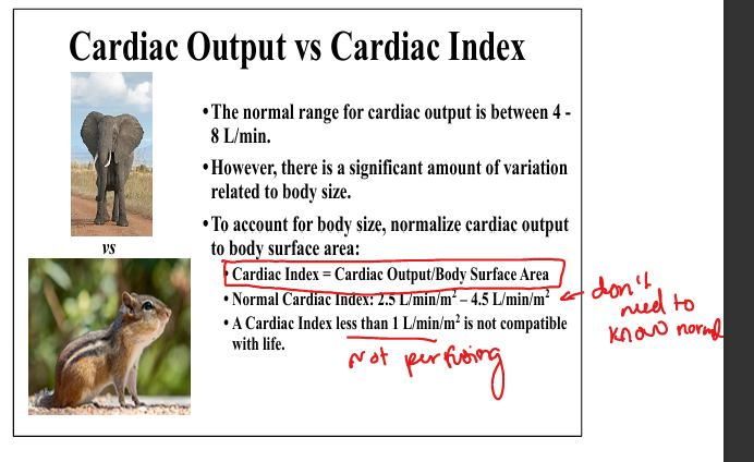
Cardiac index.
Cardiac output/body surface area.
What is stroke volume?
The amount of blood ejected from the ventricle in one heartbeat: SV = EDV – ESV.
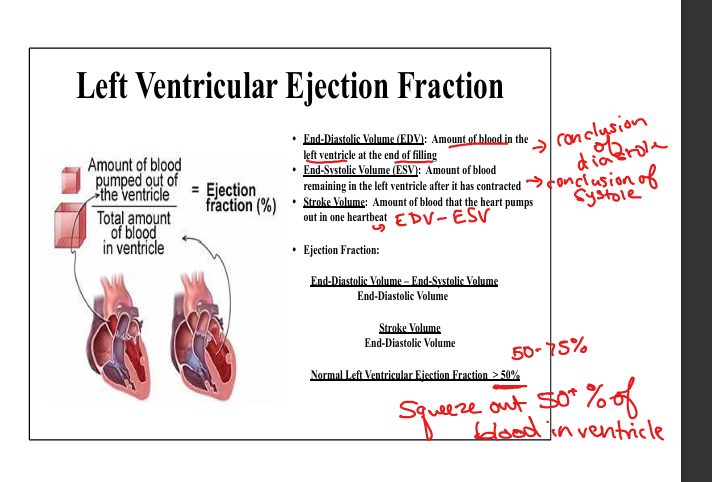
Define ejection fraction.
EF = (EDV – ESV) / EDV. Normal EF > 50%. EF reflects global LV systolic function.

Why can cardiac output be normal even with a low EF?
Because CO depends on HR × SV, and compensatory increases in heart rate or preload can maintain CO despite reduced EF.

What are the main methods to measure cardiac output?
Non‑invasive imaging (echocardiography using EDV–ESV), invasive Fick method via Swan‑Ganz catheter, and thermodilution using temperature change after injectate.
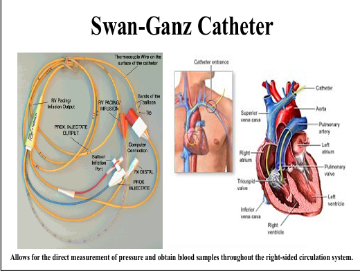
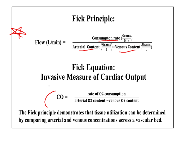
How does the Fick principle measure cardiac output?
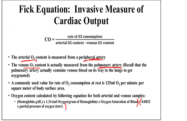
CO = rate of O₂ consumption / (arterial O₂ content – venous O₂ content). Requires arterial and pulmonary artery blood sampling.
What is the oxygen content equation used in the Fick method?
(Hb × 1.34 × O₂ saturation) + (0.0032 × PaO₂).
What is a sarcomere?
The fundamental contractile unit of cardiac muscle composed of actin (thin) and myosin (thick) filaments, titin, Z‑lines, A‑band, I‑band, and H‑band.
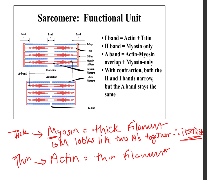
Which sarcomere bands change during contraction?
I‑band and H‑band narrow

What is the role of titin?
Acts as a spring: compressed during contraction and recoils during relaxation, contributing to LV diastolic recoil.

What determines force generation in the sarcomere?
The number of actin‑myosin cross‑bridges. More cross‑bridges = more force but slower shortening velocity.

What is the role of T‑tubules?
Deep invaginations of the sarcolemma that deliver extracellular Ca²⁺ to the interior of the myocyte.
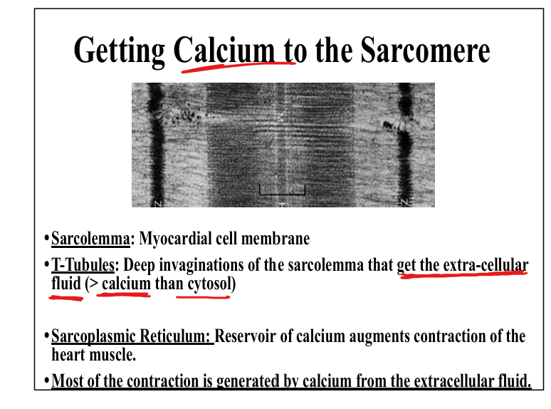
What is the role of the sarcoplasmic reticulum?
Stores Ca²⁺ and releases it via ryanodine receptors during contraction
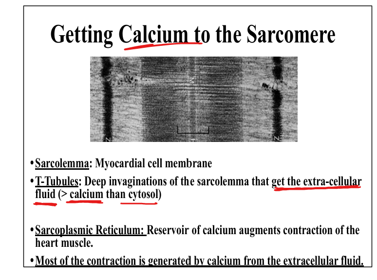
Describe calcium’s role in contraction.
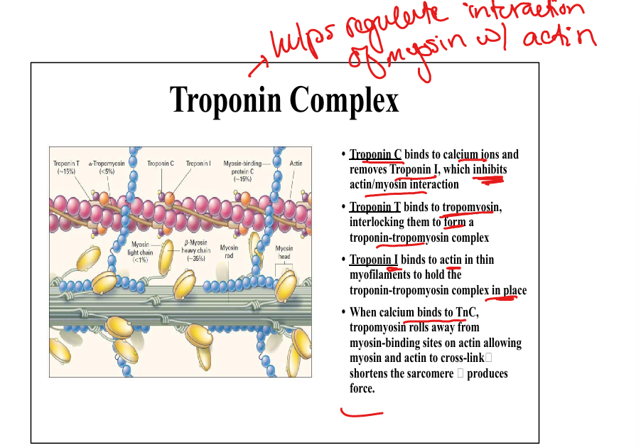
Ca²⁺ enters via voltage‑gated channels, triggers ryanodine receptor release of SR Ca²⁺, binds troponin C, moves tropomyosin, and allows actin‑myosin cross‑bridging.

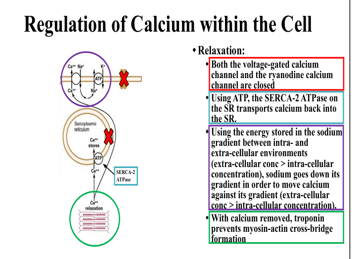
Describe calcium’s role in relaxation.
Ca²⁺ channels close
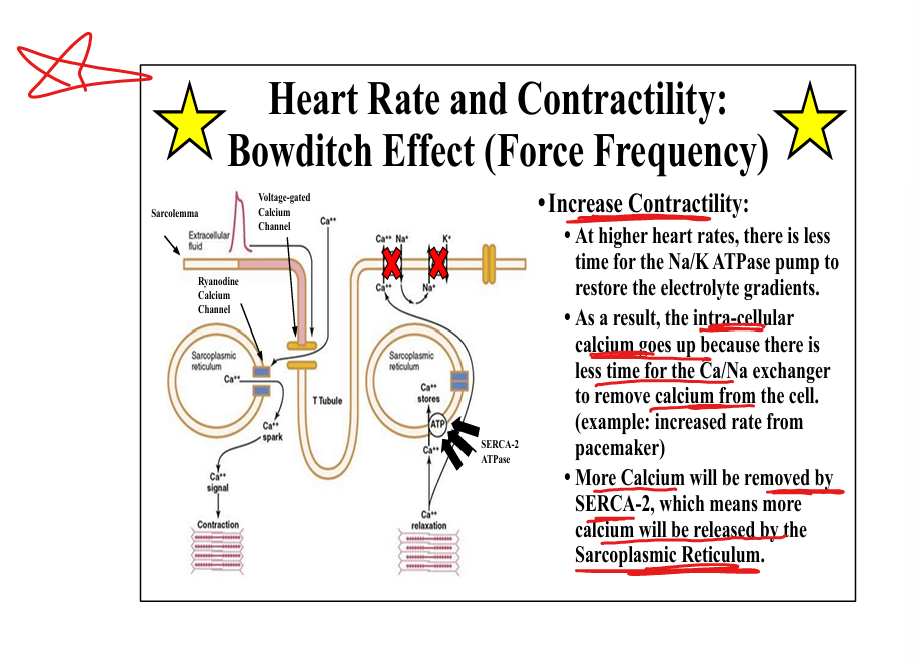
What is the Bowditch (force‑frequency) effect?
Increased HR reduces time for Na⁺/K⁺ ATPase and Na⁺/Ca²⁺ exchanger, increasing intracellular Ca²⁺ and contractility.
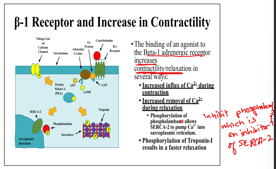
How does β‑1 receptor activation increase contractility?
Increases Ca²⁺ influx, increases SERCA‑2 activity via phospholamban(an inhibitor) phosphorylation(inhibits it), and speeds relaxation via troponin‑I phosphorylation.
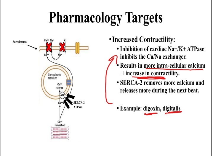
How does Na⁺/K⁺ ATPase inhibition increase contractility?
Inhibits Na⁺/Ca²⁺ exchanger, raising intracellular Ca²⁺ (mechanism of digoxin).
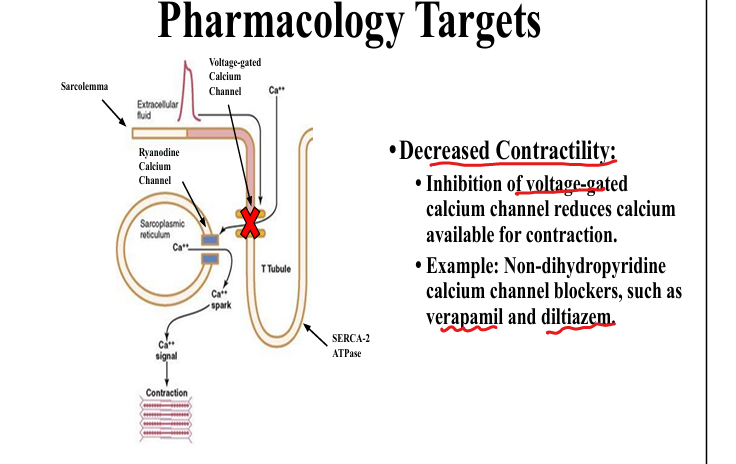
How do calcium channel blockers affect contractility?
Non‑dihydropyridine agents (verapamil, diltiazem) inhibit voltage‑gated Ca²⁺ channels, reducing Ca²⁺ entry and decreasing contractility.
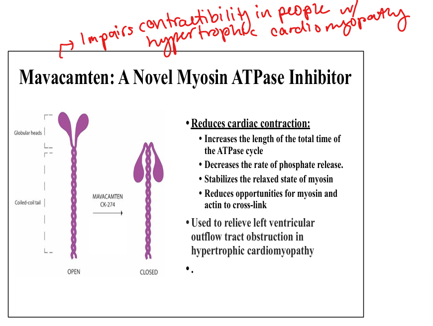
What is mavacamten?
A myosin ATPase inhibitor that prolongs the ATPase cycle, reduces phosphate release, stabilizes myosin in a relaxed state, and decreases actin‑myosin cross‑linking.
Used to releiver left ventricular outflow tract obstruction in hypertrophic cardiomyopathy
Troponin C
Binds Ca2+ ions and removes Troponin I, which inhibits actin/myosin interaction
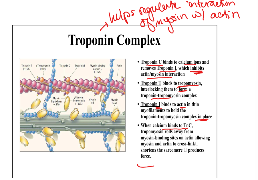
Troponin T
binds to tropomyosin, interlocking them to form a troponin-tropomyosin complex
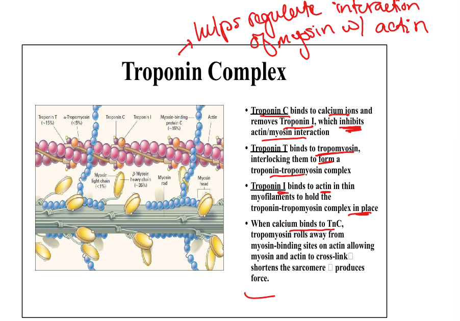
Troponin I
Binds to actin in thin myofilaments to hold troponin-tropomyosin complex in place