GI lecture 5 Large Bowel Disorders pt 2
 Call Kai
 Learn
Learn Practice Test
 Spaced Repetition
 Match
Match Flashcards
 Knowt Play
1/72

There's no tags or description
Looks like no tags are added yet.
Name | Mastery | Learn | Test | Matching | Spaced | Call with Kai | Chat |
|---|
No analytics yet
Send a link to your students to track their progress
73 Terms
one of the MC causes of acute/surgical abdomen
appendicitis
appendicitis
inflammation of the vermiform appendix located at the base of the cecum
initial inflammation of the wall if followed by localized ischemia, perforation, and the development of a contained abscess or generalized peritonitis
MCC of appendiceal obstruction
fecalith
appendicitis clinical features
early symptoms subtle and vary on location of the appendix tip
RLQ pain that begins as periumbilical pain and migrates, anorexia, N/V, fever
McBurney point, Rovsing sign, Psoas sign, Obturator sign
appendicitis diagnosis
labs- mild leukocytosis
CT abdomen- appendiceal diameter >6mm w occluded lumen, wall thickening >2mm, periappendiceal fat stranding, appendiceal wall enhancement, appendicolith
US- appendicitis with appendiceal thickening
appendicitis treatment
appendectomy w/in 24 hrs of presentation + IV pipercillin-tazobactam
nonoperative: IV ceftriaxone + metronidazole or levofloxacin + metronidazole for 1-3 days then oral abx (cefdinir + metro, cipro, amox-clav, or levo + metro) for 7-10 days
appendicitis complication
perforation
leads to localized abscess formation or diffuse peritonitis
typically seen temp >103
needs emergent sx
diverticulosis
rupture of the outer layer of the colon w protrusion of the inner layer through the defect
diverticulosis causes
increased intramural pressure and hard stools
common >50yo, western society, low fiber diet
diverticulosis is MCC of ____ in >/=60yo
hematochezia
diverticulosis diagnosis
sigmoid/colonoscopy or barium enema
what increases risk of bleed in diverticulosis
HTN and NSAID use
diverticulitis
occurs when stool or other particulate matter gets trapped in a diverticulum and abscesses
bacterial proliferation causes inflammation and microperforations of colonic tissue
diverticulitis symptoms
steady deep LLQ pain often w/palpable mass
tenesmus
constipation or diarrhea
fevers, chills, N/V, rectal bleed
diverticulosis/diverticulitis treatment
high fiber diet (mainstay)
metamucil
inpatient diverticulitis treatment
bowel rest
IV hydration
abx- piper-tazo, cipro + metro or cipro + metro
morphine
sx for perf/abscess
percutaneous drainage (abscess >4cm)
diverticulitits surgical treatment
removes diseased segment of bowel, colostomy creation, and ostomy reversal 3 months later
outpatient diverticulitis treatment
clear liquid diet 2-3 days min, amox-clav or cipro + metro for 7-10 days (tho might not be necessary)
bowel obstruction causes
neoplasm, volvulus, incarcerated hernia, stricture, obstipation, intussusception, adhesions
bowel obstructions MC occur in
elderly
bowel obstruction in neonates
imperforate anus or other anatomic abnormalities
can be secondary to meconium ileus
~Hirschsprung disease can resemble colonic obstruction
bowel obstruction presentation
mild, diffuse abdominal pain
N/V
distention
the higher the bowel obstruction
the greater the pain
bowel obstruction PE
may have diminished or absent bowel sounds, abdomen distended and poss. tender
examine inguinal and femoral regions for incarcerated hernias
digital rectal exam to reveal pathology
colonic obstruction is often caused by
left-sided inguinal hernia w sigmoid colon incarcerated in the hernia
hard stools on rectal exam
suggest impaction
soft stools on rectal exam
suggest obstipation (severe/complete constipation)
empty vault on rectal exam
suggest obstruction proximal to the level that the examining finger can reach
positive fecal occult blood on rectal exam
may suggest a more proximal neoplasm
bowel obstruction diagnosis
abdominal xray- distended loops of small bowel and colon
abdominal CT - test of choice
bowel obstruction treatment
NPO, d/c opioids if possible, NGT, IV fluids, avoid prokinetics (can worsen), sx consult
sigmoid volvulus- endoscopic reduction
MCC of bowel obstruction in children
intussusception
generally occurs in infants, often after URI
intussusception
invagination (telescoping) of the intestines
intussusception presentation
vomiting, abdominal pain, currant jelly stools (blood and mucus), lethary, sausage-shaped abdominal mass
intussusception testing
plain films, ultrasound, or contrast enema
intussusception treatment
therapeutic enemas or sx reduction
close-loop obstruction often results from
hernias/herniation, adhesions, or a twist of the mesentery
closed-loop obstruction is occluded at 2 points, causing
blood supply to be cut off = gangrene
closed-loop obstruction treatment
sx emergency - esp if small bowel d/t risk of strangulation and bowel infarction
4th MC new cancer diagnosis
colon cancer
also 2nd leading cause of cancer deaths
colon cancer is more common in people
over 50yo
colon cancer histology
adenocarcinoma (85%), mucinous (12%)
colon cancer risk factors
overweight/obese, not being physically active, processed meats, DM, smoking, alcohol, prior hx of adenomatous polyps, personal hx of UC/Crohn, family hx of inherited colorectal cancer symptoms
inherited colorectal cancer syndromes
familial adenomatous polyposis (FAP)
hereditary nonpolposis colorectal cancer (HNPCC)
Lynch syndrome
hereditary non-polyposis colorectal cancer
autosomal dominant
should suspect w/diagnosis
FAP
gene mutation causing hundreds-thousands of colon polyps beginning at age 10-12
cx usually develops by 20yo
3 subtypes of FAP
attenuated FAP-
Peutz-Jeghers syndrome (PJS)
rare, caused by genetic mutation
pts have hamartomas in digestive tract that places them at v high risk for colorectal, breast, ovarian, and pancreatic cx
diagnosed at younger age
PJS patients typically have
freckles around the mouth
sometimes on hands/feet
USPSTF colon cancer screening
everyone age 45-75
American Cancer Society colon cancer screening
everyone age 45-75, may perform up to 85yo in select populations
indicates earlier/more frequent colon cancer screening
family hx of inherited syndromes (FAP, HNPCC)
strong family hx of CRC or polyps
personal hx of CRC or polyps
personal hx of chronic IBD
screening if 1st degree relative has colon cancer hx
start at 40yo or 10 years before relative was diagnosed (whichever is earliest)
screen w colonoscopy every 5 years or more often dep. on results
FIT screening annually
colon cancer screening methods
colonoscopy- every 10 yrs
FOBT - every year
FIT- every 3 years
flexible sigmoidoscopy- every 5-10 years, usually w FOBT every 1-3 years
double contrast barium- every 5 years
if FOBT or FIT are positive
colonoscopy is needed
polyps
grossly visible mass that protrudes from the mucosal surface into the lumen of the intestine
source of rectal bleeding or partial bowel obstruction
can be non-neoplastic or neoplastic
sessile polyp
flat
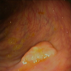
pedunculated polyp
thin stalk that supports an irregular top

adenomatous polyps increase risk for
development of adenomas and colorectal adenocarcinoma
from time of polyp formation to cancer is approx
10 years
non-neoplastic polyps
hyperplastic, juvenile, inflammatory, submucosal lesions
colon cancer mets are commonly on
liver, lungs, peritoneum
colon cancer s&s
rectal bleeding, hematochezia/melena, abdominal pain, change in bowel habits, ascites/hepatomegaly if liver is involved, weight loss
consider colon cancer in adults >50yo w/ new
iron-deficiency anemia
colon cancer diagnosis
-colonoscopy w biopsy (gold standard)
-CT scan- generalized abdominal symptoms
-flex sigmoidoscopy- most effective for palpable masses felt in rectum
-CT colonography- often used when there is an incomplete colonoscopy
colon cancer tumor markers
carcinoembryonic antigen (CEA) and carbohydrate antigen 19-9 (CA 19-9)
should not be used as screening tools
CEA levels >5ng/mL pre-op have
worse prognosis
CEA levels maintained or increased after sx indicates
aggressive disease
only curative modality for localized colon cancer
surgical resection (stage I-III)
can poss. provide curative option in stage IV disease
standard treatment for stage III colon cancer
sx + chemotherapy
role of radiation in colon cancer treatment
limited to palliative therapy for metastatic sites (bone, brain mets)
standard management for patients w metastatic colorectal cancer
chemotherapy, biologic agents w selection guided by genetic analysis of tumor
survival of colon cancer is inversely related to
stage (localized > distant)