Dr. S. Mak LAB 9: MICROSCOPIC ANATOMY OF SKELETAL MUSCLES
1/29
Earn XP
Description and Tags
Flashcards covering the microscopic anatomy of skeletal muscles, muscle fiber models, and the neuromuscular junction (NMJ) based on Dr. S. Mak's Lab 9.
Name | Mastery | Learn | Test | Matching | Spaced | Call with Kai | Chat |
|---|
No analytics yet
Send a link to your students to track their progress
30 Terms
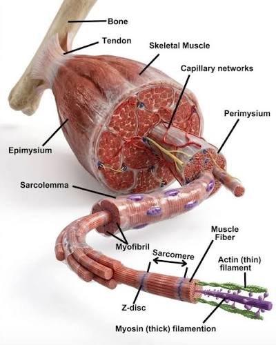
Muscle fiber
A single muscle cell which is organized into microscopic levels of structure.
Myosin
The protein that makes up thick filaments within a muscle fiber.
Actin
The protein that makes up thin filaments within a muscle fiber.
Myofibril
Long, thread-like structures found inside the muscle fiber that contain myofilaments.
Endomysium
The connective tissue layer that surrounds an individual muscle fiber.
Myofilament
The even smaller structures (actin and myosin) that make up a myofibril.
Neuromuscular junction (NMJ)
The site where a motor neuron meets a muscle fiber to communicate.
Sarcolemma
The plasma membrane of a muscle fiber.
T-tubules
Transverse tubules that carry electrical signals into the muscle cell.
Sarcoplasmic reticulum
A specialized endoplasmic reticulum in muscle cells that stores calcium.
Terminal cisternae
Enlarged areas of the sarcoplasmic reticulum found on either side of a T-tubule.
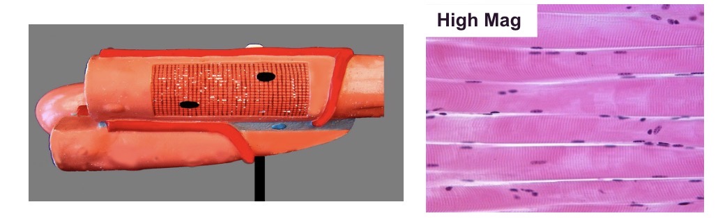
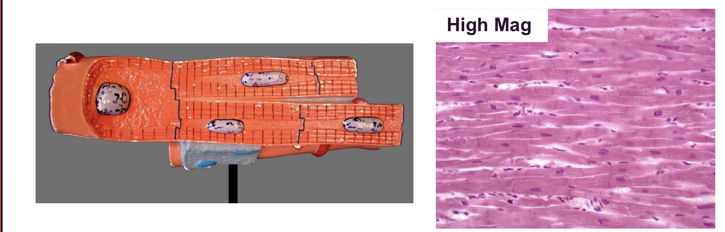
Nucleus
Organelle found in skeletal, cardiac, and smooth muscles; skeletal muscle typically has multiple nuclei per fiber.
Mitochondria
Energy-producing organelles found within the muscle fiber.
Sarcomere
The functional unit of a muscle fiber, spanning from one Z disc to the next.
A band
The dark band observed in striated muscle, containing thick filaments.
I band
The light band observed in striated muscle, containing only thin filaments.
Z disc
The boundary line of a sarcomere where actin filaments are anchored.
M line
The dark line in the middle of the H zone.
H zone
The region in the center of the A band that contains only thick filaments.
Skeletal Muscle
A type of muscle characterized by striations and multiple nuclei.
Cardiac muscle
A type of muscle found in the heart that features striations, nuclei, and intercalated discs.
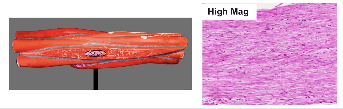
Smooth Muscle
A type of muscle that lacks striations and contains nuclei.
Intercalated discs
Specialized junctions between cardiac muscle cells.
Striations
The visible stripes seen in skeletal and cardiac muscle tissues.
Motor neuron
The nerve cell that transmits impulses from the central nervous system to a muscle.
Axon terminal
The end of the motor neuron axon that contains synaptic vesicles.
Motor end plate
The specialized region of the sarcolemma at the neuromuscular junction.
Myelin sheath (Schwann cells)
The insulating layer covering the axon of a motor neuron.
Synaptic vesicles
Sac-like structures in the axon terminal that contain chemical messengers.
Synaptic cleft
The narrow fluid-filled gap between the axon terminal and the motor end plate.