AOV - MST2
 Call Kai
 Learn
Learn Practice Test
 Spaced Repetition
 Match
Match Flashcards
 Knowt Play
1/84

There's no tags or description
Looks like no tags are added yet.
Name | Mastery | Learn | Test | Matching | Spaced | Call with Kai | Chat |
|---|
No analytics yet
Send a link to your students to track their progress
85 Terms
range of pupil diameter and retinal illuminance.
Diameter ranges from approx. 2 to 8 mm → 16-fold change in retinal illuminance, within 1-3 seconds
area is squared ---> 2 squared = 4, 8 squared = 64.... 64/4 = 16
this is where the 16 comes from
small v large pupil
diffraction v aberrations
depth of focus
small pupil
Vision through a small pupil is limited by diffraction
large depth of focus
large pupil
Vision is limited through a large pupil by aberrations
limited range. when u move off focus distance is blurred
what is the optical pupil size for limiting diffraction and aberrations
~3-4 mm
what does the posterior iris rest on?
lens
myosis vs mydriasis - pupil size
< 4 mm = “miosis” (constricted), > 4 mm = “mydriasis” (dilated)
physiological vs non-physiological anisocoria. pupil size
physiological anisocoria: normal difference in pupil sizes
less than 1 mm
most ppl have some asymmetry
non-phyiological (abnormal)
more than 1 mm
difference in brown, blue and green irises
Melanocytes (determine eye colour)
Lots → absorption → brown
None → scatter → blue
Some → brown + blue → green
does colour eyes affect drug absorption?
yep
lighter iris = more effective drops
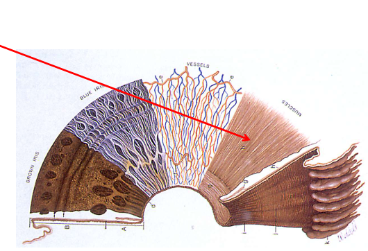
Dilator pupillae:
innervation
neurotransmitter
receptor
muscle type
action
Radial muscle (fibres oriented like spokes, towards/away from pupil)
Sympathetic innervation
Neurotransmitter: noradrenaline
Receptor: adrenergic (α1)
Action
Dilation (mydriasis): Activation of the dilator
Constriction (miosis): Relaxation of the dilator
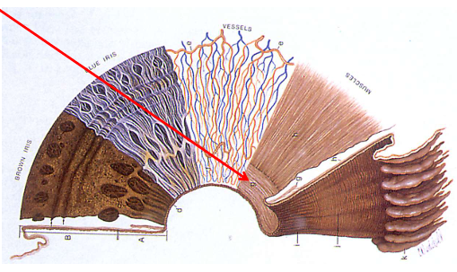
Sphincter pupillae:
innervation
neurotransmitter
receptor
muscle type
action
Circular muscle (fibres oriented around the pupil)
Parasympathetic Innervation
Neurotransmitter: acetylcholine
Receptor: muscarinic
Actions:
Constriction (miosis): Activation of the sphincter
Dilation (mydriasis): Relaxation of the sphincter
3 types of input the pupil responds to
Light reflex (both direct and consensual)
near reflex
autonomic arousal
fight or flight
fear
sex
startle
maximise retinal illumination when under pressure
pupil constricts when tired or non-aroused
is depth of field greater at near or distance?
NEAR
pupil constricts and increases depth of field
Afferent (sensory) limb - pupillary light reflex pathway
This is the pathway for:
“Shine light in the eye → pupils constrict.”
light enters retina:
retina
retinal ganglion cells
optic nerve (CN II)
optic chiasm (decussation)
optic tract
pretectal nucleus (midbrain)
this tells the brain: light has entered this eye
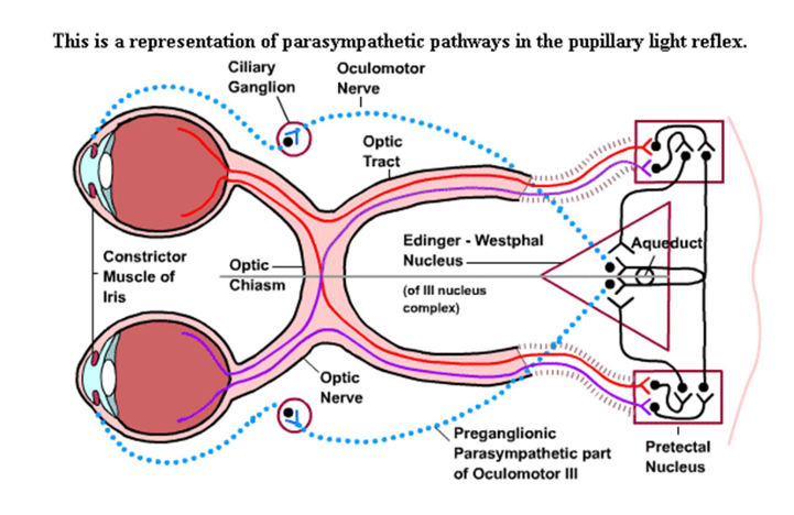
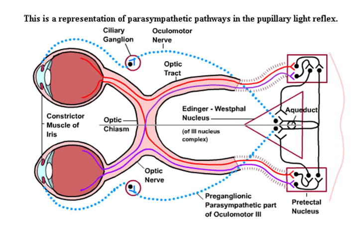
Efferent (motor) limb - motor output pathway from brain back to eye
This is the pathway for:
“Shine light in the eye → pupils constrict.”
from central pathway (pretectal nucleus)
what reflex is this? What causes the direct and concensual response
central pathway:
Pretectal nucleus → both Edinger-Westphal nuclei (bilaterally)
efferent pathway:
oculomotor nerve CN III (parasympathetic fibres) → ciliary ganglion (preganglionic synapse) → short ciliary nerves → sphincter pupillae → constriction
The key point is both Edinger-Westphal nuclei are activated, so both pupils constrict.
this is due to decussation!
this is parasympathetic light reflex
what is the near triad
Accommodation
Convergence
Pupillary constriction
Pupillary near reflex pathway
Afferent (Sensory) Limb
pupil constriction
accommodation
Retina → Retinal ganglion cells → Optic nerve (CN II) → Optic chiasm → Optic tract → Lateral geniculate nucleus (LGN) of thalamus → Primary visual cortex (V1) (occipital lobe) → Superior colliculus and parietal cortex (accommodation centres)
Pupillary near reflex pathway
Efferent (Motor) Limb
pupil constriction
accommodation
what
central pathway:
Visual cortex and parietal cortex neurons → both Edinger-Westphal nuclei (bilaterally)
efferent pathway:
oculomotor nerve CN III (parasympathetic fibres) → ciliary ganglion → short ciliary nerves → sphincter pupillae → constriction
Short ciliary nerves → Ciliary muscle Lens accommodation → constriction
this is parasympathetic near reflex (ACh - mediated)
antagonistic pathway to accommodation = sympathetic innervation (noradrenaline mediated)
main difference between near and light pupillary reflex
Main difference:
Reflex | Main pathway |
|---|---|
Light reflex | Retina → pretectal nucleus → Edinger-Westphal |
Near reflex | Retina → LGN → visual cortex/parietal areas → Edinger-Westphal |
Both eventually use the same final parasympathetic output:
Edinger-Westphal → CN III → ciliary ganglion → short ciliary nerves → sphincter pupillae.
If the pretectal nucleus is damaged, what will it impact?
we will have issues with direct and consensual light reflexes but not near
the near pathway does not pass the pretectal nucleus!
passive vs volitional:
light reflex
near reflex
light reflex = passive
near reflex = volitional (but even blind ppl can do this 🙂 don’t have to be able to see)
types of pathways:
light
near
arousal
pupil is innervated by the autonomic nervous system
light = parasympathetic (constrict)
near = parasympathetic (constrict)
arousal = sympathetic (dilate)
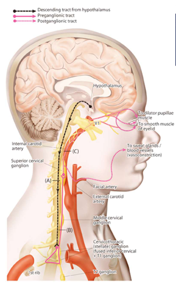
Sympathetic Pathway
EFFERENT (MOTOR) LIMB PATHWAY ONLY!
This is the pathway for:
Arousal/fight-or-flight/darkness → pupil dilates.
Hypothalamus → Sympathetic preganglionic neurons in intermediolateral column (T1-T2 spinal cord)
Preganglionic Fibres: Exit via T1-T2 spinal nerves → Superior cervical ganglion (in neck)
Postganglionic Fibres: Travel along internal carotid artery → Cavernous sinus → Ophthalmic division of CN V → Long ciliary nerves → Dilator pupillae muscle Pupil dilation
T1/T2 are near the lung
travel up the neck and synapse
what nerves are involved in:
light
near
arousal (sympathetic) pathways
Light
afferent: Optic nerve (CN II)
efferent: Oculomotor nerve (CN III)
Near
afferent: Optic nerve (CN II)
efferent: Oculomotor nerve (CN III)
Arousal
efferent: Trigeminal nerve (ophthalmic division) (CN V)
Where do the efferent pathways for parasympathetic and sympathetic innervation of the pupillary muscles meet? Where do they travel?
Ciliary ganglion
The sympathetic fibres do not synapse here, but “tag along” for the ride
Together they travel as the short ciliary nerve, to supply the iris sphincter and ciliary body (parasympathetic fibres) and the iris dilator (sympathetic fibres)
i think the dilator goes thru long fibres
Describe supranuclear inhibition of the pupil. Inhibition of pupil constriction to light (what structures are involved… 2)
give some examples of when we’d do this (3)
higher brain centres can override pupillary light reflex thru descending inhibitory pathways to the pretectal nucleus and Edinger-Westphal nucleus
inhibit parasympathetic output = reduced/absent pupil constriction
examples:
near response: accommodation - pupil constricts in dim light
arousal/attention - sympathetic activation dilates pupil overriding light reflex
ciliospinal reflex - neck pinch causes dilation, suppressing light response
Through pupil testing we can determine the function of (3)
Afferent nerves (leading away from the eye)
Efferent nerves (leading towards the eye)
Pupillary muscle function and other iris abnormalities
what target do we use when measuring pupil size
distant target
What kind of problem is it when anisocoria is greater in bright light? (diff pupil size in bright light)
what are potential diagnoses’ ?
suggests a parasympathetic problem
Bright light normally makes pupils constrict.
So if the difference is bigger in bright light, the larger pupil is suspicious because it is not constricting properly.
That suggests a parasympathetic problem.
Diagnosis
CN3 palsy
Aides tonic pupil
constricts a bit in light, but other eye constricts more due to concentric response
Traumatic iris damage
angle closure glaucoma (ACG)
pharmacologic pupil dilation
optom/med/nursing students, malingerers (pupil tyring to fake neurological condition)
What kind of problem is it when anisocoria is greater in dim light? (diff pupil size in dim light)
what is one diagnoses and three symptoms of this condition
suggests a sympathetic problem
one eye is not dilating as well when light removed
Diagnosis
HORNERS syndrome
Miosis (pupil constriction) – loss of sympathetic innervation to dilator pupillae
Ptosis (eyelid drooping) – loss of sympathetic innervation to Müller’s muscle
Anhidrosis (decreased sweating) – loss of sympathetic innervation to sweat glands
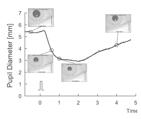
pupil reaction: speed of constriction and dilation
draw graph
pupils are slow
takes nearly a second for the pupil to constrict to about 90% of maximum
re-dilation is slower, not being able to complete even after 3 seconds
important for flashlight test!! Don’t want to go to quickly
what do u test RAPD in dim conditions?
don’t want the pupils to already be constricted
RAPD swinging flashlight test: describe the direct and consensual responses when light is removed and shone into eyes.
healthy vs diseased patient
how long do u shine light into each eye
When it first lands on one eye, there is a stimulus to dilate (consensual, due to light removed from the other eye) as well as to constrict (direct)
In a healthy patient, there is net constriction since the direct pathway is stronger
Marcus Gunn Pupil
represents a problem with the afferent system
In diseases of the optic nerve/retina, the affected side (or worse side if bilateral) produces a weak direct response (or none, if severe)
This allows the consensual response to dominate, so the worse eye dilates when the light is shone onto it! (grade 3-4+)
In less severe cases, we see a small constriction (grade 1), or a pause (grade 2), followed by a greater re-dilation
shine light into each eye for 3 seconds!
Pupil constricts to near (accommodation) but fails to constrict to light, despite intact afferent pathway
what is affected and why?
Light and near reflexes use different neural pathways after the optic nerve
Light reflex: Optic nerve → Pretectal nucleus → EWN
Near reflex: Optic nerve → LGN → Visual cortex → EWN
Light reflex: Absent/reduced
Near reflex: Normal
Pretectal pathway damage with preserved cortical pathway = light-near dissociation
Causes
Classic Causes‒ Argyll Robertson pupil (neurosyphilis) bilateral, small, irregular pupils
Dorsal midbrain syndrome (Parinaud) pineal tumour, upward gaze palsy
Diabetes, MS, midbrain stroke, trauma
Why is it important to understand pupil pathways
help interpret any abnormal pupil results
What is the visual field?
The area of one’s surroundings that is visible at one time, with a steadily fixating eye
what does the Amsler grid detect? What are two examples?
central visual disturbances
Scotomas (missing areas in vision) – severe retinal/optic nerve disease
Metamorphopsia (distorted lines) – subtle macular disorders
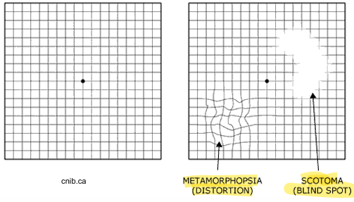
When would you use an Amsler grid on a patient?
Patients with central visual symptoms, such as:
Unexplained decreased visual acuity
Macular pathologies, (eg: age-related macular degeneration)
How much does each square subtend?
1 degree. Central is 10 degree radius from the middle.
The standard chart used in every case. Consists of a 5mm square, white grid each subtending approximately 1°, on a black background with a central, which fixation target.
Amsler grid:
distance
Rx or no?
monocular or binocular?
wear habitual reading prescription, view monocularly
held 28-30 cm away
view monocularly
Amsler grid procedure
Monocular!
Ask the patient to always fixate on the central black dot
is the central dot clear and single?
Can you see all 4 corners of the larger square?
Are any of the vertical/horizontal lines missing, wavy or distorted?
Are there any black or missing patches?
Repeat for the other eye!
What can confrontation detect? What does it test for? What is it effective and not effective for detecting?
large, absolute scotomas. tests for neglect and extinction
highly effective for post chiasmal defects (detects 9 out of 10)
less sensitive for pre chiasmal defects (detects 1 out of 3)
what is the minimum mandatory evaluation for visual field assessment in driving license requirements?
confrontation
confrontation
distance
with or without Rx?
monocular or binocular
repeat how many time?
must be at eye level with hands 50 cm from Px
habitual Rx
monocular
test both hemi-fields twice to confirm, and repeat for other eye
what is neglect and extinction: confrontation
Neglect - The patient misses one side/quadrant even when you test it by itself.
Example:
Left target alone → misses it
Right target alone → sees it
Left + right together → misses left again
Extinction - The patient can see both sides when tested separately, but when you show both at the same time, one side “loses the competition.”
Example:
Left target alone → sees it
Right target alone → sees it
Left + right together → only reports right
Red cap
distance
what it test
monocular or binocular?
40 cm
monocular
function of optic nerve
red cap procedure
normal reading?
start with good eye
Tell me what colour this cap is
On a scale of 1 to 10, with 1 being the dimmest and 10 being the brightest, if this is 10/10 (present to one eye), how would you rate the brightness of this red cap in the other eye (present to other eye)?”
An asymmetry of >2 between the eyes is a fail
What does perimetry measure? What are the 2 types of perimetry?
Measure of visual function (sensitivity) outside the fovea (i.e. across the visual field)
2 types: static and kinetic
static what we usually use
What disease is perimetry essential for detecting and managing?
Glaucoma
also useful for detection of neurological disorders and retinal disease
IF a disorder is detected using perimetry, what is the next step
Neuroimaging (MRI, CT-scan) used once disorder detected
what is the Bjerrum Screen?
a form of kinetic perimetry
mostly used in research. sometimes used in low vision cases with large areas of vision loss
useful for central scotoma
-Tangent screen (at 1 to 3 m)- Limited to central 30° to 50°- Must use 3 targets to fully evaluate scotoma
Target Size (mm)/Test distance (mm) COLOUR- e.g. 1/1000 W
3 types of perimetry that we learned in this lecture
Bjerrum screen (form of kinetic perimetry)
Goldmann perimeter
Standard automated perimetry
first one mostly used in research, second not used as much
what is the goldmann perimeter used for monitoring?
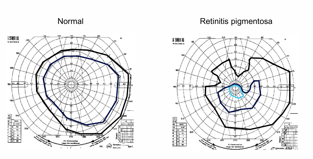
retinal degenerations
advance target of specific size and intensity inward until patient sees
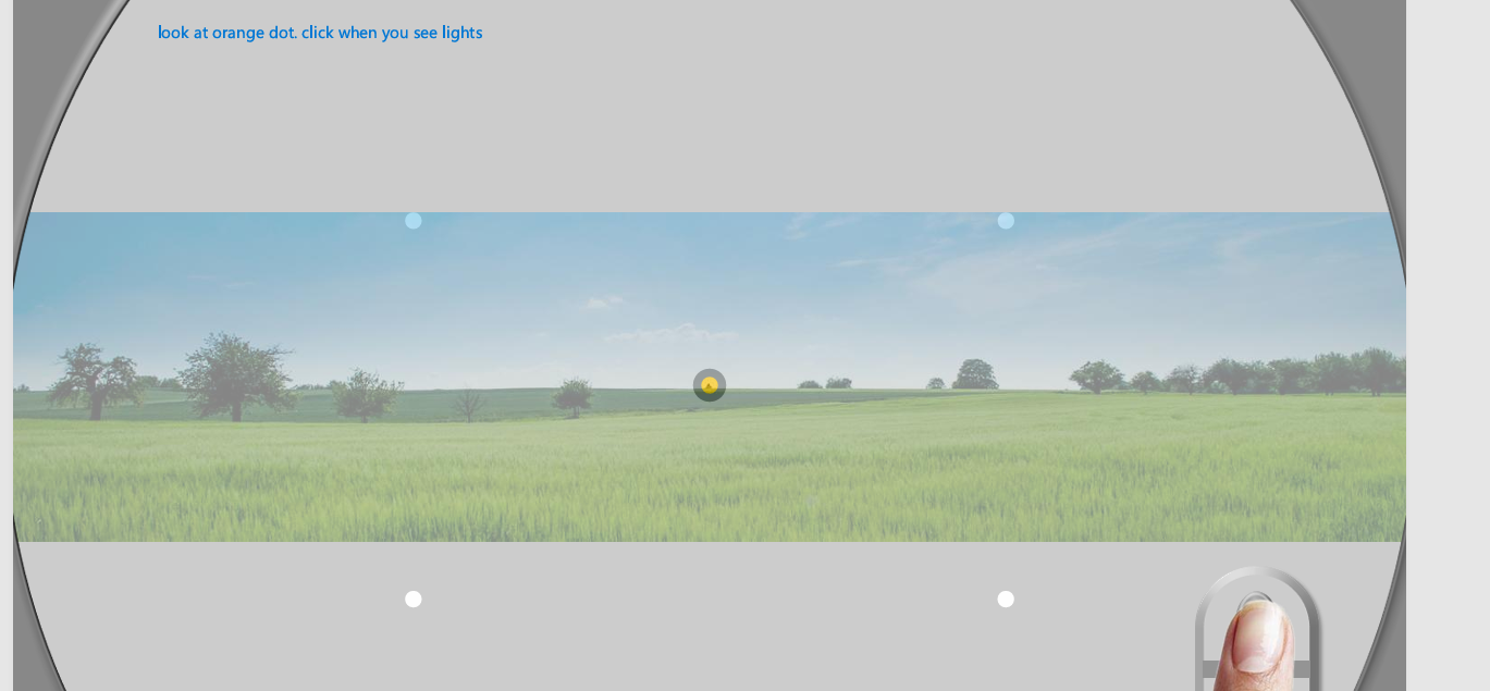
describe standard automated perimetry. What is performance compared to?
Measures sensitivity to small white lights against a dimly illuminated background
look at orange dot, click when you see lights
make them dimmer until they can’t see
Performance is compared to an age-matched normative database
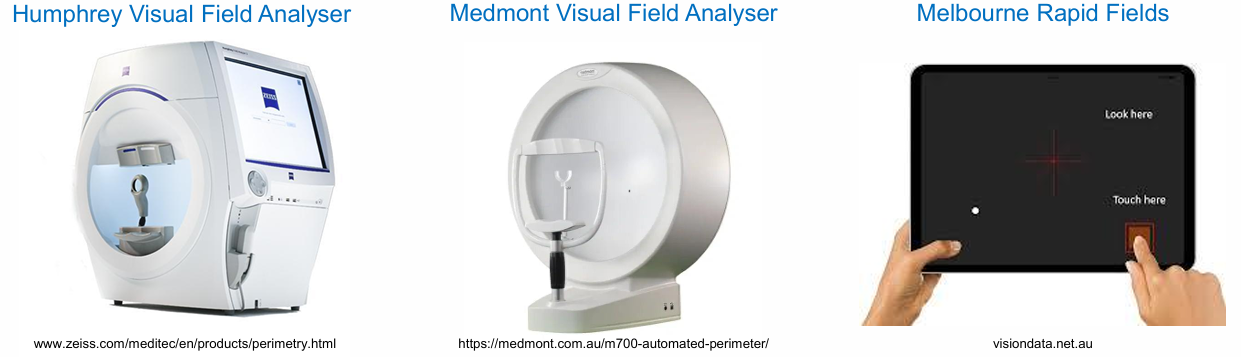
3 types of perimetry field analysers?
Humphrey visual field analyser
medmont visual field analyser
melbourne rapid fields
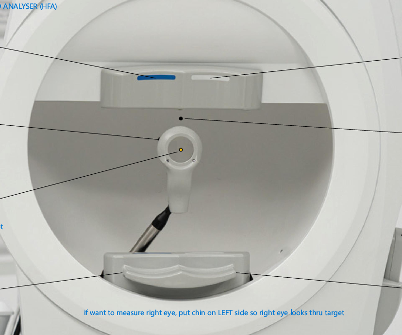
label….
if we want to measure the right eye what side would chin go on
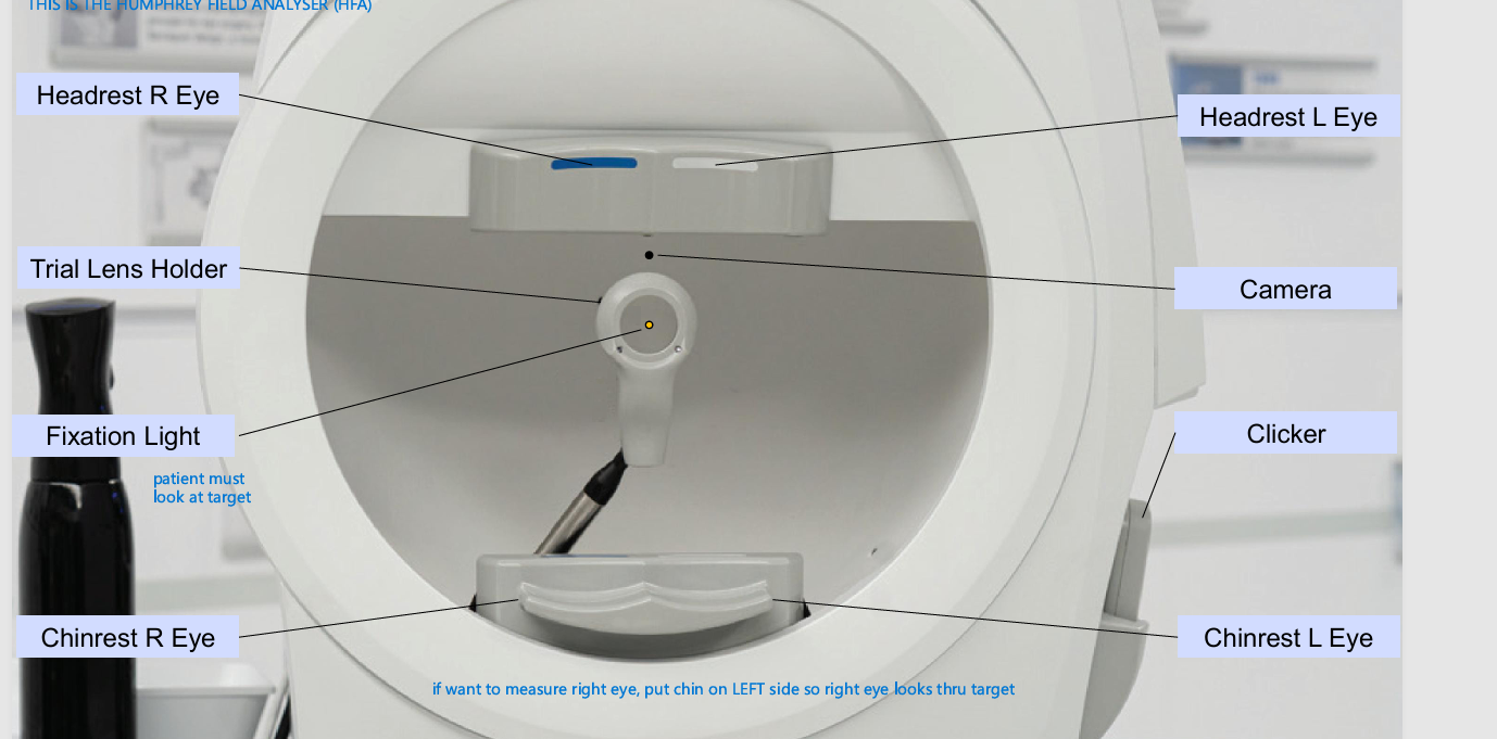
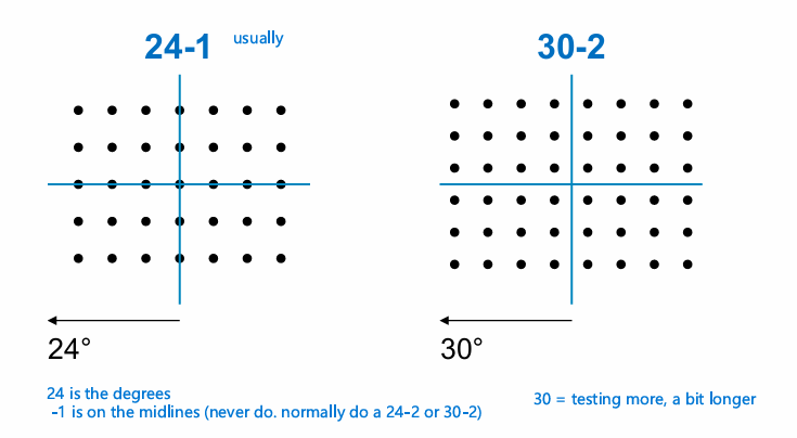
what are the 2 typical test patterns for the Humphrey Field Analyser (standard automated perimetry)
Humphrey Field Analyzer (HFA) uses a grid pattern
Patterns specified as X-Y:
where X is the extent of coverage (deg)
Y is 1 if on midlines or 2 if offset from midlines
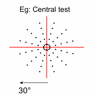
what are the 4 test patterns for the Medmont visual field analyser? What degrees are they at (standard automated perimetry)
Medmont uses a radial pattern
Patterns specified as:
Macular = 10°
Central = 30°
Glaucoma = central 30°and out to 50° nasally
Peripheral = 50°
What are the 2 types of Test Protocol for Standard Automated Perimetry?
Screening - cannot be used to diagnose
Threshold - diagnostic
4 principal aspects of eye examination. What impacts this?
refractive status
binocular vision
Ocular health discussion
Review discussion
but should be led by the patient
age helps define the examination! Also accommodation?
where do we get information on what a clinical guidelines on routine exams?
Optometry Australia
determine competency standards
medicare item number description
should be longer than 15 mins
what competency standard domains does Optometry Australia account for?
Clinical care provider (look at this particularly!)
history
plan exam based on history taking
establish diagnosis
implement an appropriate management and treatment plan. management plan can be broad!! NOT JUST SPECS
prescribe optical devices and medicines appropriately
record all relevant info
professional and ethical practitioner
communicator and collaborator
scholar and lifelong learner
quality and risk manager
Factors of condition that impact examination
how common is condition
to dilate or not to dilate? Use of OCT?
The value of early detection
Time - can’t do everything!
what do we swear by in the code of ethics?
to keep the visual welfare of the patient uppermost at all times
structure of general eye examination
History taking
Measure of VA
screening tests (OM, NPC, CT, pupils, PD, confrontation/red cap, stereo, amsler, colour vision)
Retinoscopy
Refraction - distance
more that we haven’t learned yet!
what are the 3 bodies and what they do?
AHPRA: Australian Health Practitioner Regulation Authority - Regulatory body
optometry board of Australia is part of AHPRA
determine record keeping rules of optometrist
OCANZ: Optometry Council of Australia and New Zealand - Accreditation body
OA: Optometry Australia - professional body
5 areas of history
Presenting Complaint (PC)
Patient ocular history (POH)
Family ocular history (FOH)
General Health (GH)
Visual tasks (VT)
Presenting complaint questions:
Some use LOFTSEA
Location/laterality
Onset
Frequency/progression
Type/Severity
Self treatment and its effectivity
Effect on patient
Associated signs or symptoms
Questions to ask if they are not presenting complaint
How is your vision?
distance - driving, TV
near - phone/reading
intermediate - computer
do you use glasses for these tasks
Symptoms to ask everyone
sore eyes, red eyes, itchy eyes
flashes floaters
HA related to eyes
Double vision
questions to ask in POH (patient ocular history)
Previous visits
Eye operations
Eye injuries
Eye infections
First glasses
Previous practitioners
Turned lazy eye (children maybe adults) - not a routine question
what to ask for in FOH (family ocular history) `
Glaucoma, Cat, Macular Degeneration
Turned/lazy eye/patching (if child)
diabetes, hypertension, heart disease
High rx or Blindness - not always routine
General health/Medical history questions - history taking
Start with open question
how is your general health?
history of diabetes, high blood pressure (heart disease sometimes asked but not usually)
Medications?
Allergies
Smoking - sometimes
GP name and address, how often they see - sometimes I think
Visual task questions - history taking
driving - with or without Rx
Occupation - computer use - idk most ppl are…. maybe don’t ask abt computers unless natural
Hobbies with visual demand
If patients present with particular problem (flashes/floaters, double vision), what are the MAIN important next questions:
Eg flashes and floaters
floaters = less worrysome
might be how long do flashes last?
What colour are the flashes?
Eg double vision might be
Want to be sure that you are really getting double vision vs blur so are we definitely seeing two images or one with a fuzzy second
Is the double vision two images side by side or two images once above the other
when is a good time to use open ended questions - history
at the beginning!
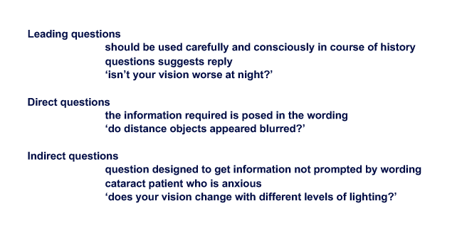
leading vs direct vs indirect questions
I think generally:
leading points you in a specific direction
direct is usually yes or no
indirect has to give some sort of other response
3 professionalism themes
Providing care that is in the best interests of patient and their carers, and warrants the respect and trust of the community
Practicing in a manner that is consistent with the regulatory and professional requirements
Engaging in reflective and evidence-based practice to continually improve knowledge and skills and providing care that prioritises effective and equitable use of health resources
5 Professionalism themes when should be able to unpack
Define a profession and describe professionalism
identify professional behaviours and how they apply to interaction with peers
Recognise the impact of bias including unconscious bias in healthcare
Define honesty, respect, integrity and responsibility in healthcare
Identify the diverse nature of optometric patients and describe additional needs that may be requeired when providing care
define unconscious bias
subconscious attitudes, stereotypes, and automatic assumptions people hold about others based on factors like race, gender, age, or appearance
what is screening (Test Protocol for Standard Automated Perimetry) good for and not good for?
not diagnostic!
good for:
elderly, cortical defects, learning/training
not good for:
subtle defects
diagnosis
monitoring disease progression
what is threshold (Test Protocol for Standard Automated Perimetry) good for and not good for?
Must be used if you are:
Confirming a defect Monitoring for stability (treatment effectiveness)
Monitoring for progression
Might not be suitable for:
Patients with limited attention spans
Patients with a disability (e.g. shaking palsy, tremor)
difference between screening and threshold - Standard Automated Perimetry
screening
stimulus of fixed intensity - lights of fixed intensity
approx 2 mins per eye
can’t be used to diagnose
dignostic
making lights dimmer and dimmer until patient doesn’t click anymore
uses a test strategy (faster = less reliable). A bit of a trade off
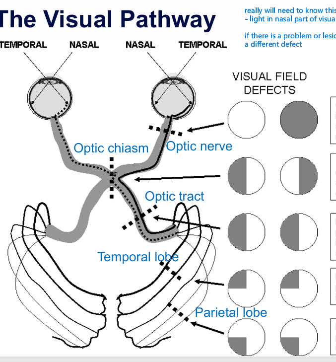
What can defects tell us about where in the visual pathway it is
Depending on the test and stuff, and failed, then did SAP to investigete. Good chance you could trace it back to part in the visual pathway depending on where the defect is!
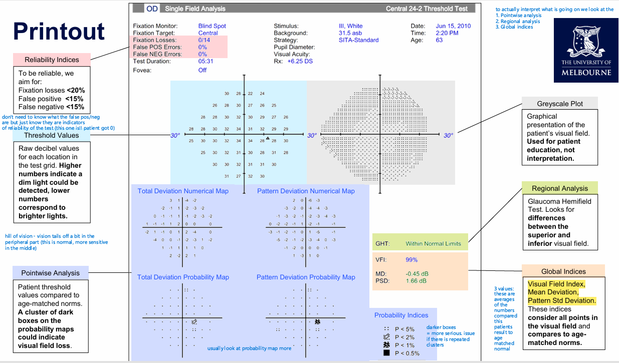
What is the most important sections of the standard automated perimetry print out? (3)
Pointwise analysis
Patient threshold values compared to age-matched norms.
A cluster of dark boxes on the probability maps could indicate visual field loss
Regional analysis
Glaucoma Hemifield Test. Looks for differences between the superior and inferior visual field
Global indices
three values:
1. Visual Field index
Mean Deviation
Pattern Std Deviation
these indices consider all points in the visual field and compares to age-matched norms
general info. Higher numbers indicate dim light could be detected, lower corresponds to brighter lights
there are reliability indices that are indicators of reliability
greyscale plot just for info
What doe