imaging in women's health (DM)_
 Call Kai
 Learn
Learn Practice Test
 Spaced Repetition
 Match
Match Flashcards
 Knowt Play
1/87

There's no tags or description
Looks like no tags are added yet.
Name | Mastery | Learn | Test | Matching | Spaced | Call with Kai | Chat |
|---|
No analytics yet
Send a link to your students to track their progress
88 Terms
Uses for Hysterosalpingogram (HSG)
Used to image uterus and fallopian tubes
Used after some sterilization procedures to verify that the tubes are blocked
Indication for HSG imaging
Submucous fibroids
Endometrial polyps
Localization of tubal occlusion
Uterine anomalies
Infertility evaluation
What are pre-op recommendations of HSG
Have procedure done during the first half of the cycle (Follicular / Ovulation) to reduce pregnancy chance
Recommend NSAIDs 1 hour prior to reduce cramping with dye
Hysterosalpingogram (HSG)
Water soluble contrast medium is injected into uterine cavity by cannula/tube
Speculum removed and films are taken
Cannula/tube removed after films are taken
What is a common symptoms to HSG
Vaginal discharge (contrast medium)
Slight vaginal bleeding
Uterine cramps
Dizziness and nausea may occur
Complications of doing HSG
Vomiting
Fainting
Heavy vaginal bleeding
Severe abdominal pain
Fever/chills
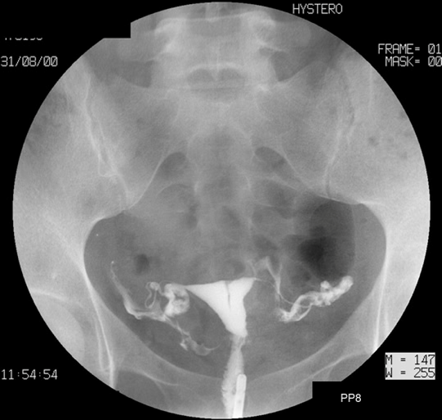
Normal HSG
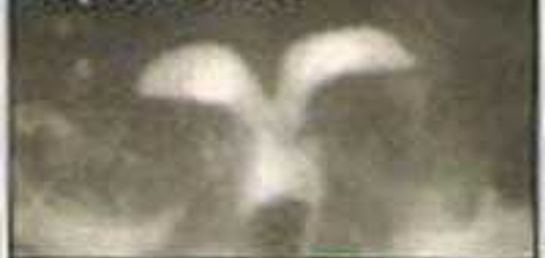
Septate Uterus
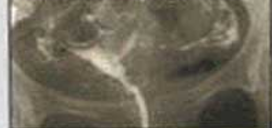
Asherman’s Syndrome

Biconate Uterus
When are abdominal XR done for Women’s Health
Calcified fibroids
IUD localization
Ovarian dermoid with calcification
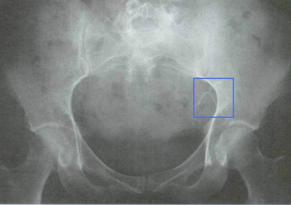
IUD
test of choice for imaging of the female pelvic organs
Pelvic ultrasound
How can US be done for women’s health
transabdominal or transvaginal
What is required to do a transabdominal ultrasound for female orgasn
Full baldder
Steps to Transabdominal US
Patient is supine, water-based gel is applied
Transducer is moved over the area of interest in order to capture desired images
What is the function of gel during a US
helps the transducer contact body and eliminate air pockets between transducer and skin
What is required to do a transvaginal ultrasound for female organs
empty bladder
Steps to Transvaginal US
Patient is supine, +/- stirrups
Transducer is placed in the vagina, with a protective cover similar to a condom, water-based lubricant is used
Transducer inserted 2-3 inches in the vagina
Images are obtained from different orientations of the transducer
Benefit of Doing a Transvaginal US over Transabdominal US
Better for an obese patient
Better for early pregnancy confirmation and evaluation
Usually more comfortable than a standard pelvic examination
What is normal endometrial thickness for follicular phase
4-8mm
What is normal endometrial thickness for luteal phase
7-14mm
What is normal endometrial thickness for menopause
< 4mm
When is the best time to perform a pelvic US of the endometrium
Follilcular as endometrium will have the most uniform linear appearance
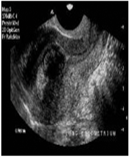
Uterine Fibroid (US)
What uterine dx may be seen on US but cannot confirm
Adenomyosis
What is the test of choice for imaging ovaries
Transvaginal ultrasound is test of choice,
then CT,
then MRI if further evaluation needed
Indications to US the Ovaries
Ovarian torsion (Doppler)
Ovarian cysts (Simple vs Complex)
Ovarican Cancer (Benign vs Malignant)
On pelvic US, you notice a Homogeneous-appearing mass with low to medium echoes. Most likely
Endometrioma
What are benign findings for pelvic US
Anechoic fluid-filled cyst
Thin walled
Homogeneous-appearing mass with low to medium echoes
Fishnet or reticular pattern of internal echoes
Markedly hyperechoic nodule with shadowing, +/- calcifications
What is considered “thin walled” for a pelvic US mass
< 30 mm in premenopausal
< 10 mm in postmenopausal
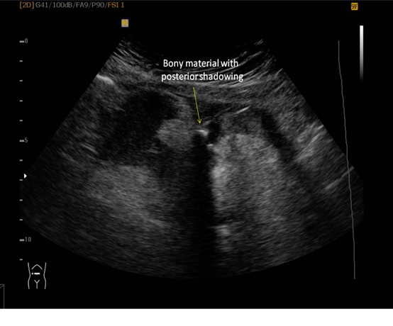
Teratoma
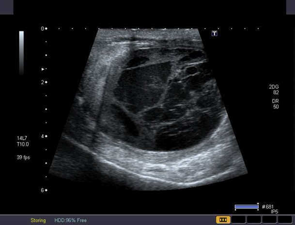
hemorrhagic cyst
Fishnet or reticular pattern of internal echoes in pelvic US is most likely
hemorrhagic cyst
Markedly hyperechoic nodule with shadowing on pelvic USis most likely
teratoma
What are malignant pelvic US findings
Solid component, not hyperechoic
Thick septations
Doppler demonstration of flow in solid component
Ascites
Peritoneal masses, enlarged nodes, matted bowel
Sonohysterosalpingogram with saline infusion (SIS)
Saline infusion is performed to distend the uterine cavity
Then the area is imaged by transvaginal ultrasound
Use of SIS
Can be used to measure the endometrium
evaluate endometrial pathology such as polyps and fibroids
Benefits to SIS
Allows for more accurate view
Will show abnormalities of the uterine lining, or uterine cavity
Provides three-dimensional view
Has the potential to replace more invasive diagnostic methods such as hysteroscopy and endometrial biopsy
Can be done in an office setting without use of anesthesia
Downside to SIS
Is painful
Indications for SIS
Evaluation of postmenopausal bleeding
Evaluation of abnormal uterine bleeding
Monitor endometrial thickness
Investigate recurrent pregnancy loss (looking for uterine anomalies)
Evaluate tubal fertility
Preoperative view of uterine fibroids
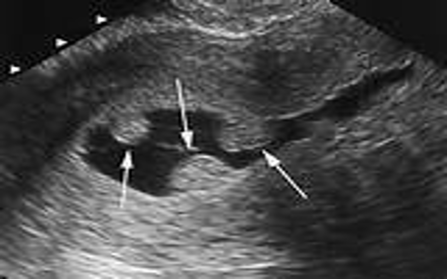
Uterine Polyps
Indications for MRI of the pelvis
Fibroids, especially malignant degeneration
Adenomyosis
Congenital abnormalities of the uterus
Endometriosis
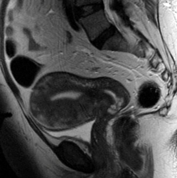
Adenomyosis
Indications for CT of the pelvis
PID
Adnexal torsion
Hemorrhagic ovarian cysts
Malignancy
When do we typically order MRI of head with coned down view of Sella
Prolactin > 100
Indications of Breast US
Patients under the age of 35
Fibroadenoma
Fibrocystic breasts
Nipple discharge
a well-defined solid mass that has benign appearance on breast US is most liekly
Fibrocystic breast changes
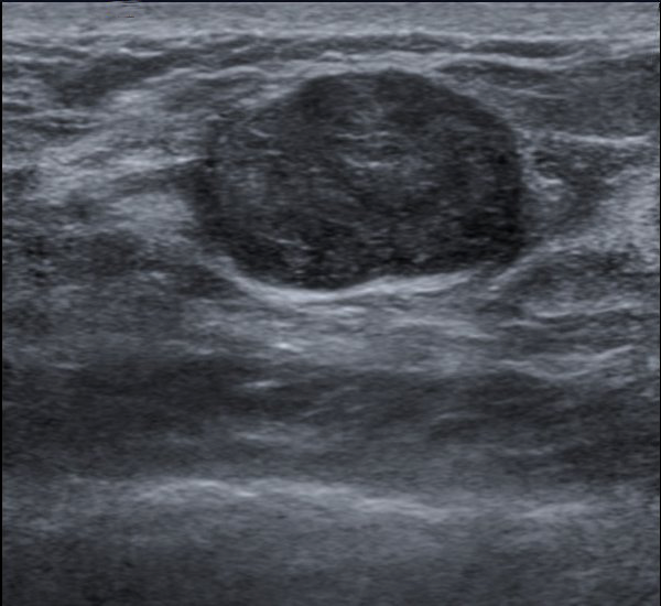
Fibroadenoma
Imaging of choice for screening for breast cancer
Mammography
When can mammography see changes suspicious for malignancy
2 years before lesion is palpable
normal mammography in a patient with a palpable breast mass needs a
Biopsy
Why is compression needed in mammography
Compression of the breasts during the procedure spreads out normal tissue of the breast
This allows the radiologist to see through the breast tissue and locate abnormalities easier
Compression also helps reduce motion artifacts (movement or breathing motions)
What views are done in screening mammography
Craniocaudad
mediolateral
When is screening mammography used
Screening in asympatomatic patients
When is diagnostic mammography used
Breast complaint
Hx of Breast Cancer
Mammography is always done
Bilaterlay
What views are done in diagnostic mammorgraphy
Craniocaudad and mediolateral done with spot compression views
Computer aided detection (CAD)
technology using a computer that will pick up mammographic patterns
Used to help radiologist identify suspicious areas
BI-RADS
A classification system for breast assessment on mammography
0 → Incomplete
1 → Normal
2 → Benign
3 → Likely benign
4 → Suspicious
5 → Highly suggestive
6 → Biopsy proven carcinoma
IBIS (International Breast Cancer Intervention Study) / Gail model and Tyrer-Cusick prevention model
risk determination tool for breast cancer
All recommendations start what age group should be screened for breast canceer
50-69
Indications for MRI of breast
Known BRCA1 or BRCA2 gene mutation
first-degree relative BRCA1 or BRCA2 gene mutation and have not had genetic testing
lifetime risk of breast cancer of 20-25% or greater
Radiation therapy to the chest between the ages of 10 and 30 years
Li-Fraumeni syndrome,
Cowden syndrome,
Bannayan-Riley-Ruvalcaba syndrome,
syndromes in first-degree relatives
Most common noninvasive diagnostic test used in pregnancy
Prenatal ultrasound
What information can be obtained from prenatal US
Confirm intrauterine pregnancy
Confirm presence of fetal heartbeat
Diagnose multiple gestation
Estimate gestational age
Screen for fetal structural anomalies
To measure fetal growth
If fetal heart tones are not auscultated by 11-12 weeks, what should be done
Prenatal US to confirm viable pregnancy
When is the best time to assess gestational age and growth by US
1st Trimester
What is the use for Doppler in prenatal US
at velocity of blood in fetal and placental vessels
Fetal anatomy best evaluated on US during
2nd trimester
Most routine US for prenatal are done during
2nd trimester
Uses of 3rd trimester US
Fetal anatomy best evaluated during
Monozygotic twins
Started from one single zygote
Identical
Dizygotic twins
Start from two zygotes
Fraternal
Monochorionic/monoamniontic twins
Share a single amnion and single chorion
Monochorionic/diamniotic twins
Have separate amnions and share a single chorion
Dichorionic/diamniotic
Have separate amnions and separate chorions
All dizygotic / franteral twins are what type of twin
Dichorionic/diamniotic
Intrauterine pregnancy can be seen with vaginal ultrasound when βhCG is
>1,500
Intrauterine pregnancy can be seen with abdominal ultrasound when βhCG is
>6,000
Concerning findings for ectopic pregnancy on US
Empty uterus with thick-walled adnexal mass separate from the ovary (95%)
Empty uterus with complex adnexal mass with solid and cystic components (70%)
Empty uterus with moderate-large amount of fluid in cul de sac (50%)
A thin-walled cyst arising from the ovary is usually a simple corpus luteum cyst
Signs of true intrauterine pregnancy
(Double Ring Sign)
Presence of an embryo in the gestational sac in the uterine cavity
Presence of a yolk sac in the intrauterine sac
Intradecidual sign
Seen before 5 weeks of gestational age,
It is a small anaechoic sac on either side of the central endometrial echo within the thickened decidua
Sign of intrauterine pregnancy
Double decidual sign
Occurs after 5 weeks
This is a hyperechoic ring (the decidual capsularis) surrounded by another hyperechoic ring (the decidual paretalis)
Sign of intrauterine pregnancy
a heterotopic pregnancy
Pregnancy in the uterus and ectopic at the same time
Risk factor for heterotopic pregnancy
IVF
Amniotic fluid index (AFI)
total linear measurement (in cm) of the largest amniotic fluid pockets in each of the four quadrants
Oligohydramnios
•reduced amniotic fluid volume (AFI<5cm)
Polyhydramnios
excessive amniotic fluid volume (AFI >20cm)