Embalming 2 Final
1/283
There's no tags or description
Looks like no tags are added yet.
Name | Mastery | Learn | Test | Matching | Spaced | Call with Kai |
|---|
No analytics yet
Send a link to your students to track their progress
284 Terms
anatomical guide
descriptive reference for locating arteries and veins by means of identifiable anatomical structures
linear guide
line drawn or visualized on the surface of the skin to represent the approximate location of some deeper-lying structure
anatomical limits
points of origin and termination in relation to adjacent structures used to designate the boundaries of arteries
linear guide
The _____________ ___________ helps you to locate the place of incision. It can and may be the actual place of incision, but that is not always the case
blunt dissection
Use _________ ________________ to get through the superficial fascia and deep fascia - as well as any adipose tissue
anatomical guide
Locate vessels by using the __________________ _________
transverse incision
what kind of incision is pictured here?

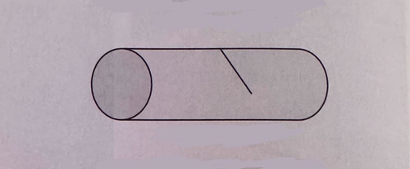
diagonal incision
what kind of incision is pictured here?
longitudinal incision
what kind of incision is pictured here?

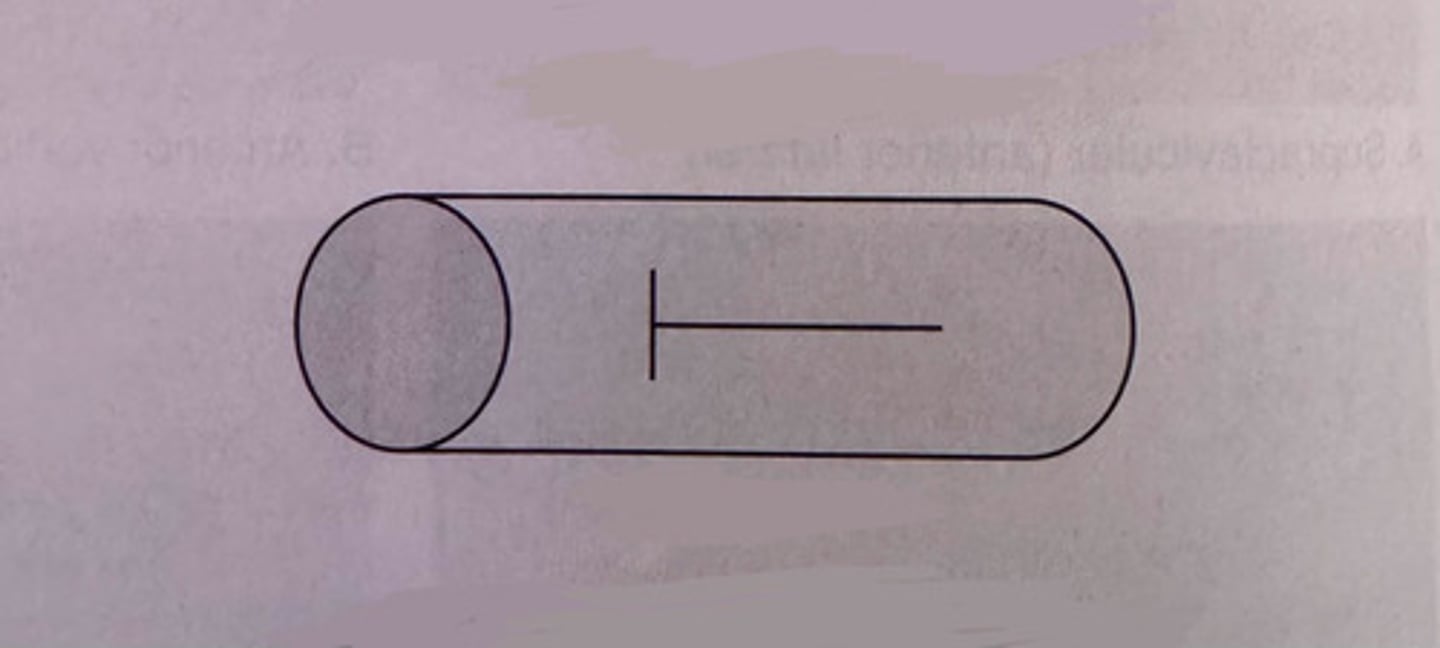
"T" incision
what kind of incision is pictured here?
double "T" incision
what kind of incision is pictured here?

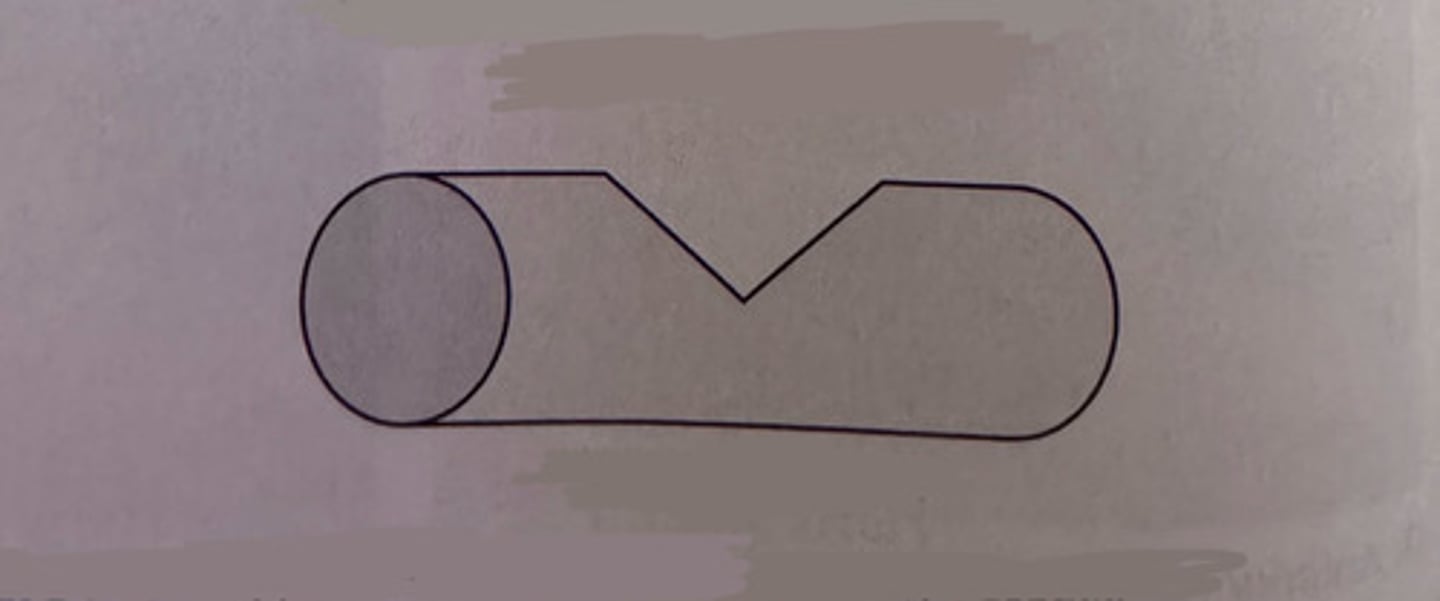
wedge incision
what kind of incision is pictured here?
transverse incision
the most common type of incision; made by cutting from the edge of the vessel to its center or slightly beyond the center
diagonal incision
more common method of incision; made by cutting at an angle from the edge of the vessel to its center or slightly beyond its center
sclerosis
the presence of ____________ in an artery makes the vessel more fragile and less elastic
longitudinal incision
begins at the center and runs parallel with the vessel; this method if favored for creating a large opening without compromising the vessel; NOT recommended for sclerotic arteries
T-incision & double T-incision
a combination of the transverse and longitudinal incisions
wedge incision
removed a wedge-shaped portion from the side of the vessel; this method allows the insertion of large diameter instruments; NOT recommended for sclerotic arteries
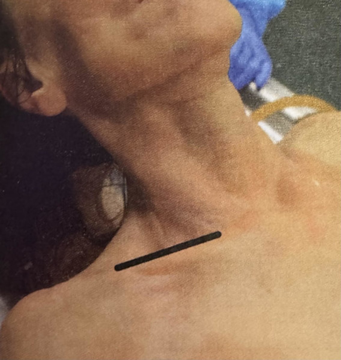
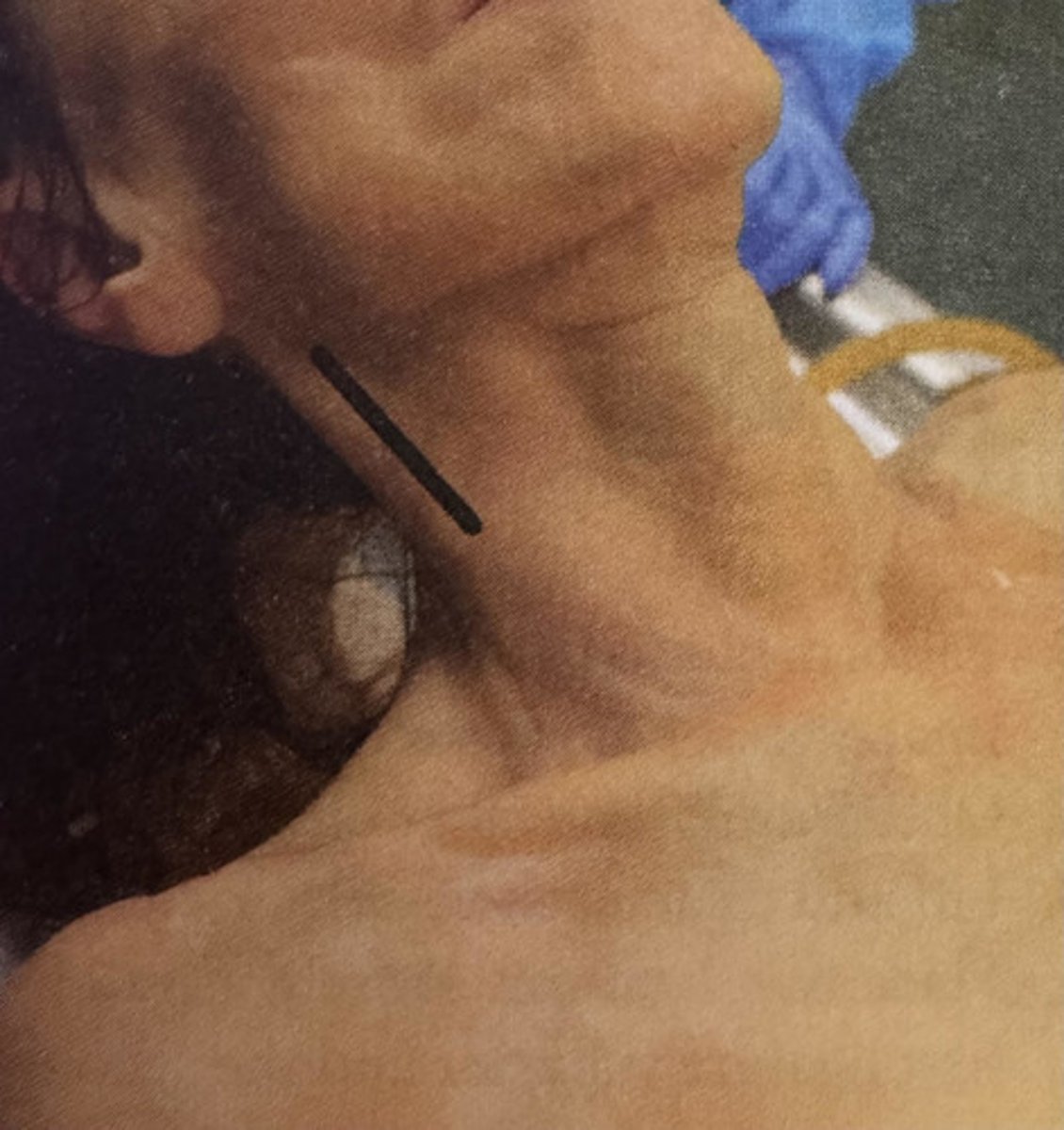
supraclavicular (anterior lateral)
what type of incision is picture here?
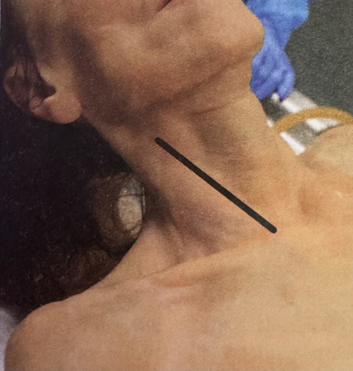
anterior vertical (parallel)
what type of incision is picture here?
posterior vertical (parallel)
what type of incision is picture here?
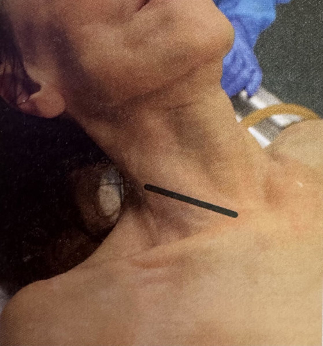
anterior horizontal
what type of incision is picture here?
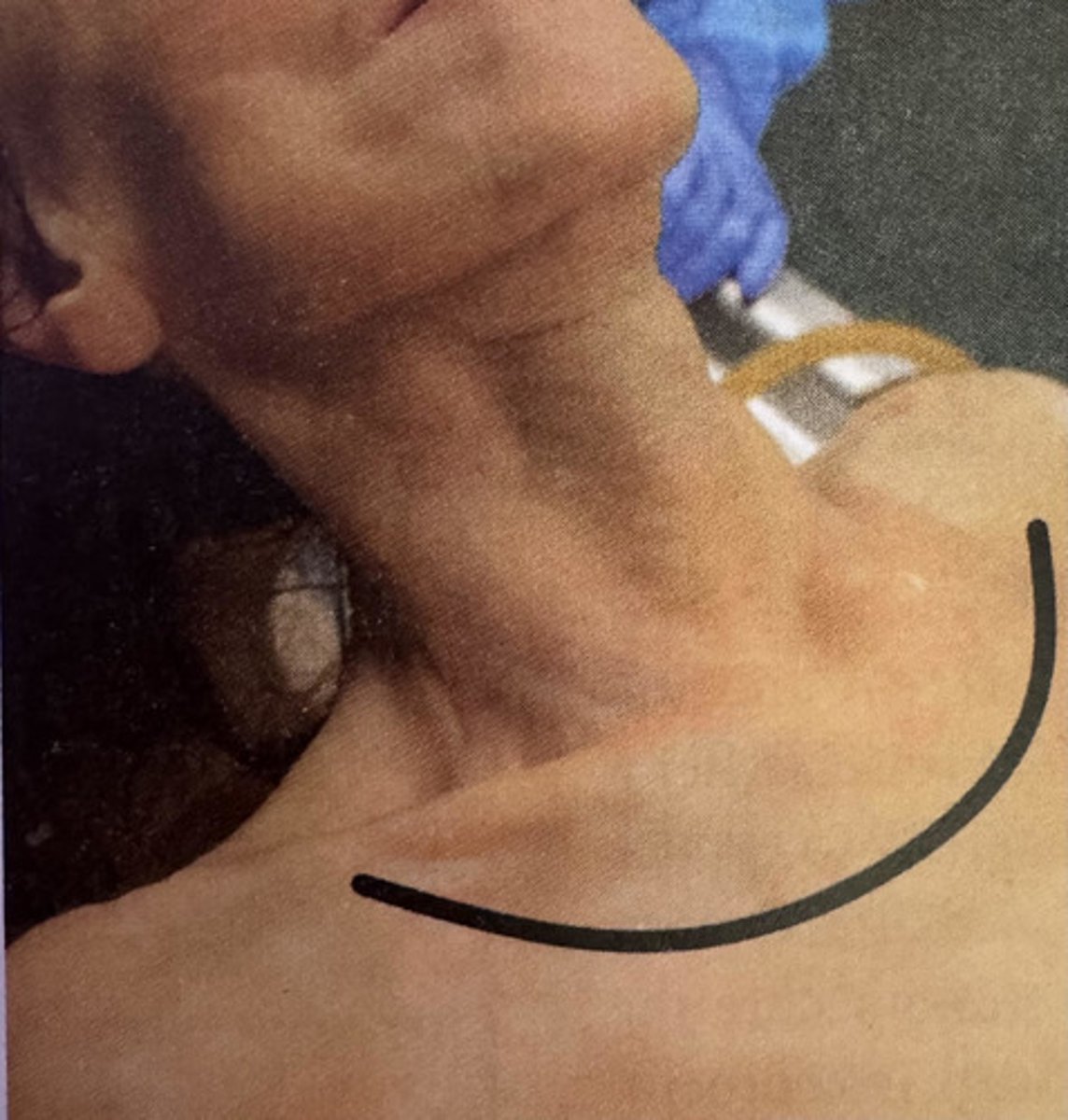
semilunar (flap incision)
what type of incision is picture here?
strap line
what type of incision is picture here?
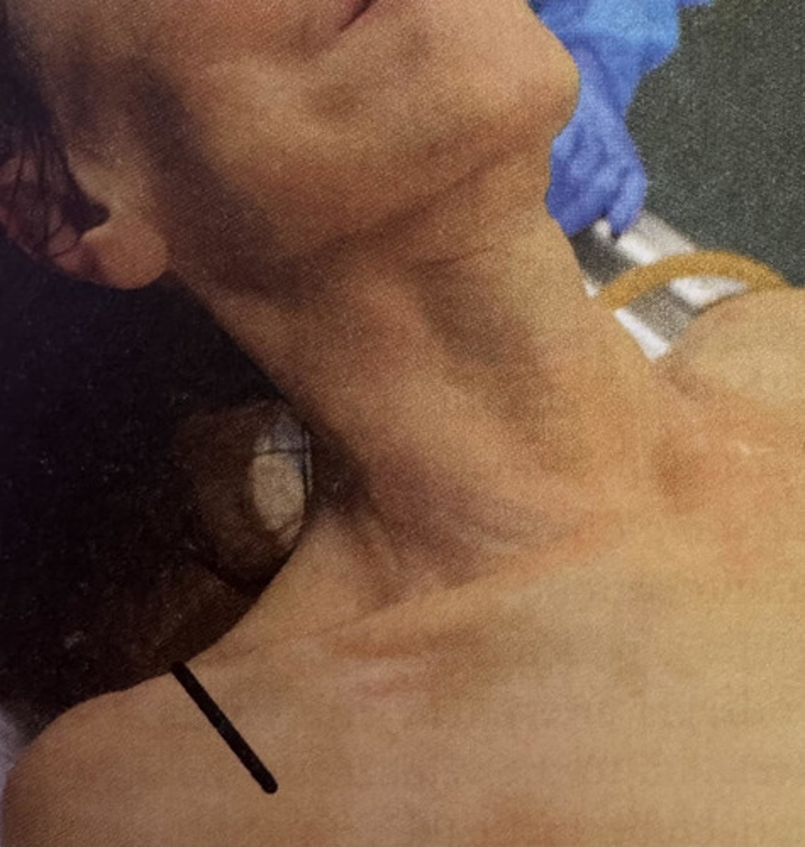
supraclavicular (anterior lateral)
incision is made along the clavicle (collar bone) from a point near the sternoclavicular articulation and is directed laterally
anterior vertical (parallel)
incision is made from a point near the sternoclavicular articulation and is directed superiorly along the sternocleidomastoid muscle
posterior vertical (parallel)
incision is made posterior to the sternocleidomastoid muscle, 2 inches below the lobe of the ear, and is directed downward toward the base of the neck
anterior horizontal
incision is made at the base of the neck from a point on the sternocleidomastoid muscle and is directed posteriorly
semilunar (flap incision)
incision extends from a point lateral and slightly superior to the sternoclavicular articulation and is directed inferiorly, crosses the upper chest in an arc, and is directed superiorly to the opposite articulation
strap line
incision in made approximately 2 inches lateral to the base of the neck on the line where the shoulder strap of a sleeveless garment crosses the shoulder
- common carotid
- femoral (or external iliac if raised by the inguinal filament)
- axillary (or brachial)
the three most common arteries raised for arterial injection:
smaller
__________ arteries are usually reserved for secondary injection sights
internal jugular vein
what vein is the companion to the common carotid artery?
femoral vein
what vein is the companion to the femoral artery?
axillary vein
what vein is companion to the axillary artery?
basilic vein
what vein is companion to the basilic artery?
internal jugular vein
which vein is most commonly used for blood drainage? (hint: it's close proximity to the right atrium of the heart makes it an ideal pairing with any artery selected)
nerves
solid structures lacking the lumen present in both arteries and veins; they have a silvery white appearance with visible striations; if incised, the edges fray similar to the ends of a cut rope
veins
thin walled vessels that contain a lumen; they have a blush appearance when filled with blood; opaque when not blood-filed; have internal valves to prevent backflow of circulating blood
lumen
the opening or inside space of the vessel or other tubular structure
vein
upon mistakenly injecting a _______, the telltale sign is that fluid will not distribute and instead flow back out from the vessel; it will collapse on itself when cut
arteries
thick walled vessels that contain a lumen; the lumen stands open and pronounced when cut; do not have valves which makes them suitable for injection in either direction of the vessel
vasa vasorum (vv)
"vessels on vessels"; tiny blood vessels that supply the large vessel walls with nutrients
case analysis
how do we determine which vessel to raise for injection?
injection; drainage
arteries are used for ______________ while veins are used for _____________
ligatures; fixation forceps
the cannula is placed into the artery and then secured with either ____________ or arterial _____________ _____________
heart; right atrium
desired drainage instruments should be inserted into the vein toward the __________; specifically the _________ ________ if possible
drain tube or angular fixation forceps
these two instruments may be used for drainage:
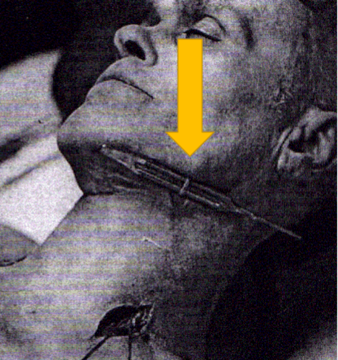
the right common carotid artery and the right jugular vein
the vessels most commonly used for arterial embalming are:
ANV
the acronym ______ is helpful to describe the relationship of the common carotid artery, vagus nerve, and internal jugular vein
opposite
when accessing the common carotid artery for arterial embalming, turn the head of the body in the direction _____________ that of the vessels bring raised
ligate
to tie off
facial artery
one of the eight branches of the external carotid artery; usually reserved for injection of the facial tissues when the common carotid artery is either damaged or unavailable
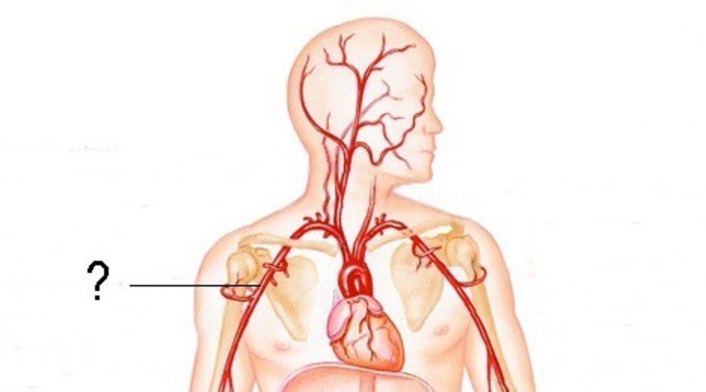
axillary artery
in the early years of embalming the __________ ________ was the preferred site to inject the entire body; now it's used principally as a secondary point of injection
right internal jugular vein
this vein is often used for blood drainage as the axillary vein is relatively small and may not evacuate clotted materials
90
when accessing the axillary artery for arterial embalming, the arm must be raised slightly less than ____ degrees from the trunk
brachial artery
continuation of the axillary artery and used principally as a secondary point of injection to reach the forearm and hand
radial artery
originates at the bifurcation of the brachial artery and supplies solution to the lateral side of the hand (thumb)
ulnar artery
originates at the bifurcation of the brachial artery and supplies solution to the medial side of the hand (little finger)
femoral artery
the second more frequently used vessel for arterial embalming; continuation of the external iliac artery and located superficial and lateral to the femoral vein
VAN
the acronym ______ is helpful to describe the relationship of the femoral vein, femoral artery, and femoral nerve
popliteal artery
continuation of the femoral artery and can be used as a secondary injection site when solution has not distributed below the knee
popliteal
the location and size of the _____________ vein makes it unsuitable as a drainage site
anterior and posterior tibial arteries
supply arterial solution directly to the portion of the leg below the knee into the foot
aorta
the largest artery in the body, traveling the length of the torso along the midline; may be selected for infant embalming when commonly used arteries are too small for injection
abdominal and thoracic aorta
either of these can be used for injection following partial autopsy or organ donation
external iliac artery
passes beneath the inguinal ligament and lies on the lateral side of the external iliac vein; supplies solution to the lower extremity and anterior abdominal wall
internal iliac artery
supplies embalming solution to the external genitalia, gluteal muscles and the peroneal regions; following a complete trunk autopsy, it may be accessible from within the pelvic cavity
inferior vena cava
the largest vein in the body; located to the right of the aorta at the posterior abdominal wall; primarily used for blood drainage in cases of partial autopsy or organ donation
right atrium of the heart
the center for venous drainage in the dead human body; routinely (during embalming of a nonautopsied body) a drainage device is inserted into the right jugular vein and guided into this
direct heart drainage
historic method first practiced when embalming was done at the residence of the deceased; today it is recommended for infectious cases to eliminate contact with blood and bodily fluids
- ascending aorta
- arch of the aorta
- descending aorta
- accompanying veins
the parts of the aorta:
brachiocephalic artery
the first branch off of the arch of the aorta; only exists on the right side of the body
right subclavian artery and the right common carotid artery
the brachiocephalic artery bifurcates (splits) into:
- common carotid
- facial
arteries used for injection of the head and neck:
- subclavian
- external iliac
arteries used for injection of the trunk:
- axial
- brachial
- radial
- ulnar
arteries used for the injection of the upper extremities:
- femoral
- popliteal
- anterior tibial
- dorsalis pedis
arteries used for the injection of the lower extremities:
aorta and brachiocephalic
almost all of the arteries we discussed are paired, with the exception of the:
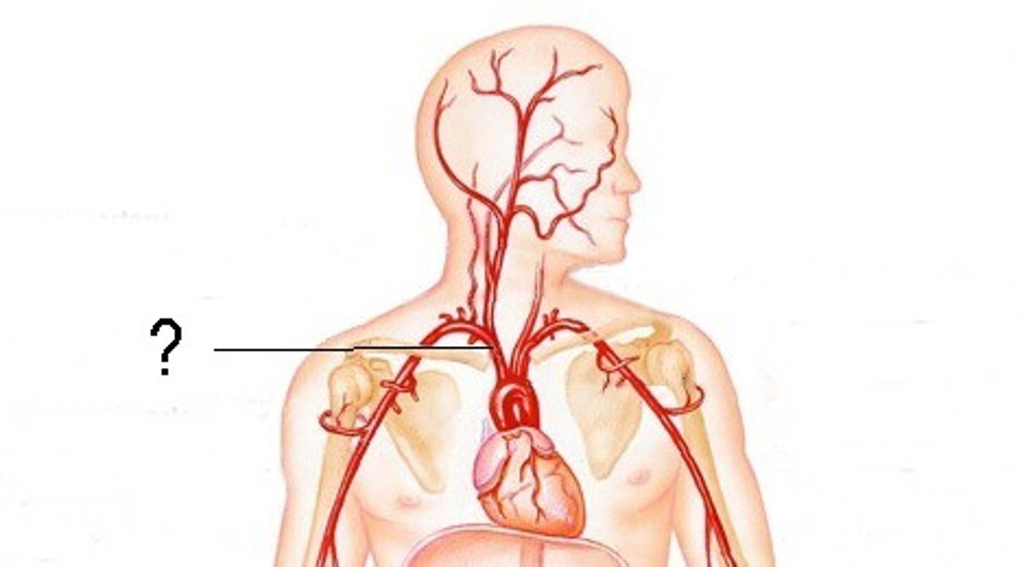
common carotid artery
most common artery used for injection (right); provides direct distribution to the face and is close to the center of circulation (heart); incisions may be visible after dressing; facial swelling may occur (over-injected)
anatomical guide of the common carotid artery
along the medial border of the sternocleidomastoid (SCM) muscle
linear guide of the common carotid artery
from the sternoclavicular articulation to the anterior surface of the lobe of the ear
anatomical limit (right) for the common carotid artery
begins at the level of the sternoclavicular articulation and terminates at the level of the superior border of the thyroid cartilage
anatomical limit (left) for the common carotid artery
begins at the level of the second costal cartilage and terminates at the superior border of the thyroid cartilage
accompanying vein for the common carotid artery
located lateral and superficial in relation to the common carotid artery
• Supraclavicular - along the superior border of the clavicle
• Parallel - along the posterior border of the inferior 1/3 of the sternocleidomastoid muscle
place(s) of incision for the common carotid artery:
internal jugular vein
what is the accompanying vein for the common carotid artery?
anatomical guide for the facial artery
along the inferior border of the mandible, just anterior to the angle of the mandible
anatomical guide for the subclavian artery
the clavicle bone
place of incision for the subclavian artery
along the upper margin of the clavicle, about 1/3 the distance from the shoulder and the root of the neck
anatomical limit (right) for the subclavian artery
begins at the sternoclavicular articulation and terminates at the lateral border of the first rib
anatomical limit (left) for the subclavian artery
begins at the level of the second costal cartilage and terminates at the lateral border of the first rib
linear guide of the axillary artery
through the center of the base of the axillary space and parallel to the long axis of the upper extremity when abducted
anatomical guide of the axillary artery
posterior to the medial border of the coracobrachialis muscle
anatomical limit of the axial artery
begins at the lateral border of the first rib and terminates at the inferior border of the tendon of the teres major muscle
place of incision for the axillary artery
along the anterior margin of the hairline of the axilla
accompanying vein for the axillar artery
located medial and superficial in relation to the axillary artery
axillary artery
artery that is close to the face, center of circulation (heart), and center of venous drainage; vessels are relatively superficial; care must be taken when positioning the arms during injection
linear guide of the brachial artery
from the center of the base of the axillary space to the center of the forearm just below the bend of the elbow
anatomical guide of the brachial artery
artery lies posterior to the medial border of the belly of the biceps brachii muscle