DDMC SPINAL W1
 Call Kai
 Learn
Learn Practice Test
 Spaced Repetition
 Match
Match Flashcards
 Knowt Play
1/112

There's no tags or description
Looks like no tags are added yet.
Name | Mastery | Learn | Test | Matching | Spaced | Call with Kai | Chat |
|---|
No analytics yet
Send a link to your students to track their progress
113 Terms
The most common cervical joint to be affected by instability caused by rheumatoid arthritis is:
C1/2
In RA, what can instability be associated with?
Neck pain
Compression of adjacent structures such as brainstem, spinal cord and spinal nerve roots
What conditions can instability in the case of RA cause?
Radiculopathy
Myelopathy
What is happening in RA?
Synovitis develops in the facet joints
Erosion of adjacent ligaments, the annulus, disc spaces and bone
Progressive instability
What are the 3 patterns of instability which occur?
The atlas shifts forward on the axis
One vertebral body shifts forward on the body of another, producing stepladder deformity (subaxial subluxation)
Axis telescopes (pushes up) into the atlas, driving the odontoid upwards
What is the symptomology of RA?
Suboccipital pain (C2 nerve root involvement due to C1/2 subluxation)
Subluxation causing C2 radicular pain is the most common radicular pain seen in RA
Myelopathy (gait disturbance, loss of fine motor control in hands, numbness in hands, balance disturbance, hyper-reflexia, extensor plantar response, + Hoffman', clonus, motor/sensory loss)
Brainstem compression less common (facial sensory disturbance, dyshagia, abnormalities in lower cranial nerves
Sudden death RARE
What is the management of RA?
Prophylactic surgery to prevent myelopathy from developing where subluxation has reduced canal diameter
Posterior stabilisation of AA subluxation good for C2 nerve root pain
Decompressive procedures to halt neurological deterioration
What are patients with AS at risk of?
Atlantoaxial subluxation
Cervical spine fractures
Cervical spine stenosis
What is the symptomology of AS in the cervical spine?
Reduced neck motion
Progressive neck kyphosis
Unable to look forward
Chin-on chest deformity
What is the commonest radiological feature in AS?
Vertebral squaring
Syndesmophytes
At what 2 cervical segments do spinal fractures in AS most often occur?
C5-C6
C6-C7
What mechanism do spinal fractures in patients with AS often result from?
Low-energy mechanism such as a fall from standing height
Why are people with AS more susceptible to fractures?
Ossification of spinal ligaments
Calcification of the annulus fibrosis
Why might it be hard to distinguish an acute fracture from a chronic inflammatory form of AS?
Fracture may not be detectable with plain radiographs due to kyphotic deformity and distorted anatomy
High riding shoulders can obscure the lower cervical spine
What is the incidence of spinal cord injury in AS patients?
Eleven times higher than the general population
What is the most frequent cause of death in AS?
Respiratory complications such as pneumonia
What percentage of the general population do minor bony malformations of all types occur in?
12%
What anatomical planes do congenital spinal deformities affect?
Sagittal plane (kyphosis/lordosis)
Coronal plane (scoliosis)
Rotational
What three categories are origins of anomalies grouped into?
Malformation
Failure of the embryologic differentiation or development of a specific anatomic structure, causing it to be absent or improperly formed before fetal period (week 8 onwards)
Disruption
Subsequent destruction of an anatomic feature that formed normally during the embryonic period (weeks 3-8)
Deformation
Alteration in the shape or structure of an individual vertebra or of the entire spine during the fetal or postnatal period, e.g. progressive, idiopathic scoliosis in patients with structurally normal spines at birth
What is the aetiology of congenital spinal deformities?
Possibly multifactorial
Inherited genetic susceptibility
De novo alteration in molecular mechanisms (exposure to teratogens such as smoking)
What is occipitilisation?
Developmental anomaly where there is partial or complete fusion of the atlas to the occiput
What is the mechanism of cause for occipitalisation?
Failure of segmentation between the fourth occipital sclerotome (proatlas) and the first cervical sclerotome during embryonic development
What is the most common aspect of the atlas to assimilate into the occiput?
Anterior arch
What can occipitilisation be associated with?
Atlantoaxial instability
Basilar invagination
→Both can result in compression of the medullary cord
Define Klippel-Feil syndrome
Congenital fusion involving two segments, congenital block vertebrae or entire cervical spine
What causes congenital cervical fusion?
Failure of normal segmentation of cervical somites during weeks 3-8 of life
What is the classic clinical triad of Klippel-Feil syndrome?
Low posterior hairline
Short neck
Limitation of neck motion (mostly lat bend and rotation)
What are the complaints of people with Klippel-Feil syndrome?
Cosmesis of the neck or cosmesis resulting from associated anomalies (Sprengel deformity)
Neck pain
Radicular pain with or without weakness
Slowly progressive or acute paraparesis or quadriparesis
What physical features are seen in KlippelFeil syndrome?
Low posterior hairline
Short neck
Torticollis
Restricted neck movements
What features is KlippelFeil syndrome associated with?
Anomalies of the jaw
Incomplete cleft of nasal cartilage
Cleft palate
Sprengels deformity
Congenital deformities of external ear
Cervical ribs
Hypospadias
Urinary tract anomalies
When is the typical onset of features in KlippelFeil syndrome?
3rd or 4th decade
In KlippelFeil syndrome, how does neurological involvement occur?
Through compression caused by laxity of the transverse ligament, alteration of course of vertebral artery, medullary compression of odontoid
What are the common anomalies associated with Killep-Feil syndrome (Decreasing order of frequency)
Scoliosis
Renal abnormalities]
Sprengel deformity
Deadness
Congenital heart disease
Ptosis
Facial nerve palsy
Hypoplastic thumb
Upper extremity hypoplasia
Where is McRae’s line drawn and what does it assess?
Drawn across the foramen magnum
Odontoid process should not project above this
What headache symptomology is seen in occipitilisation?
Dull ache
Located over posterior two thirds of skull
Precipitated by coughing or neck movements
Long tract signs associated with dysfunction to the lateral corticospinal tract, that may be present in upper and lower extremities in the form of hyperrelexia, spasticity, hoffmans sign and babinski’s sign
Cranial nerve findings including tinnitus, visual disturbances, lower cranial nerve palsies leading to dysphagia/dysarthria/downbeat nystagmus
Horners syndrome
What is the management in occipitilisation?
In patients with minor trauma or infection, can be treated conservatively with immobilisation in plaster, traction or cervical orthosis
In patients with neuro involvement, cervical spine fusion or decompression is indicated
What should patients with congenital cervical fusion avoid?
Contact sports and activities that put them at risk for head trauma
What is the management of Klippel-Feil syndrome?
Activity modification
Bracing
Traction
Must consider spinal fusion if neuro lesion is present (avoid contact sports)
Manual therapy contraindicated
Define Atlanto-Axial (AA) instability
Excessive movement at the junction between the atlas and axis due to bony or ligamentous abnormality
What can AA instability be caused by?
Congenital conditions
Trauma
Degenerative changes due to inflammatory pannus of rheumatoid arthritis
Infection
What two structures are often involved in AA instability?
Transverse ligament
Odontoid process
What structure is the primary restraint against anterior translation of C1 on C2?
Transverse ligament
What structure is the primary restraint against posterior translation of C1 on C2?
Odontoid process
State if the transverse ligament is intact in these distances:
<3.5mm
3.5-5mm
>5mm
Yes
No
No, alar ligaments also likely involved
What is vertical displacement of the atlas and what is the cause of it?
Widening of the C1-2 facet joint
Due to disruption of the alar ligament, superior longitudinal band of cruciate ligament, tectorial membrane or combination of these structures
What are the 4 types of atlantoaxial rotatory subluxation?
Type 1 (Stable)
Simple rotary subluxation without anterior shift
Odontoid acts as a pivot
Type 2 (Stable)
Rotary displacement with anterior displacement of 3-5mm
Lateral articular process act as pivot
Type 3(Not stable)
Rotary displacement with anterior displacement of >5mm
Type 4 (Not stable)
Rotary displacement with posterior displacement
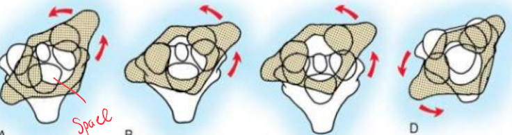
How can AA instability cause neurological signs?
Odontoid process or posterior arch of atlas can impinge on the spinal cord
Motion of C1-2 segment can cause compression of nerve roots
What severe consequences are there for individuals with RA that have AA instability?
Cervicomedullary compression
Vertebral artery insufficiency
What is the primary cause of AA instability in individuals with down syndrome?
Laxity of transverse ligament
What congenital conditions other than down syndrome can cause AA instability?
Osteogenesis imperfecta
Neurofibromatosis
Morquio syndrome
Larsen syndrome
SED congenita
Chondrodysplasia punctata
Metatropic dysplasia
Kniest syndrome
What odontoid anomalies cause AA instability?
Aplasia
Hypoplasia
Duplication
Third condyle
Os terminale
Os odontoideum
What is down syndrome characterised by?
Hypotonia
Flat face
Slanted palpebral fissures
Small ears
What spinal abnormalities are seen in down syndrome?
Incomplete fusion of vertebral arches of lower spine
AA instability
Abnormal odontoid process
Hypoplastic posterior C1 arch
What is Grisel syndrome?
Occurrence of atlantoaxial subluxation following inflammation of adjacent soft tissues after pharyngeal infection or surgical intervention
Primarily seen in children aged 5-12
What is the common presenting feature of AA instability in the case of trauma?
Acute torticollis
What is the clinical presentation of AA instability?
Pain (vague neck pain or headache)
Torticollis
Myelopathy
Vascular occlusion
Neurologic manifestations
Occipital pain
Vertigo
Brainstem signs
Lower cranial nerve palsies
When infection present→ torticollis, tenderness over SP of C2
Reduction in size of nasopharynx and increased nasal resonance due to forward displacement of arch of atlas
What is the management of AA instability?
Unless spinal cord compression symptoms present, no treatment
IF symptoms, Cervical spine stabilisation until surgery
For those with down syndrome and ADI greater than 5mm, avoid sport, or activities with high risk flexion injury
IF ADI>10mm or neuro findings develop, fusion recommended
What are the 4 grades of cervical spine injuries and what examples do they consist of?
Mild
SJD, contusions, strains
Moderate
Medical subluxations with no neuro involvement, sprains, sp fractures
Severe
Stable fractures without neuro deficit
Dangerous
Unstable fracture/dislocation, spinal cord/nerve root injury
Motor vehicle accidents account for what percentage of cervical spine injuries?
20%
Falls account for what percentage of cervical spine injuries?
50%
Women over the age of 80 account for what percentage of the 700,000 yearly spinal fractures?
40%
What is the M:F ratio of cervical spine injuries?
4:1
Sports related activites account for what percentage of spinal fractures?
15%
What percentage of cervical fractures have associated neurologic deficit?
40%
What are the possible neurological findings you can see with spinal fractures?
Cranial nerve palsy
Upper/lower limb symptoms/signs
Long tract signs such as proprioception and vibration
Spinal shock
Flaccidity
Areflexia
Fecal incontinence
Priapism (Sustained incontinence)
Loss of bulbocavernosus reflex
Hypotension
Paradoxical bradycardia
Flushed, dry and warm peripheral skin
Urinary retention
What are the two important things to consider in your history when looking at trauma?
The demographic and mechanism of trauma
What different aspects of mechanism of injury do you need to know?
Direction
Speed
Stationary/moving
Other possible injuries at the time
What should your clinical exam look like if you suspect cervical fracture?
Observe for torticollis and limited ROM (important)
Palpation from nuchal lines to T1, midline and laterally
Gentle pressure on SP and TP’s
Observe and palpate for deformities and step-offs
Neuro exam
What form of imaging has a better sensitivity for detecting spinal injury, X-Ray or CT?
CT
Plain film has sensitivity of 70%
CT has sensitivity of 99%
What signs and symptoms does the NEXUS criteria state that allow you to clear suspected cervical spine injury?
No posterior midline cervical spine tenderness present
No evidence of intoxication present
Normal level of alertness
No focal neuro deficit present
No painful distracting injury
Able to rotate neck 45 degrees left and right
What are the cervical spine injuries in order of most to least unstable?
Rupture of transverse ligament of atlas
Burst fracture with posterior ligamentous disruption (flexion teardrop fracture)
Bilateral facet dislocation
Burst fracture without posterior ligamentous disruptiomn
Hyperextension fracture dislocation
Hangman fracture
Fracture of dens
Extension teardrop (stable in flexion)
Jefferson fracture (burst fracture of ring of C1)
Unilateral facet dislocation
Anterior subluxation
Simple wedge compression fracture without posterior disruption
Fracture of posterior arch of C1
SP fracture (clay shoveler fracture)
What are the cervical fractures that occur with a flexion mechanism?
Simple wedge fracture
Flexion teardrop fracture
Anterior subluxation
Bilateral facet dislocation
Clay shoveler fracture
What is the most common cause of a simple wedge fracture?
Osteoporosis
Can be traumatic
What is the mechanism of injury in a simple wedge fracture?
Longitudinal pull exerted on the nuchal ligament complex
Anterior vertebral body compresses
No posterior disruption

What is the mechanism of injury in a flexion teardrop fracture?
Flexion with vertical axial compression causing fracture of anteroinferior aspect of vertebral body
Significant posterior ligamentous disruption must occur
Most common at C5/6
Usually indicates spinal cord injury

When does anterior subluxation of the cervical spine occur?
When posterior ligamentous complexes (nuchal ligament, capsular ligaments, ligamenta flava, posterior longitudinal ligament) rupture

What is the mechanism of injury for bilateral facet dislocation?
Significant degree flexion and distraction causing ligamentous disruption to extend anteriorly
Extremely unstable condition
75% occur between C3 and C7
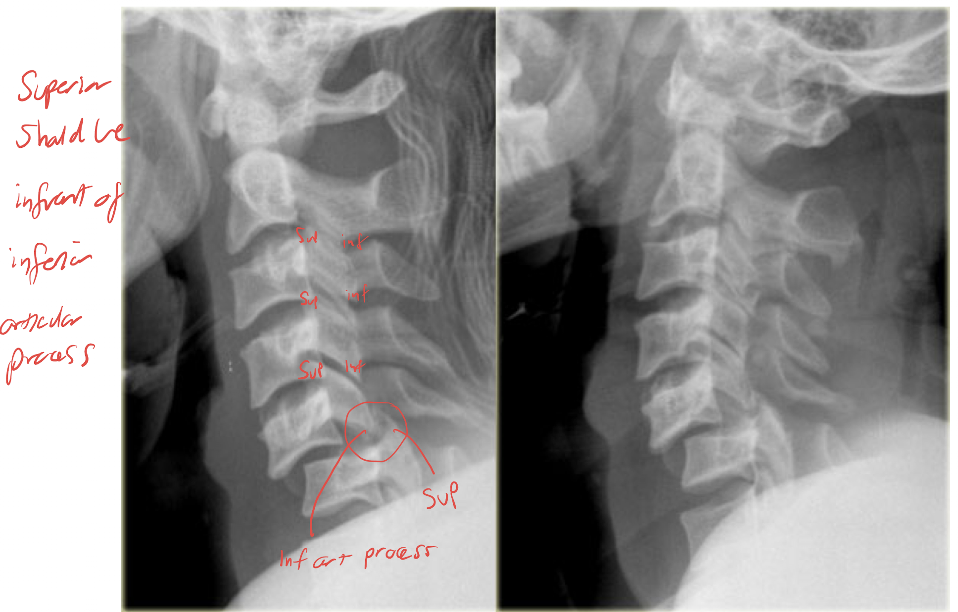
What is the mechanism of injury for clay shoveler fracture?
Abrupt neck flexion combined with a heavy upper body and lower neck muscular contraction, causing avulsion via the supraspinous ligament

What are cervical fracture occurs with a flexion and rotation mechanism?
Unilateral facet dislocation
What is the mechanism of injury for a Unilateral facet dislocation?
Flexion and rotation
Forces an inferior articular facet of an upper vertebra to pass superior and anterior to the superior articular facet of a lower vertebra
Rests in the IVF
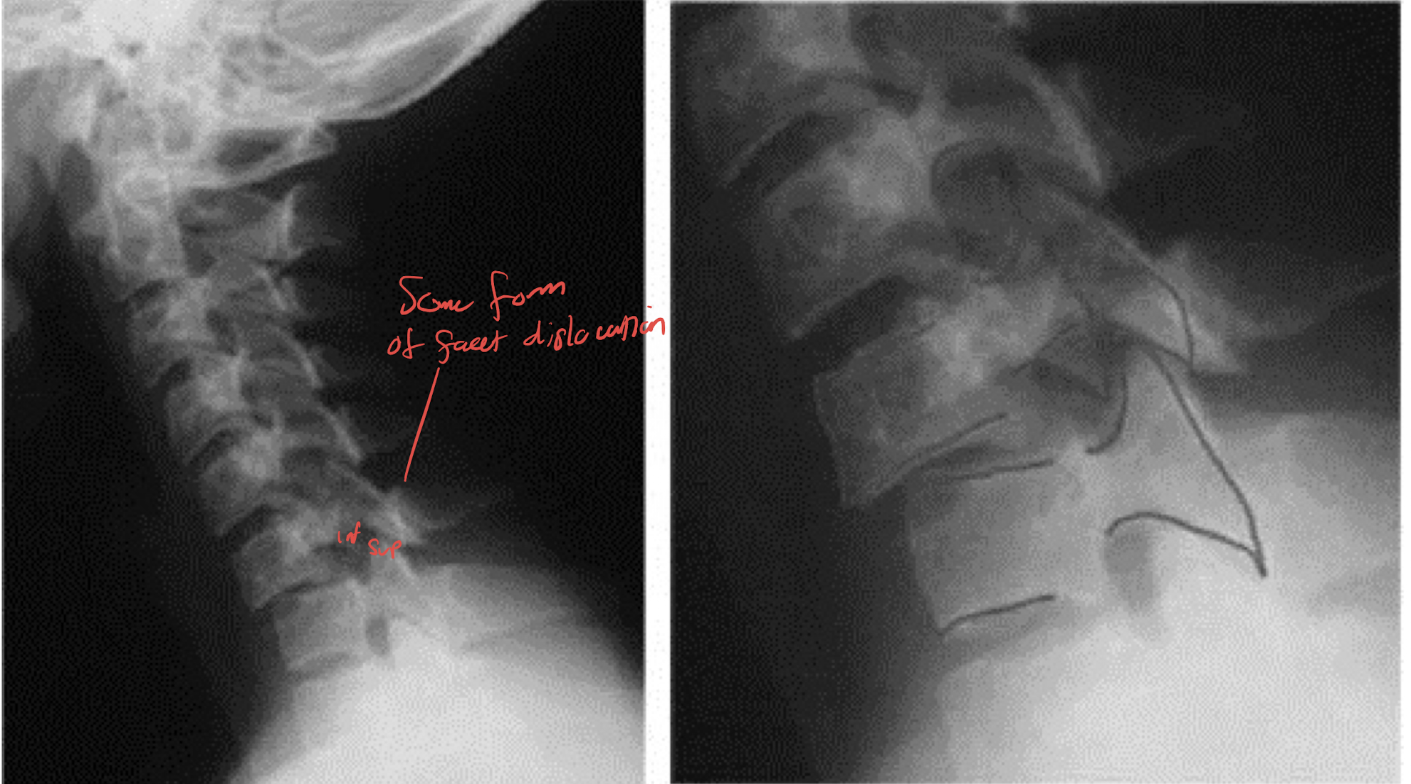
Is a unilateral facet dislocation a stable injury?
Yes, while the posterior ligament is disrupted, the vertebra are locked in place
What are the cervical fractures that occur with an extension mechanism?
Hangman fracture
Extension teardrop fracture
Posterior arch of C1 fracture (Posterior neural arch fracture)
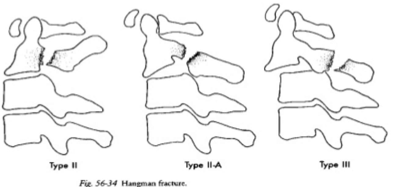
What change is there to C2 in a Hangman fracture?
Bilateral fracture through the pedicles due to hyperextension
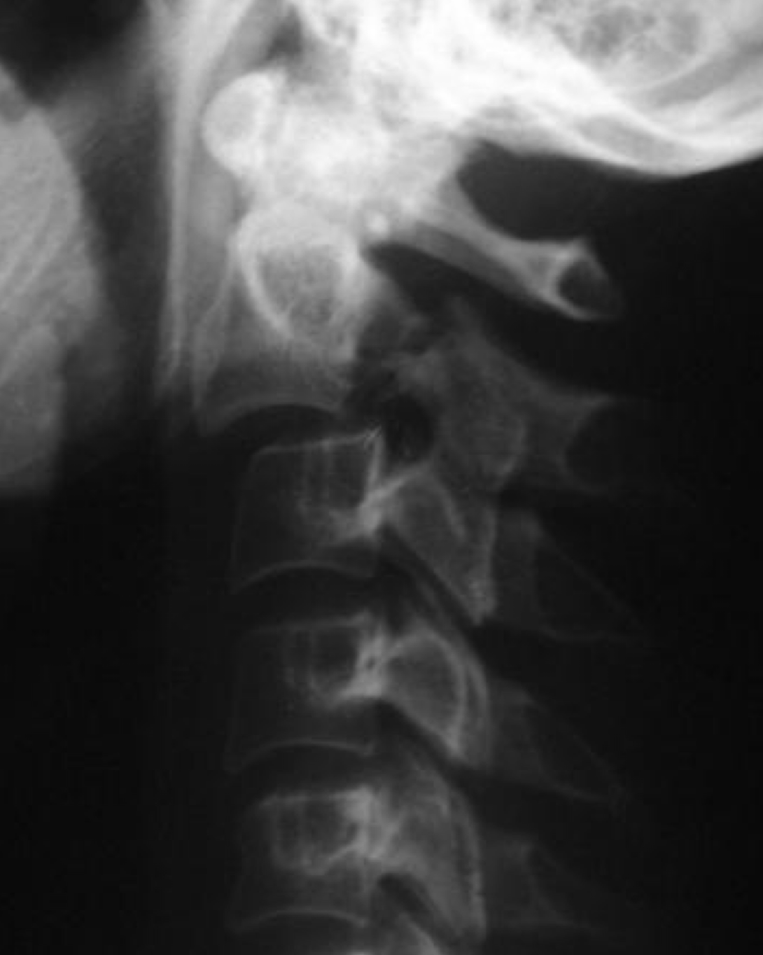
Are there neural complications in a hangmans fracture?
Yes, when associated with a unilateral or bilateral facet dislocation
Is a hangmans fracture considered stable or unstable?
Unstable
Describe the Levine and Edwards classifications of Hangmans fractures?
Type 1
<3mm displacement with no angulation of anterior aspect of fracture
Type 2
Greater forward angulation than type 1
Cervical traction with halo vest required
Type 3
Severe angulation and/or facet dislocation
Requires surgical restoration
What is the mechanism of injury in an extension teardrop fracture?
Anterior longitudinal ligament pulls fragment away from inferior aspect of vertebra due to sudden hyperextension
Causes a displaced anteroinferior bony fragment
Common after diving accidents and tends to occur at C2
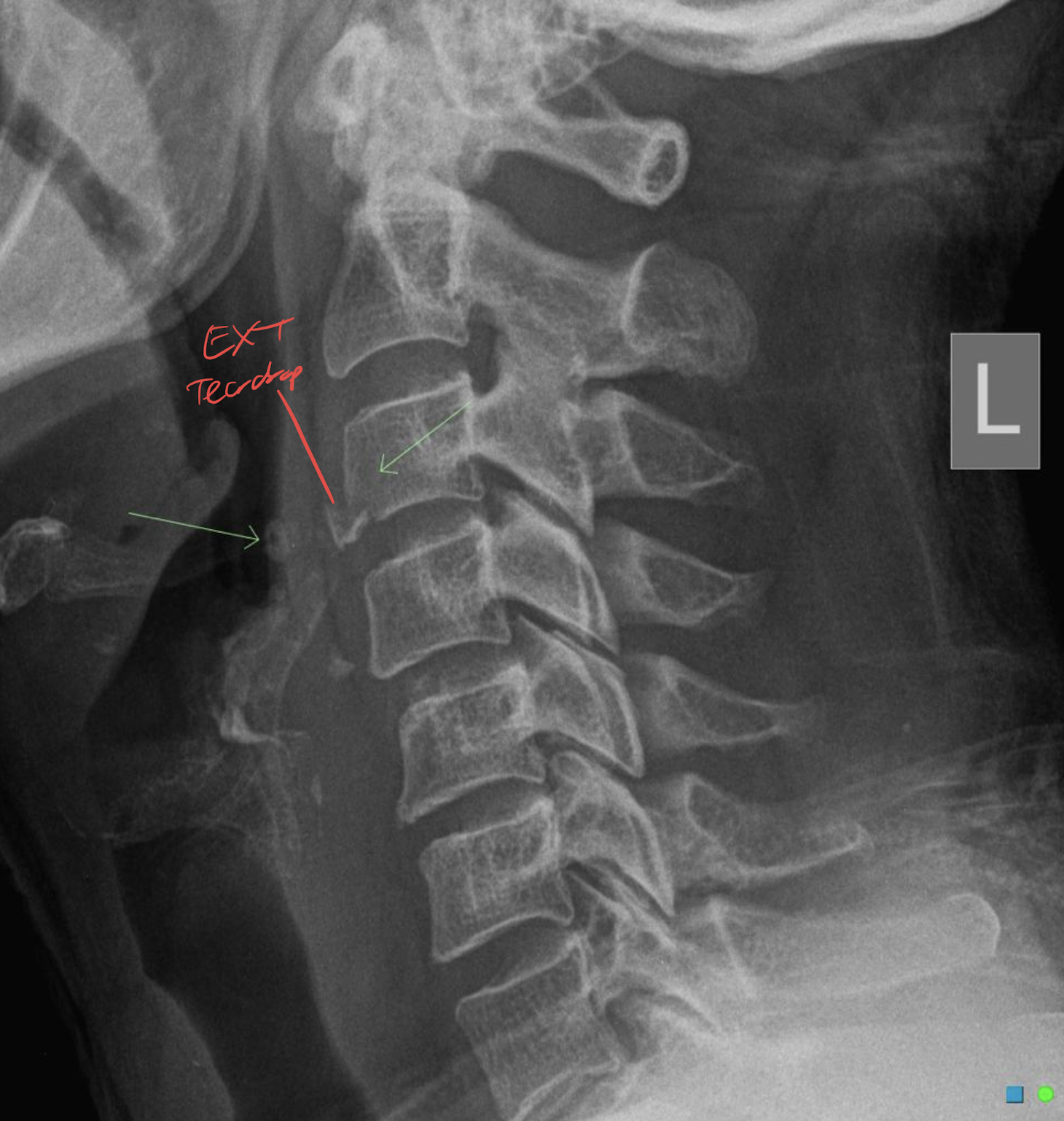
Is an extension teardrop fracture considered stable or unstable?
Usually stable, but if extreme is associated with central cord syndrome due to buckling of ligamenta flava into spinal canal
In Central cord syndrome, will you see more motor involvement in the upper limbs or lower limbs? What other signs will you see?
Upper limbs
Bladder dysfunction and variable sensory dysfunction
What is the mechanism of injury in a Posterior arch of C1 fracture (Posterior neural arch fracture)?
Head is hyperextended and posterior neural arch of C1 is compressed between the occiput and the spinous process of C2
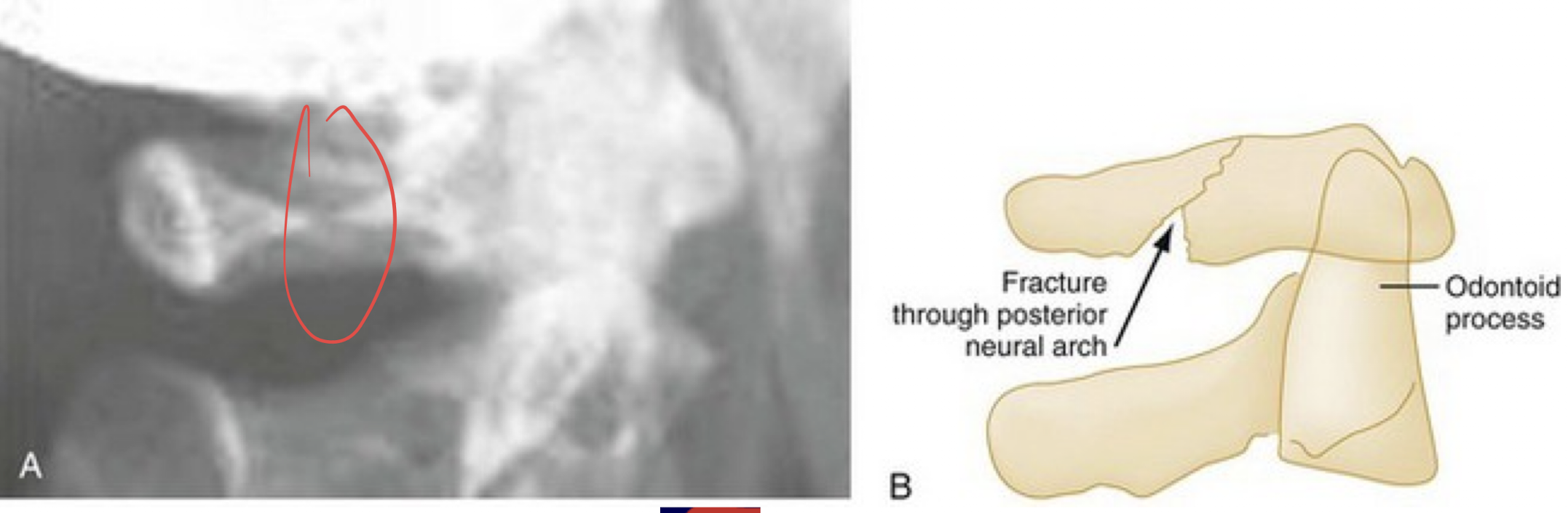
Is Posterior neural arch fracture considered stable or unstable?
Stable, as transverse ligament and anterior arch of C1 are not involved
What is another possible differential to consider when you suspect a posterior neural arch fracture?
Jefferson fracture
What are the cervical fractures that occur with an compression mechanism?
Jefferson fracture (burst fracture of ring of C1)
Burst fracture of lower cervical vertebral body
What is the mechanism of injury in a Jefferson fracture?
Compressive downward force transmitted evenly through occipital condyles to superior articular surfaces of lateral masses of C1
This displaces masses laterally and causes fractures of anterior and posterior arches
Possible disruption of transverse ligament (displacement of lateral masses is >6.9mm) (consider instability)
→ If less than 6.9mm, transverse ligament still intact and neurologic injury unlikely
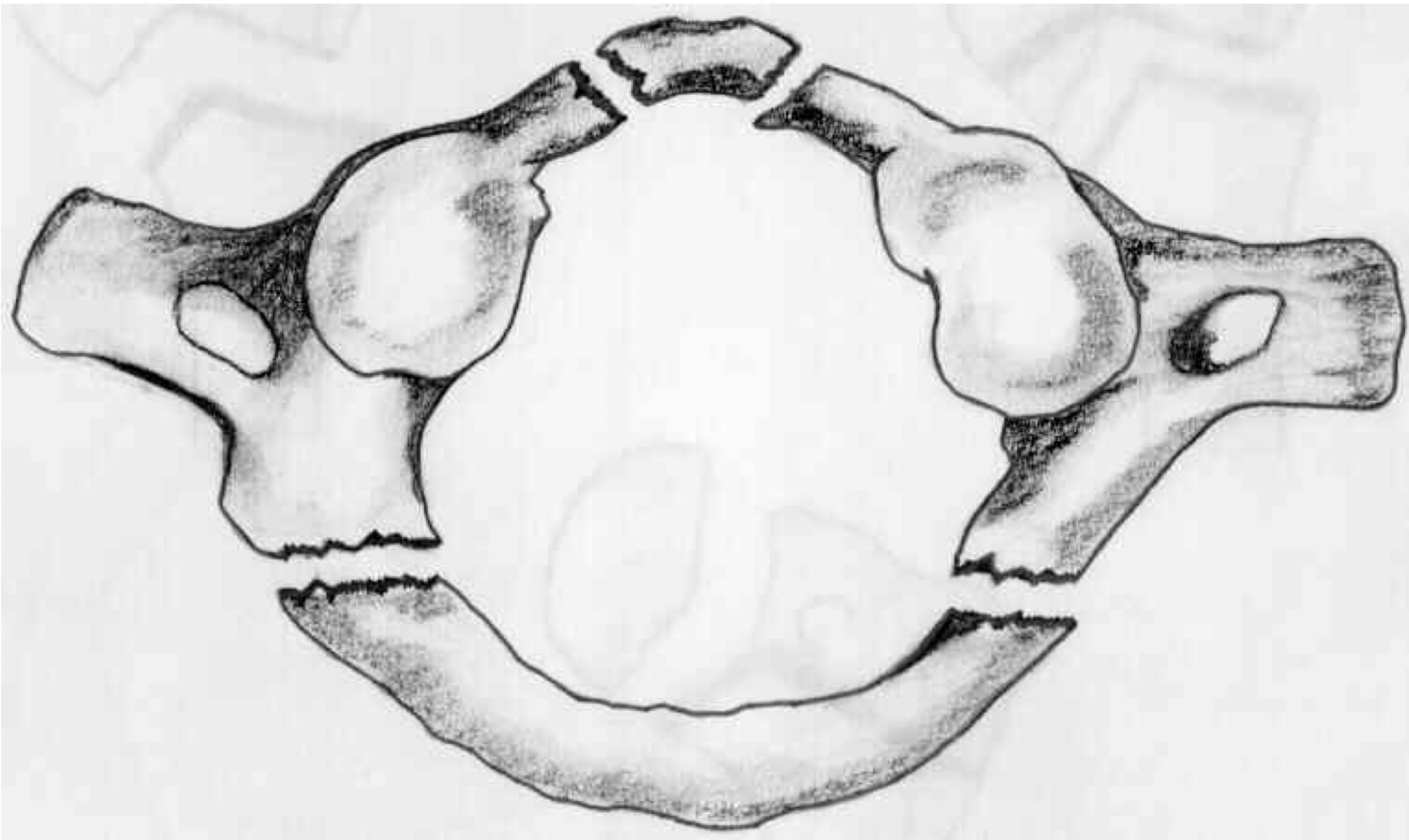
Describe the 3 types of lateral displacement of C1
Type 1
Intact transverse ligament
ADI <3mm
Lateral displacement <7mm
Type 2
Transverse ligament torn
ADI >3mm
Lateral displacement <7mm
Cord damage possible
Fusion may be required
Type 3
Lateral displacement >7mm
What is the management of the 3 types of lateral displacement of C1?
Type 1 and 2 treated with cervical orthosis
Type 3 may require internal fixation
What is the mechanism of injury in a burst fracture?
Downward compressive force transmitted to lower levels in cervical spine causing cervical vertebra to shatter
Posterior protrusion may extend into spinal canal, can be associated with anterior cord syndrome
Internal fixation required if neurological deficit is present

Is a burst fracture considered stable or unstable?
Yes, in the absence of neurological deficit
What spinal tract is involved in anterior cord syndrome where there is complete motor paralysis below the level of the lesion?
Corticospinal tract
What spinal tract is involved in anterior cord syndrome where there is loss of pain and temperature at and below the level of injury?
Spinothalamic tract