GMH endocrine
 Call Kai
 Learn
Learn Practice Test
 Spaced Repetition
 Match
Match Flashcards
 Knowt Play
1/59

There's no tags or description
Looks like no tags are added yet.
Name | Mastery | Learn | Test | Matching | Spaced | Call with Kai | Chat |
|---|
No analytics yet
Send a link to your students to track their progress
60 Terms
endocrine glands
epithelial tissue derivatives specialised for secreting
what are the four chemical messengers
autocrine; local either on neighbouring cells or itself
neurotransmitter; short range chemical messengers diffusing across the synapse
neurohormones; released into blood by neurosecretory neurons and travel through blood
hormones long range messengers secreted into blood by endocrine gland
what are hormones changing
rarely change cellular function; alter rate or normal functioning
what are the two categories of hormones
hydophillic=peptide and catecholamines; surface receptors
hydrophobic=thyroid and steroid hormones; nuclear receptor
surface versus nuclear receptors
surface receptors=hydrophilic hormones=peptide hormone binds which activates G protein and induced intracellular signals→includes prolactin, ACTH, vasopressin, oxytocin, and insulin
nuclear receptors=hormone binds inducing heat shock protein detachment which uncovers the nuclear localization sequence and then the hormone enters the nucleus acting as a TF
-oestrogen, cortisol, testosterone, aldosterone
primary versus secondary endocrine glands
primary=thyroid, parathyroid, pituitary, adrenal and pineal
secondary=liver and heart
hyposecretion and hypersecretion
primary hypo=within gland abnormality, secondary due to tropic hormone deficiency
primary hyper=too much hormone due to within gland abnormality, secondary do to excessive gland stimulation
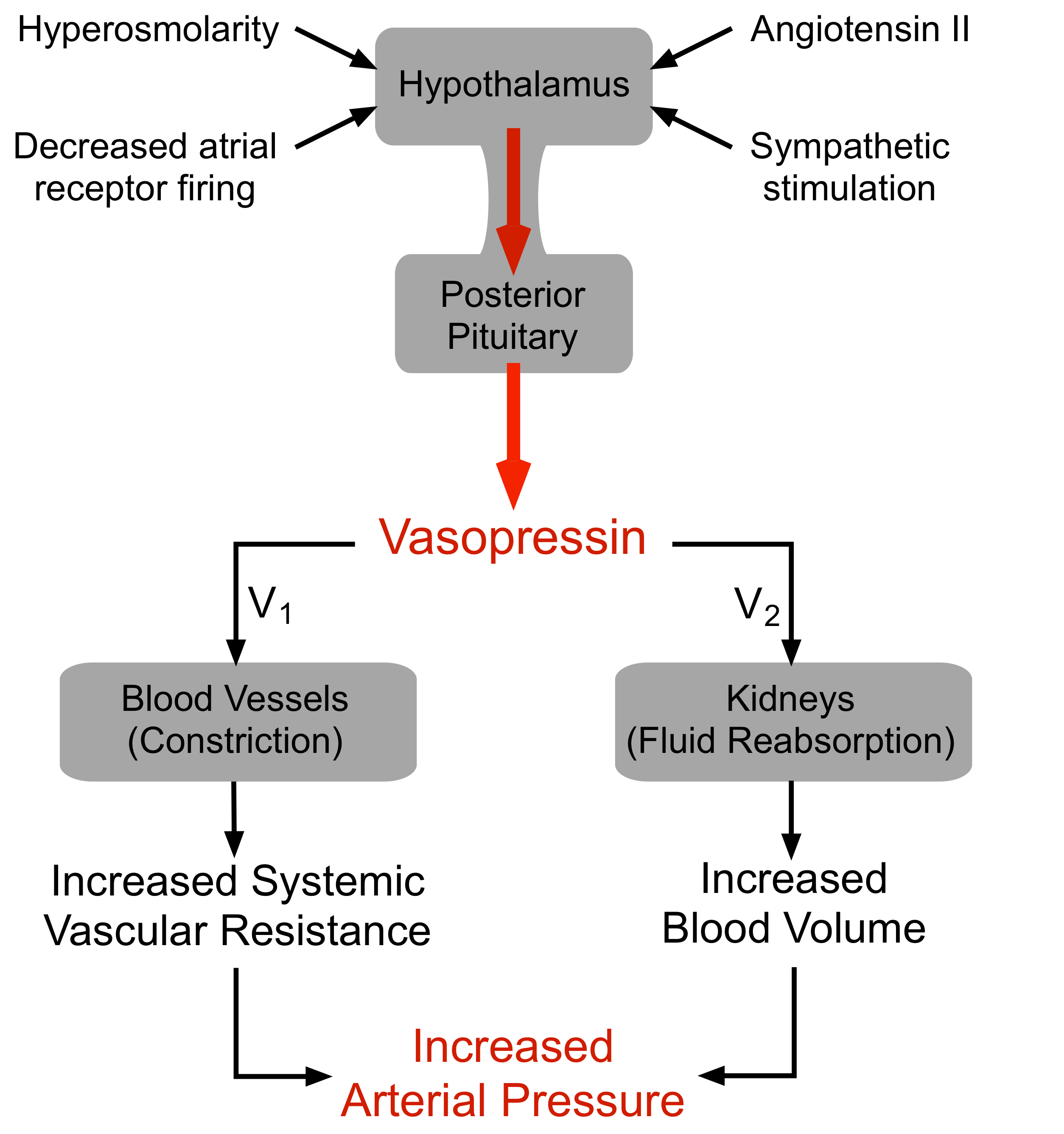
two functions of vasopressin
binds V2 and promotes H20 reabsorption
binds V1 causing arteriole vasoconstriction
noreepinephrine
hormone from adrenal medulla but NT from NS
pituitary gland location veruss hypothalamus
is 1cm and lies at the base of brain below hypothalamus and is connected via nerve fibers and blood vessels
hypothalamus lies at the base of the brain and the pituitary connected to the base of the brain via the infundibular stalk
anterior versus posterior pituitary
anterior=epithelial struture developing from rathkes pouch
posterior=neural structure developing from midbrain floor consisting of nerve fibers running from hypothalamus to anterior pituitary (oxytocin and vasopressin=peptide hormones stored in axonal ends)
how are vasopressin and oxytocin produced
nerve cell bodies in the hypothalamus and then are packaged into vesicular granules and then are transported down axons into posterior lobe and are released from terminal similar to neurotransitters
Vasopressin (ADH)
controls variable water reabsorption in final tubular segments of kidney
65% of water reabsorption is obligatory in the proximal tubule while in the distal tubule and collecting duct it is variable (ADH dependent)
Increases tubule cell permeability to water via aquaporin 2 channel insertion
water deficiit increases ADH and vice versa
ADH binds with receptor sites on basolateral membrane of a principal cell in distal or collecting tubule which activates cyclic AMP increasing H2O permeability by AQP-2 water channel insertion
membrane is impermeable to water in the absence of ADH
oxytocin
bonding hormone→increases during birth as uterus increases receptors; synthetic oxytocin can induce labour
aids in smooth muslce contraction so milk ejection
anterior pituitary
epithelial derived upgrowth from roof of oral cavity; regulated by hypothalamic hormones
unlike posterior pituitary→anterior pituitary itself synthesizes the hormones it releases into the blood
somatotropes makes growth hormone
thyrotropes secrete TSH
corticotropes produce ACTH to stimulatee cortosil section by adrenal gland
gonadotropes secrete FSH and LH
lactotrophs secrete prolactin
anterior hormone pituitary secretion regulated by
hypothalamic hormones and feedback by target gland hormones
factors influencing growth
growth hormone, genetics, protein in diet, freedom from stress (cortisol can promote protein breakdown, blocking GH secretion
GH, somatomedins (IGFs), estrogen and testoserone, thyroid hormones, calcitonin, PTH and vitamin D
growth hormone release
is not continous→featal growth is promoted by placenta hormones so GH plays no role however first two years of life is the postnatal growth spurt and puberty growth spurt is promoted by teste androgen production and in women androgen from adrenal gland less potently promote growth
prepuberty sex differences in growth are not apparent
testoserone and estrogen affect on height
both act on bone to halt its further growth so full height attained by end of puberty
growth hormone release
from AP in several bursts; high in morning before waking and low at night; stimulation occurs during deep sleep
growth hormone releasing hormone (GHRH) produced by hypothalamic neurons
GHRH receptor is 7 transmembrane domain G-PCR
GH bursts initiated by bursting secretion of GHRH and terminated by somatostatin
GH releases IGF1
acts on bone and soft tissues to promote growth; protein synthesis and lengthening and thickening of bones
GH stimulates liver to produce IGfs which stimulatee bone and soft tissue growth→prevent apoptosis and cause hypertrophy
IGF can overcome GH defects
metabolic effects of growth hormone
increase fatty acid in blood due to fat breakdown and increase blood glucose due to decreased glucose uptake by muscles
pituitary gigantism
GH excess due to AP tumour
somatostatin medication stop GH production
Dwarfism
GH defiency, low GH is treated with GH; hereditary=low GH, Laron Dwarf=defectivee GH receptor and African Pygmy=defective IGF-1 receptor
overall GH metabolic effect
mobilising fat stores as major energy substrate while conserving glucose for glucose dependent tissues such as the brain since it can only use glucose as its metabolic fuel, yet it cannot store glycogen
GH effects only manifest when sufficient TH is present
thyroid gland
located on front upper part of trachea developing from small outgrowth of the tongue
follicular cells arranged in hollow spheres form the functional units; colloid is the substance serving as extracellular storage for TH
three types of thyroid hormone
T3 and T4 in follicates and calcitonin in C cells
T3 and T4 function to accelerate metabolism and increase substrate turnover
tyrosine based hormones meaning T3 as three iodine and is more effective but T4 as four but is more abdundant
how are T3 and T4 levels controlled
by TSH from AP; thyroid hormone is the main determinant of BMR and increases target cell responsivess to catecholamines, increases HR and contraction force
TH negatviy feedback loop with AP production TSH
Goiter
low iodine uptake=no T3 or T4 made, iodide uptake increases and thyroid can grow by 10X
Hashimotos
autoimmune where autoantibodie destroy thyroid follicular cells
hypothyroidism
; primary is failure of thyroid gland itself and secondary is due to a deficit of TRH, TSH or decrease in iodide
generalized fatigue, weight gain, deep voice
hyperthyroidsim
graves disease=autoimmine where TSH is mimicked by antibodies→treated with beta blockers and antithyroid medication
weight loss and bulging eyes
adrenal glands
embedded above each kidney in capsule of fat and each has two division
cortex(outer part)=makes steroid hormones, aldosterone, cortisol, androgen sex steroids (DHEA)
medulla(inner part)=chromaffin cells from sympathetic NS→catecholamines, epinephrine, and norepinephrine
cortex layers
zone glomerulsoa=outermost
zona fasciulata=middle and largest
zone retiularis=inner most zone
aldosterone
mineral corticoid that maintains electrolyte balance; low Na+ or high K+→RAAS→angiotensin II activated which increases aldosterone relase from adrenals→acts on istal renal tubule to increase Na+ and water retention, and increase K+ and H+ ion excretion
Low Na+ or high K+→angiotensin II→adrenal cortex→aldosterone
cortisol
secreted in diurinal rythym playing a role in stress to increase blood glucose and blood fatty acids, control water and electrolyte balance and is also antiinflammatory and immunosuppressive
negative feedback on hypothalamus(CRH) and anterior pituitary(ACTH)
CRH→ACTH→adrenal cortex→cortisol
Catecholamines
adrenal medulla, stimulated by SNS activation; adrenaline for fight or flight responses
glucocorticoid therapy
adrenal fatigue/failure in addisons disease and is also antiinflammatory to reduce T cell proliferation in asmtha, eczema or cancer as anti emetic
benefits must outweight cost due to cushings symptoms of decrease immune system osteoporosis weight gain
Addisons disease
low corticoids (corticoids and aldostrone)
primary=damaged adrenal glands via autoimmune or TB
secondary=low ACTH
weight loss, low liver glycogen
treated witth steroid replacement therapy
cushings
high glucocorticoids→ACTH secreting tumour in pituirty
obesity, hyperglycemia, moon face
treated with antiglucocorticoids, tumour removal
Conns syndrome
excessive mineralcorticoids (aldosterone)
primary=tumour, secondary=excessive RAAS in kidney disease
water and salt retention, increase ECF and hypertension
HPA axis
generalized stress response=increas epinephrine secretion from adrenal medulla chromaffin cells
increase CRH-ACTH-cortisol (HPA) mobilizing metabolic resources while RAAS and vasopressin maintain blood volume and pressure
chronic stress causes heart disease, hypertension, depression with prolonged CRH release
long versus short cortisol loop
long=cortisol acts on CRH neurons in hypothalamus to stop CRH
short=cortisol acts on AP cells to stop ACTH release
Islets of Langerhans
beta cell secretes insuline and alpha cell secretees glucagon while D-cells secrete somatostain (glycogen breakdown)
insulin is synthesized as a prohormone then cleaved to a mature peptide
excitation secretion coupling stimulates insulin exocytosis
food intake
increases PSNS activation and beta cells release insulin
diabetes mellitus=most common endocrine disorder
T1D childhood onset loss of beta cell function
TIID=slow development and is remedied through lifestyle changes
spurs excessive eating due to cells being starved from carbohydrates and dehydration as blood glucose increases due to ketoacidosiis and brain O2 decreases
calcium
1 kg of Calcium in human body with 99% in skeleton and teeth
.9% intracellular in soft tissue and .1% in extracellular fluid; half of ECF Ca is bound to plasma proteins or complexed with PO4³- so it is not available for cellular reactions whereas the other half can pass from plasma to ICF and interact with cells
free ECF Ca2+
only this is biologically active and subject to regulation; functions to prevent aberrant neuromuscular exciteability
fall in this causes overexcitability and rise in depression
excitation contraction coupling versus stimulation secertion coupling
cardiac and smooth muscle results from increased Ca2+ permeability in response to AP
stimulation secretion coupling in B-cells→Ca2+ entry=>insulin
bone remodeling
skeleton acts as storage depot for Ca2+ and is turned over every ten years
osteoblasts=build bone via seceretion of extracellular organic matrix within which calcium and phopshate crystals precipiate
osteocytes=retired osteoblasts imporsioned with in bony call desposited around them
osteoclasts=breakdown matrix and reabsorb bone in presence of PTH to increase Ca2+ levels in plasma
signals that osteoblasts produce
RANK ligand=osteoclast.mascrophage→differentiate to osteoclasts and prevent apoptosis→increas osteoclast action and when this is more than osteoblast→decrease bone mass
osteoprotegrin (OpG)=binds to rank ligan so it cant bind→decreases osteoclast action→osteoblast outpaces clast and there is an increase in bone mass
bone deposition
mechanical stress favours bone deposition; in children bone builders outpact destroyers (IGF1 and GH); atheletes have greater stress on bone so mass increases
bone density peaks at 30 and declines at ago 40
osteoporosis
reduced osteablast activity and an increase in osteoclast acitvity; post menoppausal women
what three hormones control plasa concetrations of Ca2+ and phopshate
calcitonin (thyroid gland C-Cells)
parathyroid hormone
vitamin D
what regulates calcium
hormonal control between ECF and bone (short term), kidneys(seceretion), and intestines (absorption)→Ca2+ is involved in the function of all cells
Parathyroid hormone
=small oval gland assoicated witht he thyroid and PTH is a major adjustor whereas calcitonin is for fine adjusments.
Principal chief cells secrete PTH in repsonse to low blood Ca2+ leading to an increase in calcium via increase bone calcium release promoting tubular reabsopration and increase absorption from the small intestine via vit D activiation
PTH 2 major effects on bone
uses bone as a bank to maintain Ca2+ plasma levels with two major effects on bone.
induces fast Ca2+ influx into plasma from small labile pool of Ca2+ in bone fluid
stimulates bone dissoultion promoting transfer into plasma of Ca2+ and phosphate from stable bone mineral pool within the bone
osteocytic-osteoblastic bone membrane
entombed osteocytes and surface osteoblasts are interconnected via long cytoplasmic processes extending from these cells and connect to one another within caniculli
seperates mineralized bone from plasma in central canal
bone fluid lies between the membrane and mineralized bone
calcitonin
comes from C cells of thyroid gland and respond to high plasma Ca2+; PTH antagonist lowering plasma calcium and phosphate via inhibiting osteoclast activity and stimulates calcium secretion in kidneys
vitamin D
cholesterol derivative when exposed to the sun and is activated by the liver then the kidneys
essential for GI calcium and phopshate absorption, increases renal calcium reapsorption and reugulates osteoclast and blast activity; kidney enzymes are stimulated by PTH in response to fall in calcium plasma levels
calcium metabolism disorders
PTH hypersecretion(hyperparathyroidism)= hypersecreting tumour in PTH gland→hypercalcemia and hypophospahtemia-reduced nerve excitiably and muscle weaknesss-excessive calcium and phopshate from skeletal stores leads to bone thinning and an increase in kidney stones
PTH hypsecretion (hypoparathyroidism)→autoimmine attack or removal during thyroid removal surgery-hypocalcemia and hyperphosphatemia
vitamin D deficiency=impaired intestinal absoprtion of Ca2+ and PTH maintains plasma calcium levels at thee expense of bone