Unit 1 PP
 Call Kai
 Learn
Learn Practice Test
 Spaced Repetition
 Match
Match Flashcards
 Knowt Play
1/54

Earn XP
Description and Tags
Nervous system intro
Name | Mastery | Learn | Test | Matching | Spaced | Call with Kai | Chat |
|---|
No analytics yet
Send a link to your students to track their progress
55 Terms
Stem cells
undifferentiated cells that have the ability to proliferate and become diff cell types
Neurons
type of stem cells, responsible for transmitting electrical signals in the nervous system.
Glial cell
type of stem cells, provide metabolic support, protection, and insulation for neurons
Ependymal cell
The pink cell in pic
Responsible for SECRETING CSF
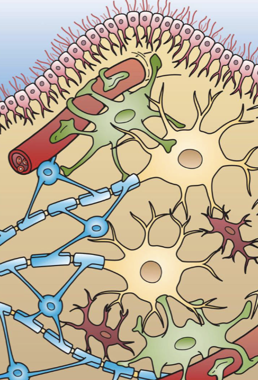
Microglia
Red cell in pic
Defensive function : performs phagocutosis ; provides immune system function
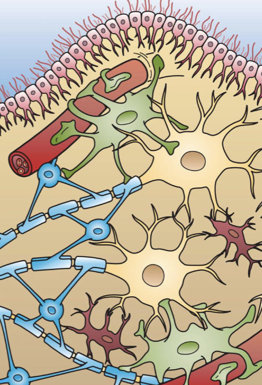
Astrocyte
Green cell in pic
Provides structural support, maintains ionic/chem enviornment ; nutritive role; phagocytocis
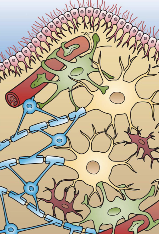
Oligodendroglia
Blue cell in pic
Mylenates multiple axons in CNS

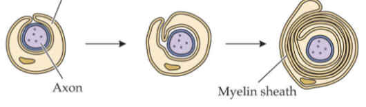
Schwann cell
mylenates one axon in PNS

Motor neuron
direct biobehavioral responses appropriate for the situation.

Sensory neurons
convert physical stimuli into electrical signals.

Interneurons
neurons in brain and spinal cord;
form interacting neural circuits; responsible for conscious sensations, recognition, memory, decision-making, cognition.
Soma
cell body; contains nucleus and other organelles
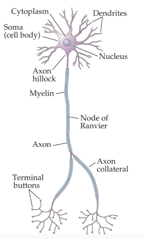
Dendrites
projections from the soma that receive information.
Effect on receiving cell may be excitatory or inhibitory

Axon
extension that conducts electrical signals from the cell body to the terminal
buttons

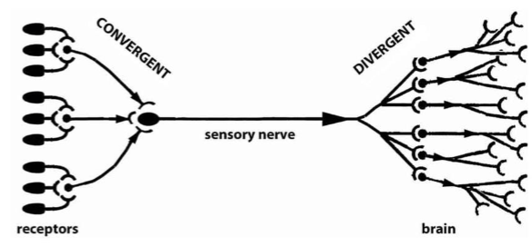
Convergence
a neuron receives and integrates a vast amount of information from many cells.

Divergence
the integrated information can be transmitted to a few or thousands of other
neurons.
Nodes of Ranvier
Breaks in the myelin sheath
the sites where action potentials are regenerated.
The myelin sheath increases speed of conduction along the axon; the thicker
the myelin, the faster the conduction.
receptors
Initial sites of action of neurotransmitters, hormones, and drugs
enzymes
that catalyze biochemical reactions.
ion channels and transporters
for charged molecules such as amino acids, glucose, and
metabolic products.
Ligand-gated channel
opens when a ligand binds to a receptor.

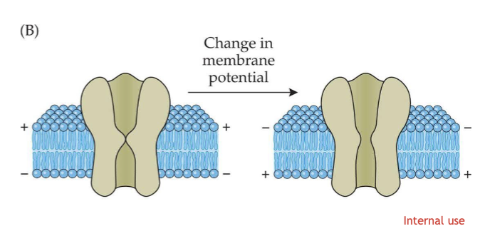
Voltage-gated channel
opens when the electrical potential across the membrane is altered.
ATP
Brains energy source - mitochondria
Rate of ATP synthesis reflects neuron
activity; this is used in techniques that
allow visualization of brain function
is also an extracellular signaling
molecule between neurons and glia
Resting membrane potential:
not sending a signal = –70 millivolts (mV)
inside the cell = it is polarized.
There are more negative ions (and amino acids) inside the cell, and more positive ions outside.
K+ movement
moves into the cell because it is attracted to the negatively charged
particles (electrostatic pressure).
moves back out of cell when its concentration rises (concentration
gradient
Na+–K+ pump
moves 3 Na+ to the outside for every 2 K+ moved to the inside. (against their concentration gradient)
Action potential (AP)
rapid change in membrane potential
THRESHOLD = –50 mV.
Voltage-gated Na+ channels open, generating a rapid change in membrane potential
all or none
Local potentials
Ion channels open momentarily, causing small, local changes in ion
distribution and potential differences
If Na+ channels open
local depolarization
If Cl– channels are stimulated to open
hyperpolarization, which is inhibitory.
n myelinated axon, onduction seems to jump along the
axon—saltatory conduction.

This neuron is at what state?
Resting (polarized)
An action potential will be propagated only if a stimulus reaches a
Threshold value
The two most important characteristics of the action potential's transmission are
Depolarization and hyperpolarization
In myelinated axons, regeneration of the action potential occurs only at
the nodes of ranvier
SNS
Pregang = Ach
Postgang = Ne

PNS
Pregang = Ach
Postgang = Ach

SNS division
thoracic - lumbar
PNS division
Cranial nerves + sacral spinal cord
Spinal cord:
gray matter contains neuron cell bodies; white matter is myelinated axons.
Sensory afferent nerves
Carry information to the spinal cord;
soma are in the dorsal root ganglia.
Motor efferent nerves
result in voluntary movement; soma are in ventral horn.
Meninges
layers of tissue that cover and
protect the brain and spinal cord
ORDER OF LAYERS (thoughest to thinnest)
Dura matter
Arachnoid mattter (has subarachnoid filled with csf)
Pia matter
CSF
surrounds brain and spinal cord, fills the
cerebral ventricles and the central channel in
the spinal cord
CSF is formed in
choroid plexus
Telencephalon
neocortex, basal ganglia, Limbic system
Diencephalon
Thalumus, hyptohalamus
Mesencephalon
Midbrain
Metencephalonn
Cerebellum, pons
Myencephalon
Medulla
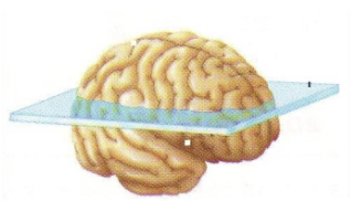
Horizontal

sagital
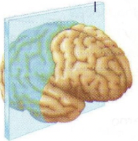
coronal