Lecture 10 - Lipid metabolism
 Call Kai
 Learn
Learn Practice Test
 Spaced Repetition
 Match
Match Flashcards
 Knowt Play
1/37

There's no tags or description
Looks like no tags are added yet.
Name | Mastery | Learn | Test | Matching | Spaced | Call with Kai | Chat |
|---|
No analytics yet
Send a link to your students to track their progress
38 Terms
Main function of cytosol in lipid metabolism
Lipid building
Main function of mitochondria in lipid metabolism
Lipid breaking
How does the location of triglyceride synthesis differ between the fed and fasted states, and why?
Fed state → glucose is available, so it’s converted to glycerol 3-phosphate → triglyceride is made in the adipose tissue itself
Fasted state → no glucose available, so adipose tissue can’t make glycerol 3 phosphate → the body breaks down already stored triglycerides instead into glycerol + fatty acids → fatty acids go to the liver, which DOES have glycerol 3 phosphate → triglyceride is made there instead
Nomenclature of fatty acids
number of carbon atoms:number of double bonds
Δ → which carbon the double bond is at (THE CARBONS ARE NUMBERED STARTING FROM THE COOH END OF THE FATTY ACID)
Fatty acids
Unbranched, long hydrocarbon chains with a carboxylic acid end (COOH + R)
Lipid
molecule that is not soluble in water
Neutral lipids
uncharged lipids
Why can’t fatty acids be stored on their own, and what do you have to do to them to store them?
Fatty acids are reactive on their own, so you need to add a glycerol cap to neutralize it so that it can be stored
Saturated fat
No double bonds (C-C)
Unsaturated fat
Has double bonds (C=C)
Monoglycerol, diaglycerol
One fatty acid chain, two fatty acid chains
Trans fatty acids
unsaturated fatty acid with hydrogen atoms on the opposite sides of the double bond (C=C)
cis → hydrogens on same side
Hydrogenation turns cis double bonds into trans double bonds → elevated LDL (bad cholesterol), can’t be metabolized
Glycerophospholipid + structure
Type of phospholipid
hydrophilic head (phosphate+attached group), hydrophobic fatty acid tails
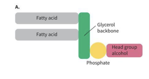
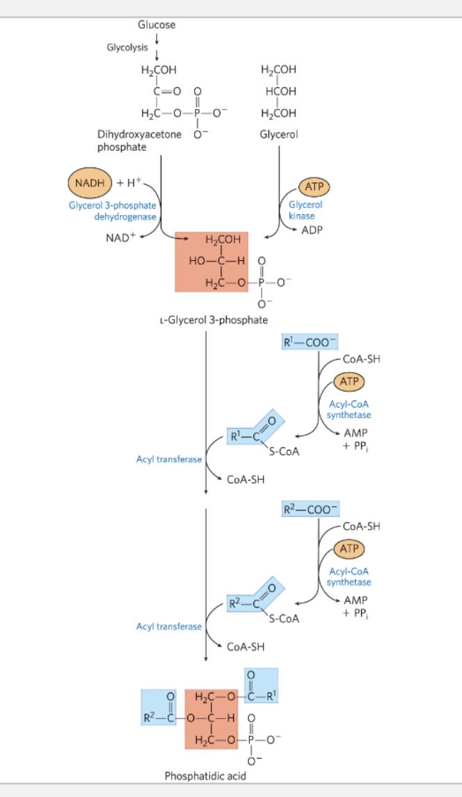
Explain this image (NOTECARD)
Step 1: Get to glycerol 3-phosphate (two possible routes)
Route A (from glycolysis): Dihydroxyacetone phosphate (DHAP) is reduced by glycerol 3-phosphate dehydrogenase, using NADH + H⁺ (oxidized to NAD⁺) → produces L-glycerol 3-phosphate.
Route B (from free glycerol): Glycerol is phosphorylated by glycerol kinase, using ATP (→ ADP) → also produces L-glycerol 3-phosphate.
Both routes converge on the same molecule — glycerol 3-phosphate, which has a free OH on C1 and C2 (the phosphate is already on C3).
Step 2: Activate the first fatty acid
A free fatty acid (R¹—COO⁻) reacts with CoA-SH, using acyl-CoA synthetase and ATP (→ AMP + PPᵢ) → produces fatty acyl-CoA (R¹—C(=O)—S-CoA).
Step 3: Attach the first fatty acid to glycerol 3-phosphate
Acyl transferase transfers the R¹ acyl group from CoA onto glycerol 3-phosphate (releasing CoA-SH) → produces a monoacylglycerol phosphate (lysophosphatidic acid), with R¹ esterified at one hydroxyl.
Step 4: Activate the second fatty acid
A second free fatty acid (R²—COO⁻) is likewise converted to fatty acyl-CoA by acyl-CoA synthetase + ATP (→ AMP + PPᵢ).
Step 5: Attach the second fatty acid
Acyl transferase transfers the R² acyl group onto the remaining free hydroxyl (releasing CoA-SH again) → now both R¹ and R² are esterified to the glycerol backbone.
Step 6: Final product — Phosphatidic acid
The result is phosphatidic acid: a glycerol backbone with R¹ esterified at C1, R² esterified at C2, and the phosphate group remaining at C3.
Cholesterol
A type of lipid. acetate provides the carbon for cholesterol
4 hydrocarbon ring structure (hydrophobic)
OH group (hydrophillic)
*maintains fluidity of lipid bilayer!!!! (this makes sure it won’t burst from osmotic pressure)
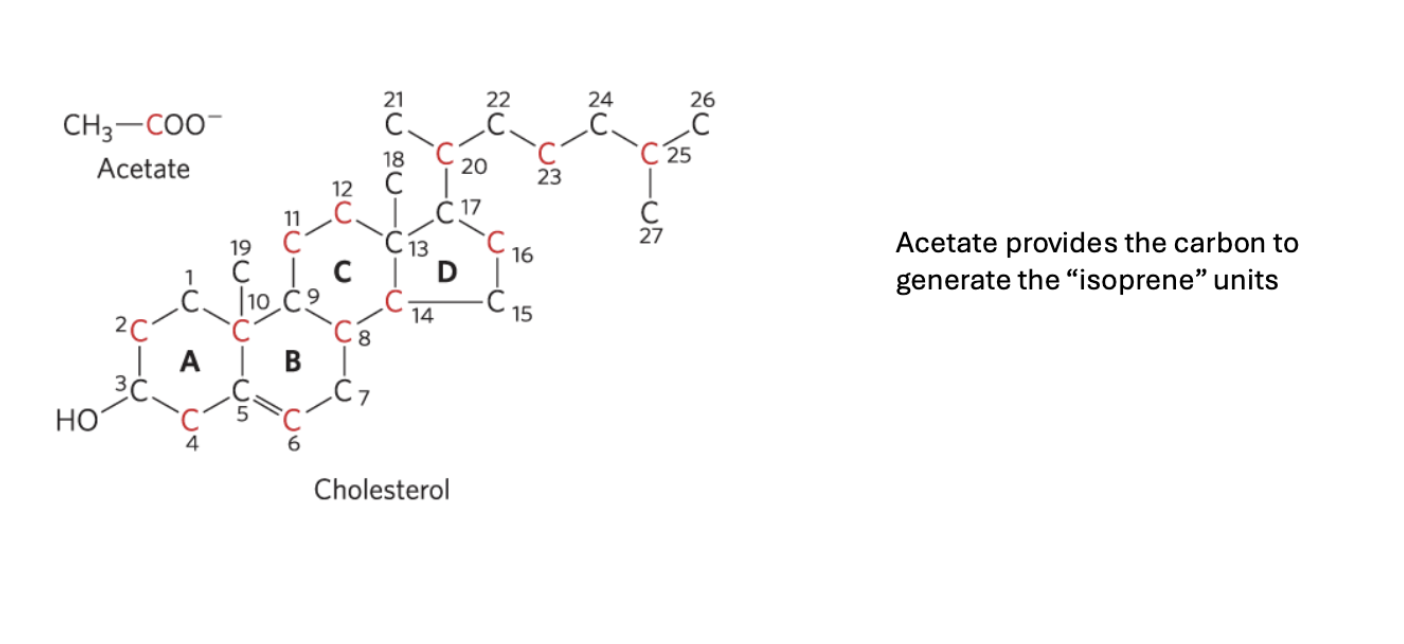
Cholesterol generation steps (NOTECARD;MAYBE)
3 acetates → Mevalonate, a 6 carbon intermediate (via 2 NADPH → 2 NADP+)
Mevalonate → 6 isoprene units (via 3 ATP → 3 ADP)
6 Isoprene units → squalene (via polymerization)
Squalene cyclization (forming a ring structure) + modifications → cholesterol
What is the rate-limiting step in the process of generating cholesterol?
acetyl-CoA → mevalonate
this is because this is where statins (medications used to lower cholesterol) target with mevalonate mimics
Chylomicrons
type of lipoprotein (unhealthy)
large, low density
moves dietary triglycerides to the liver
VLDL
type of lipoprotein (unhealthy)
made by the liver when there is excess triglyceride
transports body-made triglycerides to tissues
low density
LDL
type of lipoprotein (unhealthy)
low density
made from VLDL → IDL → LDL
packaged in the liver, distributed to muscle and adipose
HDL
lipoprotein (healthy)
high density
Made by the liver
collects excess cholesterol from tissues and transports it back to the liver
Apo B
lipid is loaded onto protein in the liver (“package”)
backbone of “bad cholesterol”
ApoB-100: (VLDL, LDL)
ApoB-48: chylmicrons, intenstine
ApoE
sits on the surface, hopping between particles (“label on different packages telling them where to go”)
Chylomicrons, HDL, VLDL
3 alleles of ApoE4 → predictor of Alzheimer’s
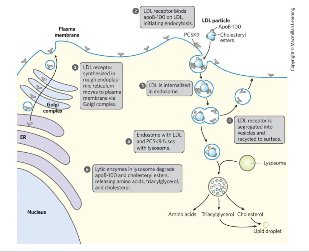
Process of cholesterol being transported into cells (NOTECARD)
LDL receptor moves to plasma membrane via Golgi complex
LDL receptor binds to apoB-100 on LDL → endocytosis initiated
LDL goes into endosome
LDL receptor is recycled to the surface
Endosome with LDL and PCSK9 fuses with lysosome
Enzymes in lysosome degrade apoB-100 and cholesteryl esters → amino acids triglyceride, and cholesterol released
Process of cholesterol disease (NOTECARD)
Monocyte attracted to lipoproteins that stick onto the extracellular matrix
Monocyte → macrophage
Macrophage ingests lipoproteins → becomes foam cell
free cholesterol accumulates → cholesterol-rich plaque forms

How does insulin regulate cholesterol synthesis?
Activator (activates HMG-CoA reductase by signaling fed state)
How does glucagon regulate cholesterol synthesis?
Inhibitor (inhibits HMG-CoA reductase by signaling fasted state)
How does ATP regulate cholesterol synthesis?
Inhibitor if LOW (low ATP → AMPK activated → HMG-CoA reductase inhibited)
How does oxysterol regulate cholesterol synthesis?
Inhibitor (if there is a lot of cholesterol, there are a lot of oxysterols, which are derivatives of cholesterol → inhibits cholesterol synthesis)
Acetyl-CoA carboxylase (ACC)
Catalyzes: acetyl-CoA → malonyl-CoA
expressed in adipose, liver
Fatty acid synthase (FAS)
Produces fatty acid chain
What is the co-factor for ACC?
Biotin
biotin covalently linked to lysines in the active site
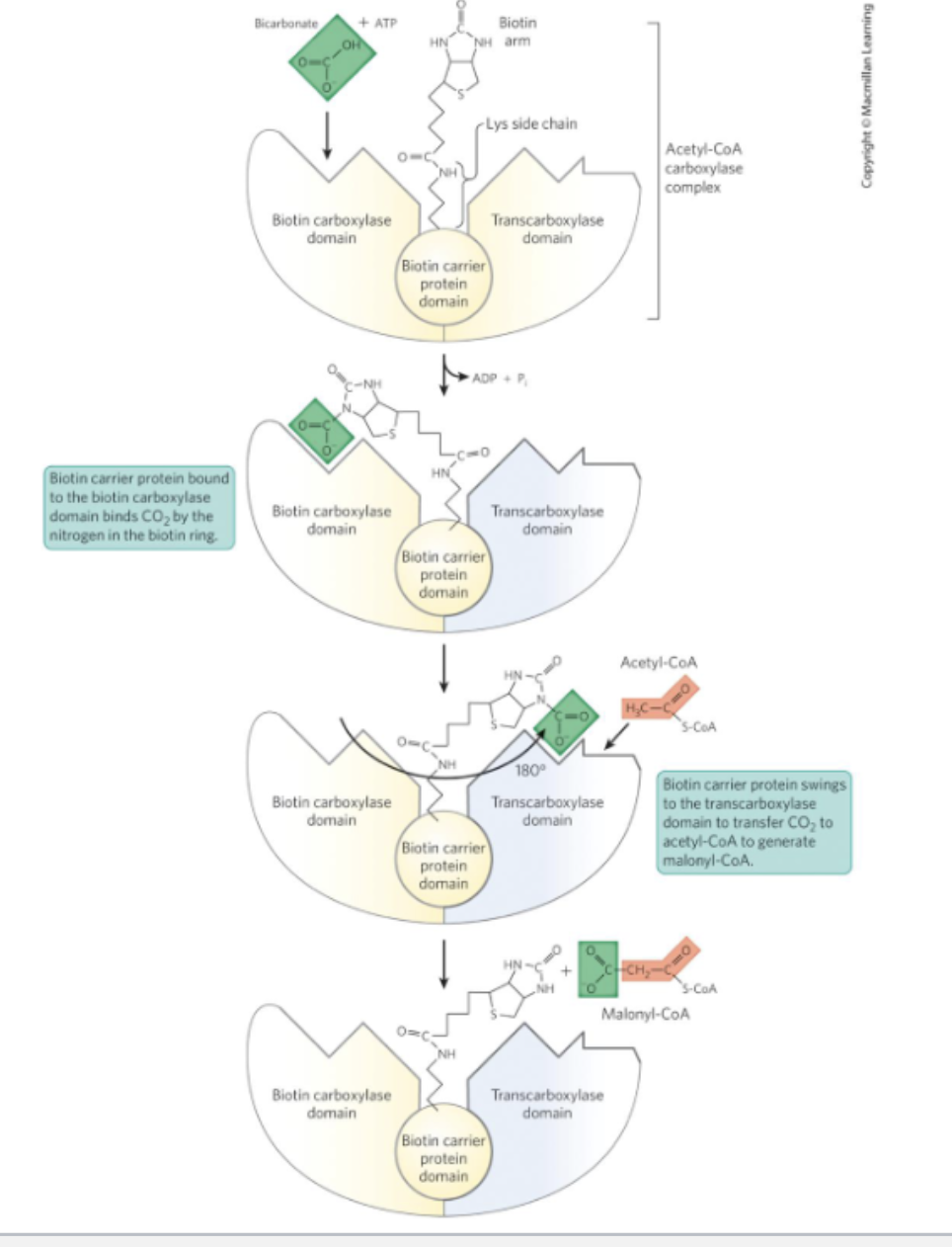
ACC mechanism (NOTECARD)
1. Biotin carboxylase domain (left) loads CO2 onto biotin.
Bicarbonate + ATP react at the biotin carboxylase domain. ATP is spent (→ ADP + Pi) to activate bicarbonate and attach a carboxyl group (CO2) onto the nitrogen of the biotin ring. Biotin is covalently tethered to the enzyme via a lysine side chain — this is the same biotin-lysine "biotinylation" you may have seen elsewhere (e.g., pyruvate carboxylase uses the same trick).
2. The biotin arm physically swings 180° to the other domain.
This is the conceptually important part: biotin isn't just a static cofactor sitting in one active site — it's attached to a long flexible arm that can rotate/swing the CO2-loaded biotin ring roughly 180 degrees, carrying the activated CO2 group away from the biotin carboxylase domain and over to the transcarboxylase domain on the other side of the same enzyme complex.
3. Transcarboxylase domain (right) transfers CO2 to acetyl-CoA.
Now that the CO2-biotin has arrived at the transcarboxylase domain, acetyl-CoA binds there, and the CO2 group is transferred from biotin onto acetyl-CoA's methyl carbon, forming malonyl-CoA.
4. Biotin is released, regenerated, and ready to reload.
The now-empty biotin swings back, free to pick up another CO2 from bicarbonate and repeat the cycle.
FAS mechanism (NOTECARD)
1. Acetyl CoA starts the chain by binding to ACP
2. Acetyl domain is transferred to KS domain
3. Malonyl CoA is brought in to donate two carbons to the chain
4. KS domain is catalysis
5. KR domain utilizes NAPDH
6. DH domain catalysis
7. ER domain utilizes second NADPH
8. REPEAT 3-7
9. Palmitate (16:0) is released from ACP
ACP domain
smal protein covalently linked to a coenzyme A-like molecule called 4’-phosphopantetheine
connects growing fatty acid chain
Mobilization of fatty acids/lipolysis mechanism (NOTECARD)
Glucagon triggers a phosphorylation cascade/hormone signaling cascade
1. Glucagon binds its receptor on the adipocyte membrane, activating the Gs protein.
2. Adenylyl cyclase converts ATP → cAMP.
3. cAMP activates PKA (protein kinase A).
4–5. PKA phosphorylates perilipin (on the lipid droplet surface) and phosphorylates HSL (hormone-sensitive lipase).
Perilipin's phosphorylation is the key switch here: unphosphorylated perilipin normally guards the lipid droplet, blocking lipase access. Once phosphorylated, it flips function entirely — it becomes an "usher" that recruits and chaperones CGI-58 over to ATGL, activating it.
6. ATGL (activated by CGI-58) catalyzes the first cleavage: TAG → DAG (removes the first fatty acid).
7. Phosphorylated HSL travels to the lipid droplet and catalyzes the second cleavage: DAG → MAG (removes the second fatty acid).
8. MGL (monoacylglycerol lipase) catalyzes the third and final cleavage: MAG → glycerol (removes the last fatty acid), releasing free glycerol.
9. Free fatty acids exit the adipocyte into the blood.
10. In the blood, fatty acids bind serum albumin for transport, and enter the myocyte via a fatty acid transporter.
11. Inside the myocyte, fatty acids undergo β-oxidation → citric acid cycle → respiratory chain, ultimately generating ATP and releasing CO2.
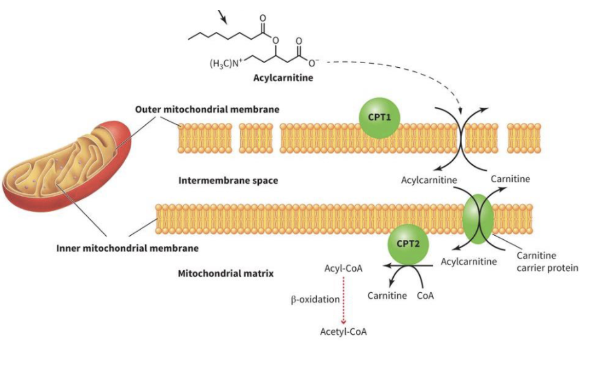
Explain the process of how fatty acids are transported into the mitochondria
Acyl group transferred from CoA’s thioester onto carnitine’s hydroxyl group → acyl carnitine made, CoA-SH released (via CPT1)
Acyl-carnitine crosses the mitochondria membrane via a translocase (carnitine-acylcarnitine translocase)
CPT2 reverses the rxn by transferring acyl group back onto a mitochondrial CoA → acyl-CoA regenerated + free carnitine release
β-oxidation
Breaking down lipids (named after the breaking of the β carbon → comes off in 2 carbon chunks)
acyl-CoA formed in cytosol, delivers β carbons to the TCA
water not needed (bc lipids are hydrophobically stored)
ATP required, HIGHLY favorable rxn