Unit 2 - the CNS
1/74
There's no tags or description
Looks like no tags are added yet.
Name | Mastery | Learn | Test | Matching | Spaced | Call with Kai |
|---|
No analytics yet
Send a link to your students to track their progress
75 Terms
explain the evolutionary trends in nervous systems
Nerve net (jelly fish)
Bilateral symmetry (anterior and posterior)
cephalization
Consolidation of peripheral nervous system to create nerves (as opposed to a loose network like with the nerve net)
Ventral nerve cord(s) (starts at front)
Dorsal nerve cord (goes back)
Spinal cord (both routes protrude into spinal cord)
Increasing role of forebrain (more power there throughout evolution)
which nerve is responsible for most parasympathetic innervation in the CNS
what do dorsal and ventral mean
dorsal = back
ventral = front
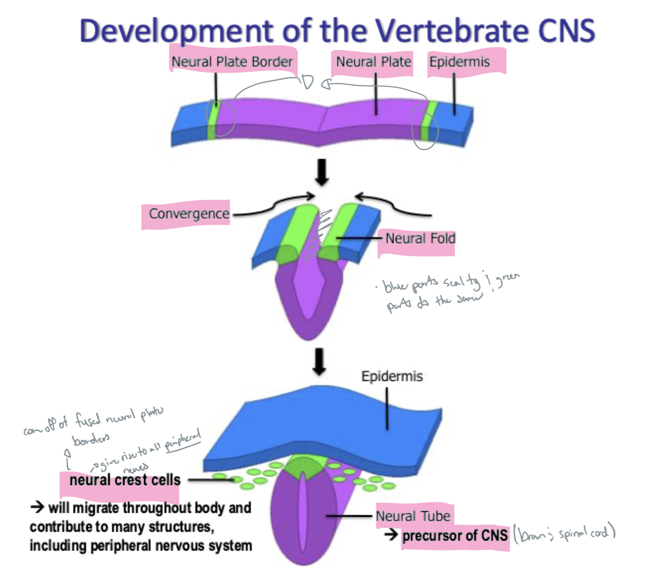
explain the development of the vertebrate CNS (how the neural fold is made)
Neural plate Borders come together and create the neural fold
the neural plates converge to make the neural tube (which will then become the CNS)
neural crest cells shed off the neural tube and migrate throughout the body → contribute to many structures, including the peripheral nervous system
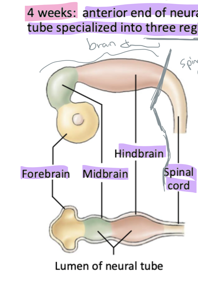
explain the development of the human CNS (4 weeks)
anterior end of neural tube specialized into three regions (the forebrain, midbrain, and hindbrain)
explain the development of the human CNS (6 weeks)
neural tube differentiated into major brain regions present at birth
Hindbrain
Medulla oblongata
Cerebellum and pons
Forebrain
Diencephalon
Cerebrum
explain the development of the human CNS (11 weeks)
growth of the cerebrum much more rapid than that of other regions
explain the development of the human CNS (birth)
cerebrum covers most of other brain regions; convoluted surface due to rapid growth in confined space
what provides protection and support for the CNS
surrounded by a bony cage → cranium (brain) and vertebrae (spinal cord)
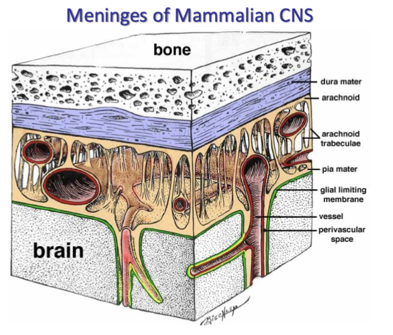
three layers of connective tissue → meninges (in both the brain and spinal cord)
fluid between layers → cerebrospinal fluid (in central canal (spinal cord) and in ventricles (brain)
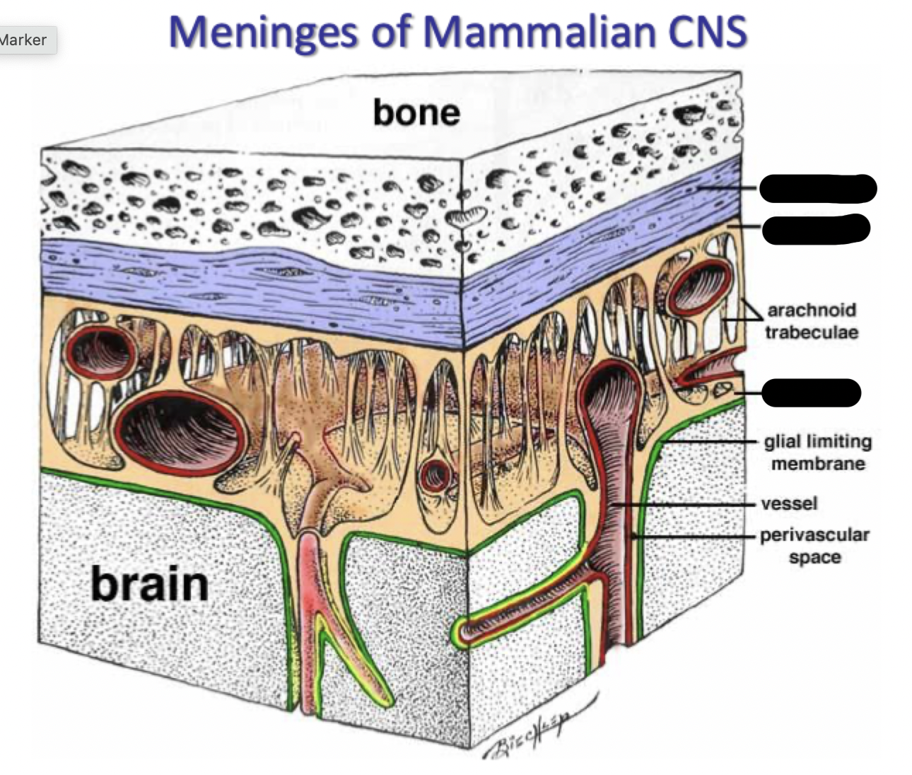
what are the meninges
the cura mater
arachnoid
pia mater (closest to brain, follows grooves on it, thin)
what is a subdural hematoma
when blood leaks into the subdural layer (between dural and arachnoid layer)
where is cerebral spinal fluid found
in ventricles within brain and the hollow central canal within spinal cord
fluid is continuous with both
in ventricular system and bathes external surfaces of brain, between meninges
what are ventricles
fluid-filled cavities in the brain
two lateral ventricles, two descending ventricles that extend through in the brain stem
what are ependymal cells? how do they work
A type of glial cell
line the inner surfaces of the brain ventricles and the central canal of the spinal cord → create barriers between compartments
act as a barrier between the cerebrospinal fluid (CSF) and the brain tissue, regulating what flows in and out of the brain and maintaining overall brain health
they allow CSF to escape into the subarachnoid space so ventricles don’t expand into the brain → bathes CNS from outside as well (cleanses brain)
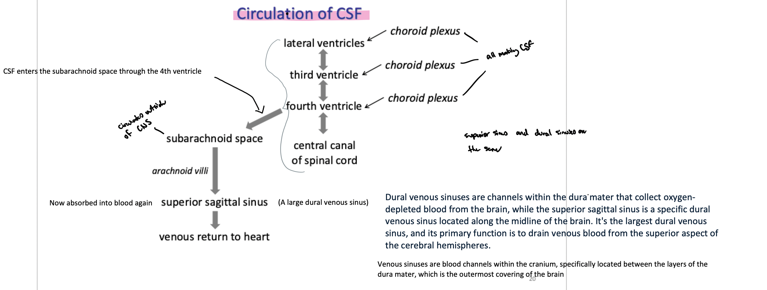
specialized ones in the choroid plexus create CSF
what allows CSF to escape into the subarachnoid space so ventricles don’t expand into the brain → so they can bathe CNS from outside as well (cleanses brain)
ependymal cells
what produces CSF
the specialized ependymal cells within the choroid plexus within each ventricle (each ventricle has them)
blood from the capillaries in the choroid plexuses is filtered through ependymal cells producing CSF (water and some ions, nutrients, and vitamins passed through)
what is interstitial fluid
surrounds neurons and glial cells
what is plasma
within cerebral blood vessels
how does CSF compare to plasma
low protein, no blood cells
presence of RBCs or elevated protein in CSF collected via lumbar puncture (sampling of fluid from subarachnoid space between vertebrae) suggests infection
what are oligodendrocytes
form myelin sheaths within the CNS (white matter)
what are microglia
immune cell lineage - phagocytic
what are astrocytes
numerous in brain
regulate local extracellular fluid by releasing chemicals
lots of projections (look like a star)
provides another layer around capillaries in the brain (a part of the the blood brain barrier)
what type of cells are ependymal cells
non-neuronal cells (type of glial cell → NOT epidermal cells)
how is CSF reabsorbed into venous blood
through arachnoid villi (finger-like extensions of the arachnoid membrane into the dural venous sinuses)
→ CSF moves from arachnoid villi in the subarachnoid space into venous blood (it’s reabsorbed)
explain the circulation of CSF
what is a hydrocephalic brain and how does it compare to a normal brain
ventricles are enlarged due to CSF build-up (when it can not drain)
what do astrocyte foot processes do
secrete paracrine factors that promote tight junction formation → aids in tight junction formation → key in the brain (and blood-brain barrier)
what do tight junctions do
prevent solute movement between endothelial cells
determine how leaky/prone to exchange blood vessels
VERY tight in brain, not as tight in spinal cords
what is the blood-brain barrier and what can/can not cross it
lipid soluble molecules cross readily
hydrophilic substances (ions, amino acids, peptides) only cross if specific transporters / carriers are present on endothelial cells of capillaries within the CNS
what determines if an antihistamine is drowsy or not
non-drowsy ones do not cross the blood-brain barrier
what causes parkinson’s disease
loss of dopaminergic neurons in regions of the brain
how is parkinsons treated
dopamine can not readily cross the blood-brain barrier
L-dopa (dopamine precursor) can be transported into brain because it is similar structurally to L-tyrosine (chemical that has a transporter for it)
then is transferred into dopamine after crossing to treat parkinsons
what are 2 things the brain absolutely needs to survivr
oxygen and glucose
explain neural tissue’s oxygen requirement
neurons are obligate aerobes
unable to switch to anaerobic metabolism
O2 readily crosses the blood brain barrier to compensate
explain neural tissue’s glucose requirement
capillaries of CNS express high levels of glucose transporters to promote adequate levels of glucose
brain is responsible for approx half of body’s glucose consumption
what are the sections of the spinal cord
Cervical
Thoracic
Lumbar
Sacral
what does the spinal cord do
major path for info flow between CNS and skin, joints, muscles
contains neural networks involved in locomotion
divided into 4 regions, each of which is divided into segments
each segment gives rise to pair of spinal nerves
what do parasympathetic and sympathetic nerves connect to respectively within the CNS and what do they innervate
parasympathetic:
connect to the brainstem and sacral sections of the spinal cord
innervates
which subsection of the nervous system (parasympathetic or sympathetic) does the vagus nerve fall under
the parasympathetic NS
what are the 2 types of matter that primarily compose the CNS
white and gray matter
what are white and grey matter respectively
compose the CNS
White matter:
type of brain tissue that consists primarily of myelinated neuronal bodies (the myelin sheaths giving them that white colour)
typically just myelinated axons that form tracts going to/from the brain
Gray Matter:
unmyelinated neuronal bodies (cell bodies, unmyelinated axons, dendrites)
makes up structures within the CNS such as the dorsal and ventral horns
typically considered an integration site within the CNS so it is involved in processing and passing information between neurons
what type of matter makes up the dorsal and ventral horns
grey
what type of neurons make up the dorsal root of the spinal cord
sensory neurons
what are the dorsal root ganglia
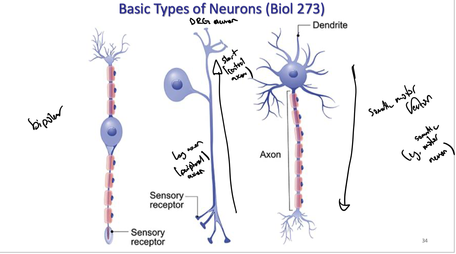
clusters of neuronal cell bodies within the dorsal root of the spinal cord. The cell bodies are typically bipolar sensory neurons
bring in sensory information from the body.
explain the flow of afferent and efferent information in and out of the spinal cord
The spinal cord is split up into 2 roots → the dorsal and ventral roots
dorsal roots (or also called the afferent root) carry (primarily sensory) information from the body to the spinal cord
dorsal root ganglia are clusters of neuronal cell bodies within the dorsal root.
These cell bodies are typically bipolar sensory neurons
bring in sensory information from the body.
sensory info is then brought from the dorsal root ganglia to the dorsal horns → so the dorsal horns are where the terminals for these sensory neurons that protrude from the dorsal roots are.
dorsal horns receive this incoming information from the terminals of the sensory neurons.
signal reaches the dorsal horns for integration then goes into the ventral horns (so this is where the ventral root begins).
The ventral root (also called the EFFERENT root) is the off-ramp for information so it carries newly integrated signals (that now typically contain motor information to go TO the body in response to the sensory information previously obtained FROM the body)
So within the ventral root the signals are carried through the ventral horns and into the ventral roots where they will propagate out of the spinal cord
because the newly integrated information is most commonly motor information now, the ventral root is typically composed of motor neurons instead of sensory neurons like in the dorsal root
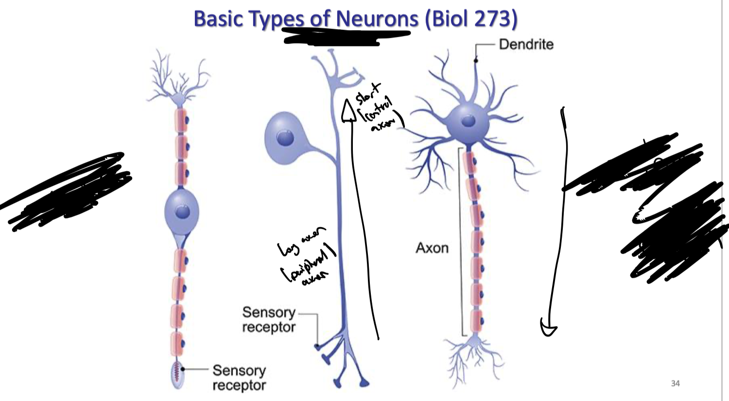
what is the difference between bipolar neurons, DRG neurons, and somatic motor neurons
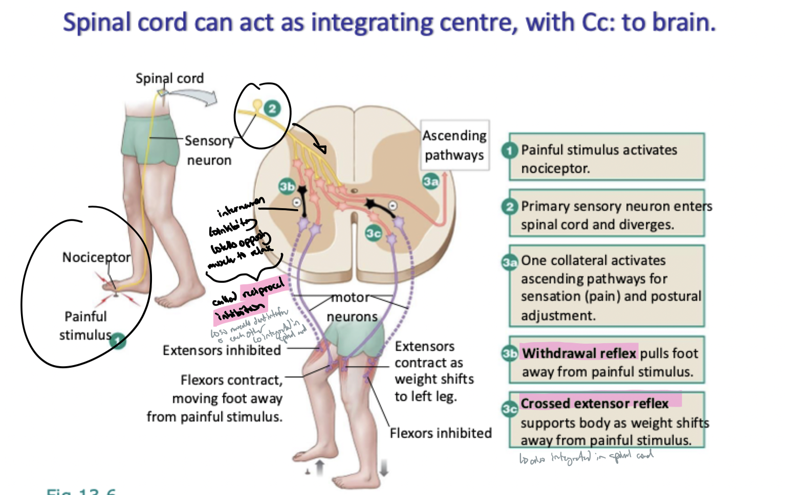
explain how spinal reflexes work
spinal reflex initiates response without input from brain
stimulus stimulates sensory neurons that send signal to spinal cord
interneurons in the spinal cord (grey matter) serve as the integration center for this info → often also sends signals up to brain so brain knows whats happening but that’s SEPARATE process from this one
interneurons send a command to muscles or glands which invokes a response from the target muscle/gland
what composes ascending and descending tracts in the CNS
white matter
what are the ascending tracts in the spinal cord and what do they do overall and specifically
sensory info going up
dorsal columns (touch/pressure and propioception) [main one we focus on]
spinocerebellar (proprioception (posture, coordination))
spinothalamic (pain, temp)
these tracts are organize the info throughout the spinal cord and pile on as they get closer to the brain
what are the descending tracts in the spinal cord and what do they do overall and specifically
descending motor info
the corticospinal tracts (voluntary movement)
what is the oldest and most primative part oft he brain
the brainstem
what is the brainstem organized a lot like? explain how
the spinal cord
most cranial nerves originate from there
most cranial nerves originate from here
carry sensory and motor info for head/neck
contain cranial nerves (notably cranial nerve X - vagus)
contains nuclei associated with reticular formation
what is reticular formation and what contains nuclei associated with it
reticular formation
network of neurons involved in processes such as arousal/sleep, muscle tone, coordination of breathing, etc
brainstem contains nuclei associated with reticular (network) formation
what are the main structures that make up the brainstem and what do they do
midbrain
eye movement coordination, visual and auditory reflexes
pons
relay station between cerebrum and cerebellum
works with medulla to regulate breathing
medulla
gray matter involved in controlling of many involuntary functions (eg. blood pressure, breathing, swallowing, vomiting, etc)
white matter is involved in ascending somatosensory tracts and descending corticospinal tracts
site of decussation (crossing over)
Notably pons and medulla
what is the site of decussation (crossing over) for most neurons in the corticospinal tract
the medulla
what is cranial nerve X and what does it do?
vagus nerve - provides sensory info to a lot of places
what composes the cerebrum
the cortex (has lobes, outside of brain)
each cerebral hemisphere is divided into 4 lobes
frontal, occipital parietal, temporal
subcortical structures (structures beneath cortex)
what is the cerebellum
second largest structure in the brain, coordinates movement
where is the diencephalon and what 4 structures composes it? explain what its structures do
in the middle of the brain, between the brainstem and cerebrum
thalamus
relays and integrates sensory info from lower parts of the CNS
hypothalamus
major centre for homeostasis
influences autonomic responses, endocrine systems
contains centers that drive behaviour related to hunger, thirst, etc)
pituitary gland
regulated by hypothalamus
pineal gland
secretes the hormone melatonin → involved in circadian and seasonal rythums
what does the cerebrum do
site of “higher” brain functions
largest and most distinctive part of brain in higher primates
t/f: degree of folding related to capacity for higher processing in a species
f → not completely determined by folding
what is a sulcus and gyrus
sulcus - forrow or groove
gyrus - convolution (ridges on surface, bumps)
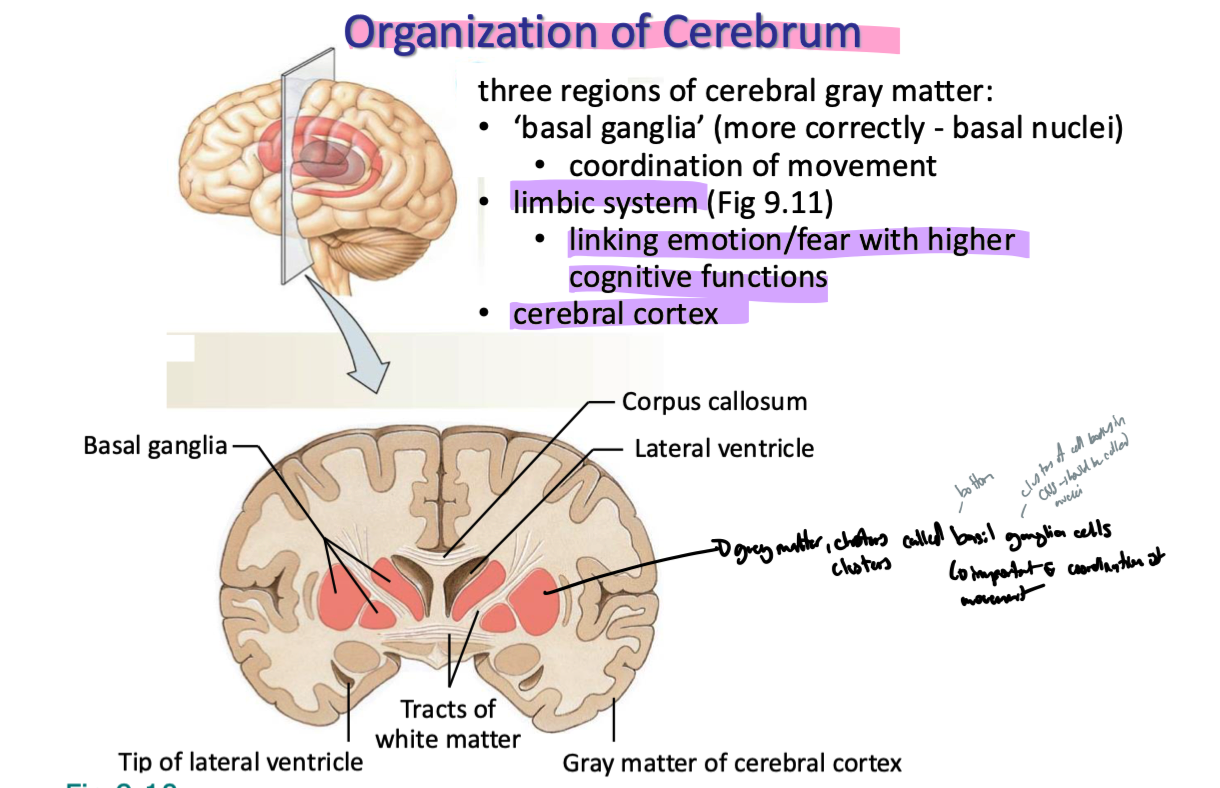
what are the 3 regions of cerebral gray matter and what do they do
basil gangliali (basil nuclei since in CNS)
coordination of movement
limbic system
linking emotion/fear with higher cognitive functiuons
cerebral cortex
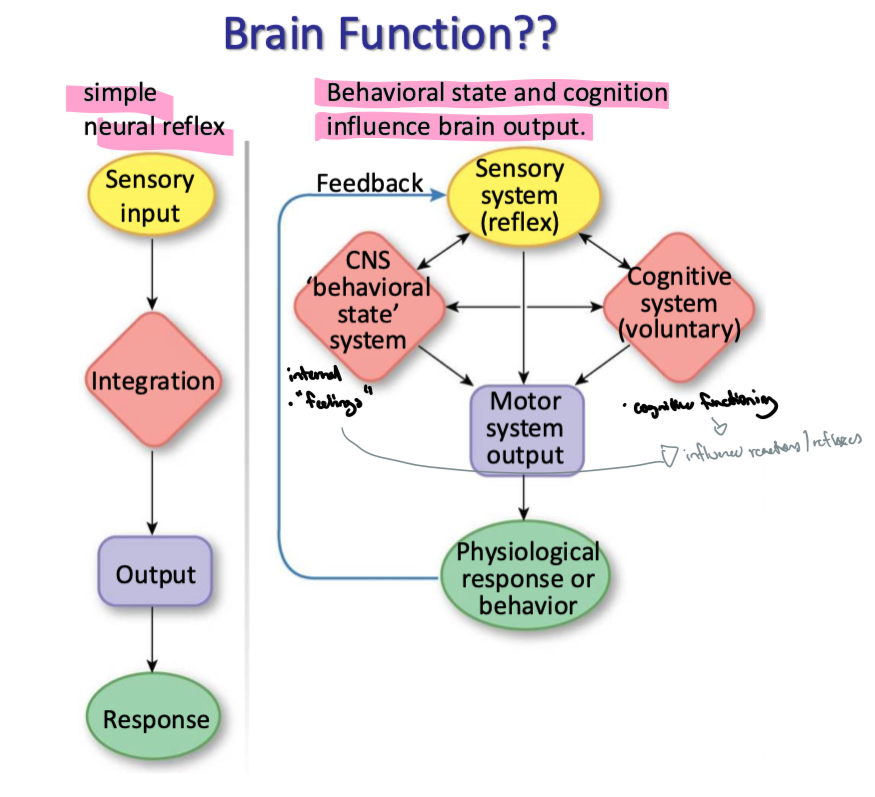
t/f: behavioural state and cognition effect motor system output in the brain
true → internal feelings and cognitive functioning influence reactions, reflexes and each other
what do sensory areas in the cerebral cortex do
translate sensory input into perception (awareness)
what do motor areas in the cerebral cortex of the brain control
skeletal muscles
what do association areas in the brain do
integrate info from sensory and motor areas → many areas involved with coordinating somatic movement and sensory info
what do the gustatory cortex and olfactory cortex interpret respectively
taste and smell respectively
what information does the parietal lobe and frontal lobe process specifically
parietal - contains somatic sensory cortex → processes sensory info from skin, musculoskeletal system, viscera
frontal - contains primary motor cortex, involved in skeletal muscle movement
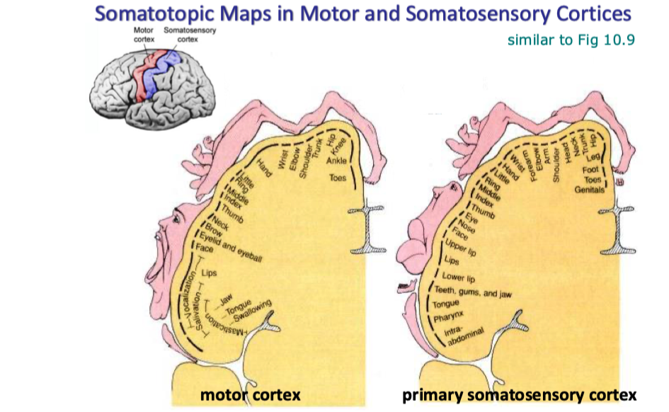
what do the somatotopic maps in motor and somatosensory cortices show
Motor: hands and face have a higher SA on the motor cortex → more control
Somatosensory: lips and hands are very big
where do ascending and descending tracts go and what type of signals are they respectively
ascending → to brain, sensory information
descending → from brain, motor information
explain the flow of motor information from the brain to right tunk muscles and right limb muscles respectively
trunk muscles:
motor signals sent from primary motor cortex of left cerebral hemisphere
signals go down nerves in the anterior corticospinal tract
crosses over to the right side of the body lower down the spinal cord
synapses with somatic motor neurons to skeletal muscles on the right side of the trunk
limb muscles:
motor signals sent from primary motor cortex of left cerebral hemisphere propogate down nerves
signals go down nerves in the lateral corticospinal tract
crosses over to the right side of the body higher up in the brainstem (specifically the medulla)
synapses with somatic motor neurons to skeletal muscles on the right limbs
trunk muscles go straight down anterior corticospinal tract, cross low,
limb muscles go down the anterior corticospinal tract, cross high (in medulla), go down lateral corticospinal tract
where do most fibers from the motor cortex cross over
the base of the brain (medulla)
how does the spinal cord act as an integrating centre with the brain