Unit 5 Beam Restricting Devices v20.pptx
1/52
There's no tags or description
Looks like no tags are added yet.
Name | Mastery | Learn | Test | Matching | Spaced | Call with Kai |
|---|
No analytics yet
Send a link to your students to track their progress
53 Terms
What is Scatter Radiation?
A change in the direction of the x-ray photon after interaction with the atoms of the patient
How does scatter affect an image?
Increasing Density (FOG), Decreases Contrast and Resolution
What is the overall affect from scatter radiation on a radiograph?
Overall gray appearance
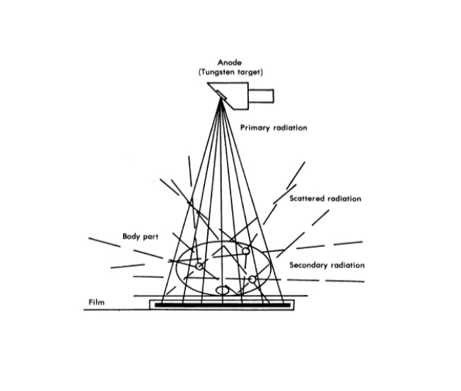
What is Primary Radiation
Radiation emitted from the x-ray tube
What is Remnant (exit) Radiation
Unabsorbed and scattered x-rays that interact with the IR
What does Remnant (exit) Radiation CARRY?
CARRIES THE AERIAL IMAGE
What types of radiation does Remnant (exit) Radiation have?
SCATTER and PRIMARY RADIATION
Factors that influence the degree of scatter radiation:
Field size
Thickness of object
Density (composition) of the object
Tube Potential (kV)

What is Field size?
Area of the x-ray beam
HOW is Field Size altered?
By the use of a beam limiting device
As Field Size INCREASES Scatter radiation….
Increases

HOW does Thickness of the Object influence Scatter?
More atoms are present in a larger object to cause more interactions
As Thickness of the Object INCREASES Scatter….
Increases due to more interactions among the atoms.
HOW does Density (composition) of the object influence Scatter
The more dense the object is, the more scatter is produced
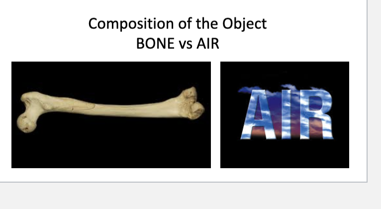
As Density (composition) of the object INCREASES Scatter….
Increases, bone=more scatter, air=less scatter
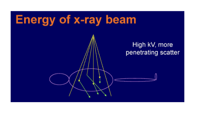
HOW does Tube potential (kV) influence Scatter
As kV is increased, the energy of scatter radiation also increases, thus scatter radiation has a better chance of reaching the IR
As Tube potential (kV) INCREASES Scatter….
Increases (FOR kV)
What Factors that affect the amount of scatter
reaching the IR can WE CONTROL
FIELD SIZE AND KV
What Factors that affect the amount of scatter
reaching the IR can WE NOT CONTROL
THICKNESS AND DENSITY (COMPOSITION of patient)
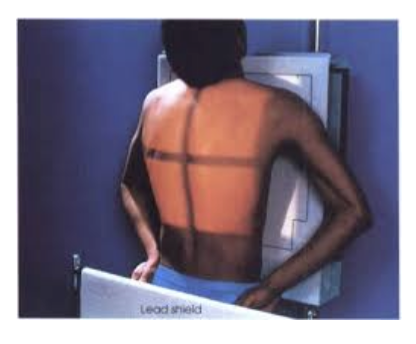
What is THE #1 ADVANTAGE of a Beam Limiting Device?
REDUCES PATIENT EXPOSURE
How does Beam Limiting Devices increase Image Quality?
By DECREASING scatter to the IR
How does a Beam Limiting Device affect Field Size, Scatter, Density (FOG), Contrast and Detail?
Decreases Field Size → decreases Scatter → decreases Density (FOG).
Which then Increases Contrast and Detail.
How does a Beam Limiting Device affect Detail
Increases visibility of Detail
What PERCENT of density on the radiograph is the result of scatter radiation
50-90%
Where is Penumbra (BLUR) greater on a x-ray beam
On the edges of the beam
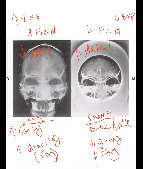
How does a bigger Field Size affect contrast and exposure to the patient?
Decreases contrast→more grays and increases exposure.

How does a smaller Field Size affect contrast and exposure to the patient?
Increases contrast→more Black/White and decreases exposure.
Types of Beam Limiting Devices
1. Aperture Diaphragm
2. Cones
3. Variable Aperture
***What material are Beam Limiting Devices made from?
***constructed of lead or lead-lined because of its characteristic to attenuate
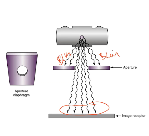
Details of Aperture Diaphragms (NOT IN USE CURRENTLY)
Simplest, Least expensive
Disadvantage: Change for each different Field size
Large amount of Blur
What types of Cones exist (IN USE)
Flared
Cylindrical

What does a Flared Cone match in x-ray imaging?
Matches the divergence of beam (NOTE: The flared cone is shaped in a way that follows the same spreading angle of the x-rays.) AND (It’s a beam because don’t forget that the Cone is in place of what would be a collimator there, so after it leaves the cone, then it’s a field)
Does the Flared portion of the cone contribute to the beam?
NO
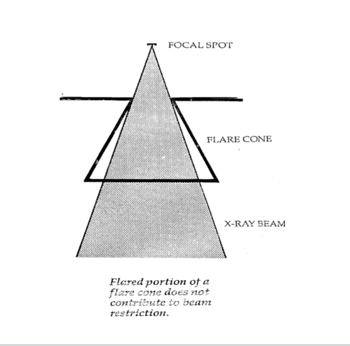
How does the size of the Flare Cone compare to the x-ray beam?
Usually larger than beam

Why does a cylinder cone reduce blur in radiographic imaging?
Blur is reduced because it truly limits the field size.
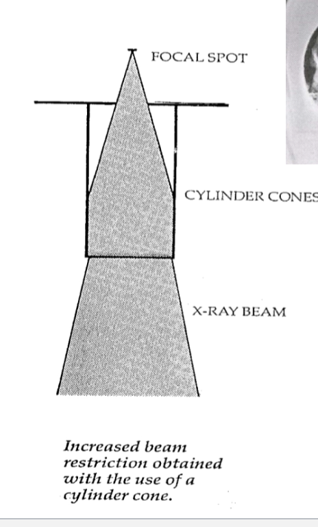
What shape of field does a cylinder cone create?
Circular field.
Disadvantage of using Cones
Excessive radiation for larger body parts, causing unnecessary patient exposure!!! (NOTE: When a large beam from a CONE is used—even on a large body part—some x-rays can miss the intended target and still enter surrounding tissues)
What Beam Limiting Device do we use TODAY
Variable Apertures: Collimator (manually controlled), Auto Collimator, PBL (Auto Colli with a safety feature)
Variable Aperture is also known as…
Collimator
What is an Automatic collimator? (NOTE:
The X-ray beam is broader — it's the full radiation emitted from the tube.
The X-ray field is the collimated portion of that beam.
The field is shaped at the collimator (after the window), not at the window itself.)
A type of Variable Aperture that automatically adjusts the x-ray field size to match the image receptor using motorized shutters.
What is the Positive Beam Limiting Device (PBL) (NOTE: IT IS AN AUTO COLLIMATOR WITH ONE IMPORTANT FEATURE)
Ensures the size of the field does not exceed the size of the IR
What does the automatic collimator use to shape the x-ray beam?
Two sets of horizontal lead shutters.
What does the automatic collimator allow for regarding field sizes?
Adjustable. Allows for infinite number of field sizes.
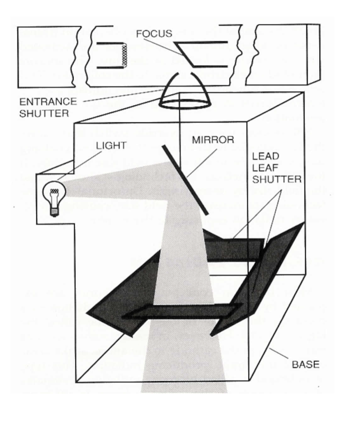
What is the function of the upper shutters in an automatic collimator?
Upper shutters control stem radiation (off-focus). (NOTE: Its the Entrance Shutters)
What is the function of the lower shutters in an automatic collimator?
Lower shutters reduce blur.
How does the automatic collimator affect filtration?
Adds to “added” filtration. (NOTE: their function is more about shaping the beam and reducing blur, not hardening it like traditional filters.)
What visible guide does the automatic collimator provide?
Provides a light field.
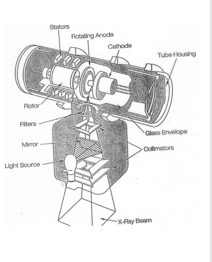
What is stem radiation also known as?
Off-focus, Extra-focal

What does stem radiation refer to?
Photons not produced at the focal spot of the anode
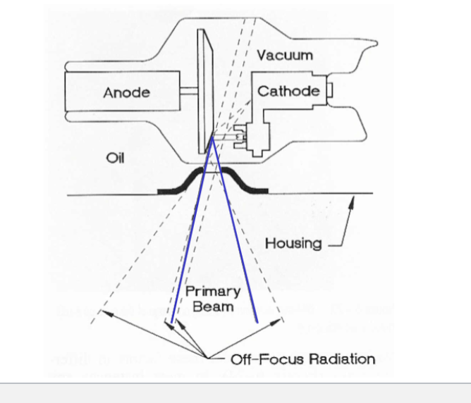
What are sources of stem radiation inside the x-ray tube?
Tube housing and evaporated tungsten on the envelope
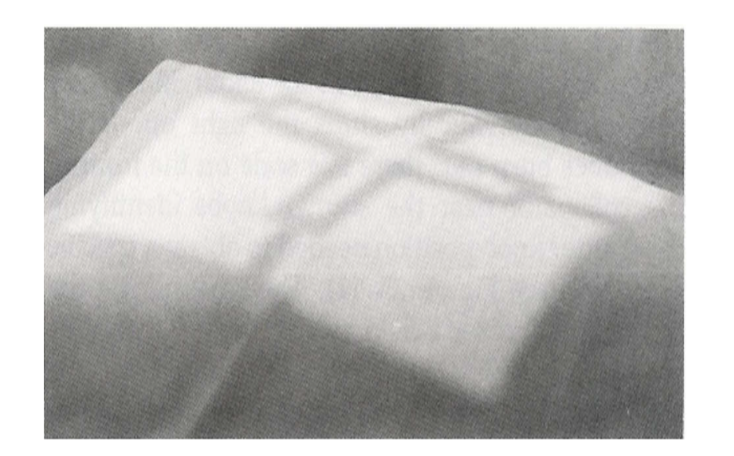
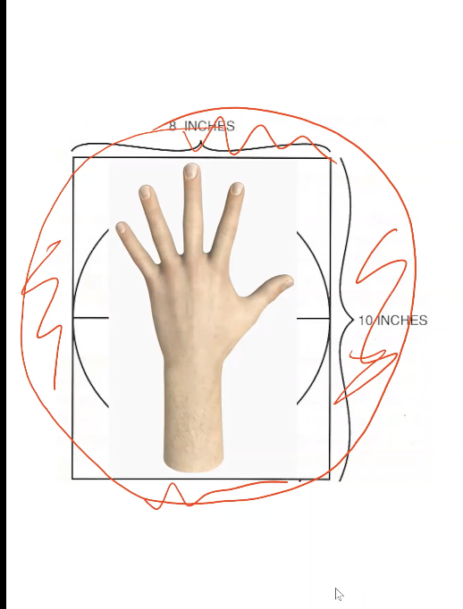
What does the Light Field and Cross-hairs represent?
Light Field shows the X-ray field, C-h shows the Central Ray
What is Field to Light Congruency?
It is the alignment between the x-ray field and the collimator's light field. (NOTE: refers to how accurately the x-ray field (the area exposed to radiation) matches the light field)
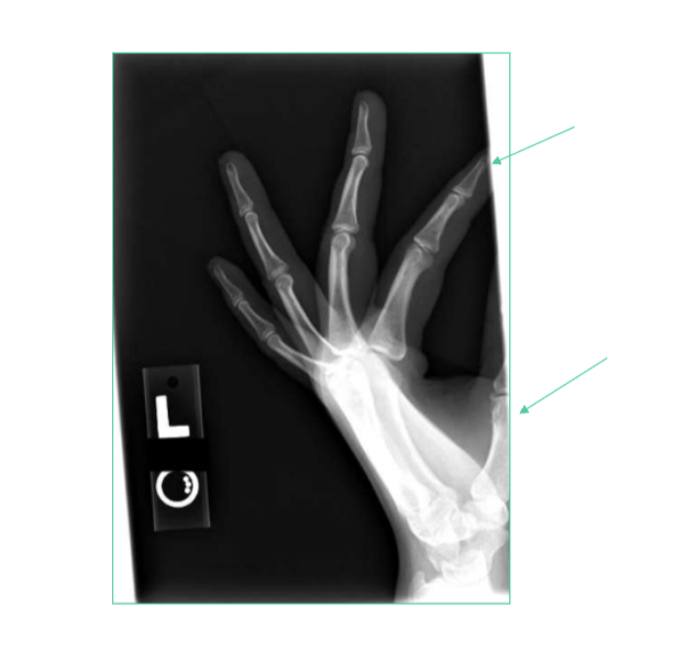
What does poor field-to-light congruency lead to?
Parts of the anatomy being cut off