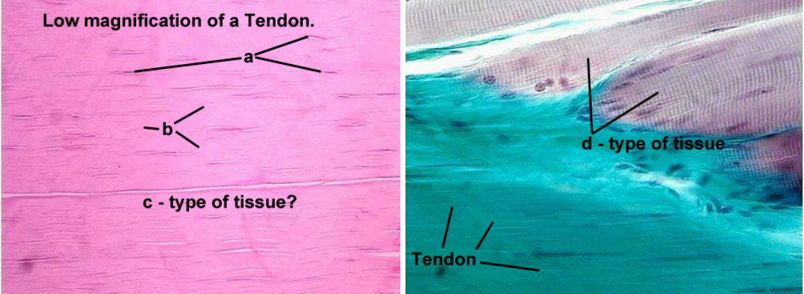
Low Magnification of Tendon
a. Fibroblasts
b. Collagen
c. Connective
d. Muscle
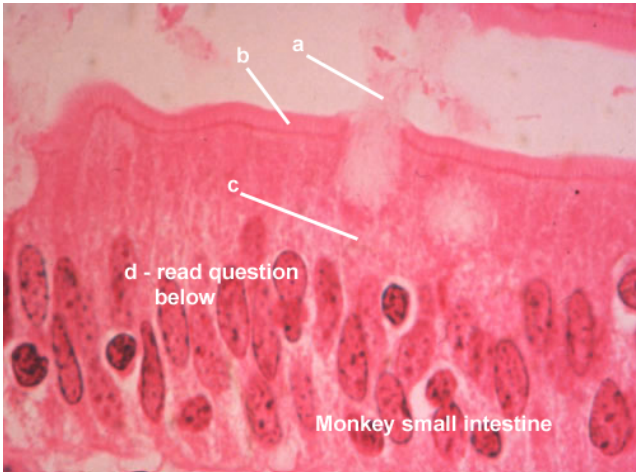
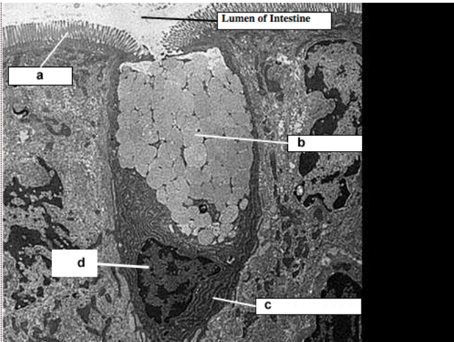
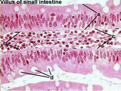
a. Mucus
b. Brush Border
c. Goblet Cell
d. Microvilli- What structures form answer "b" when viewed with the electron microscope?
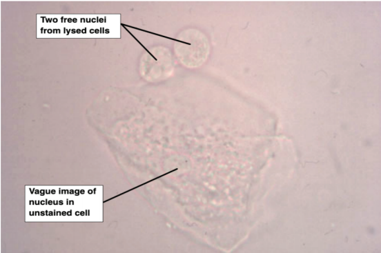
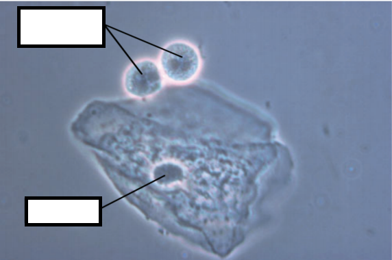
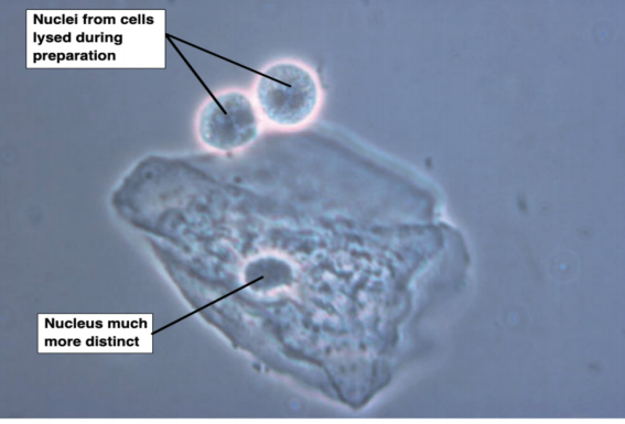
Nuclei from cells lysed during preparation
Nucleus much more distinct
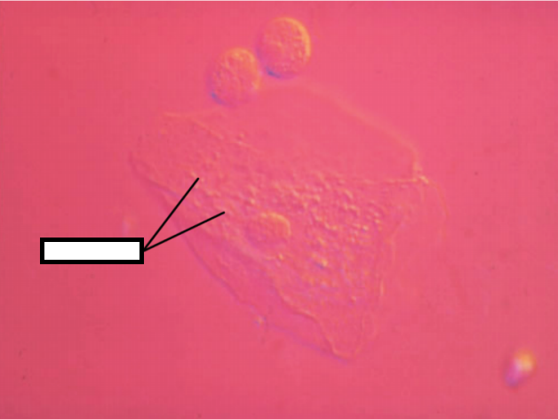
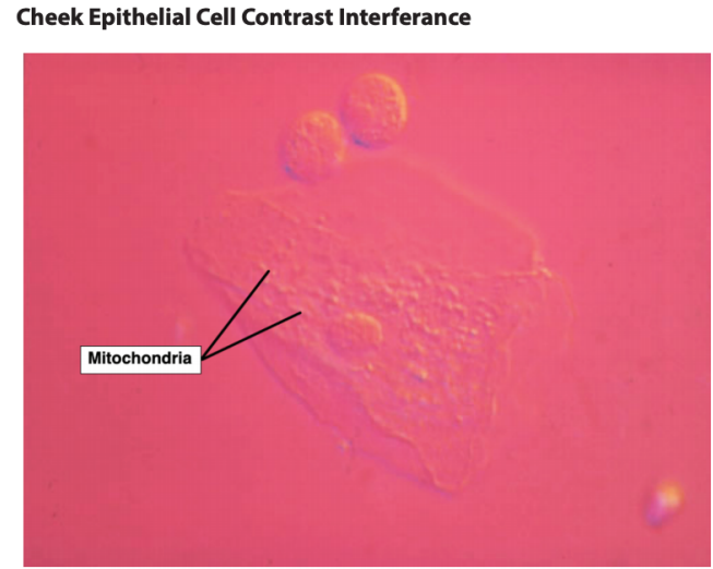
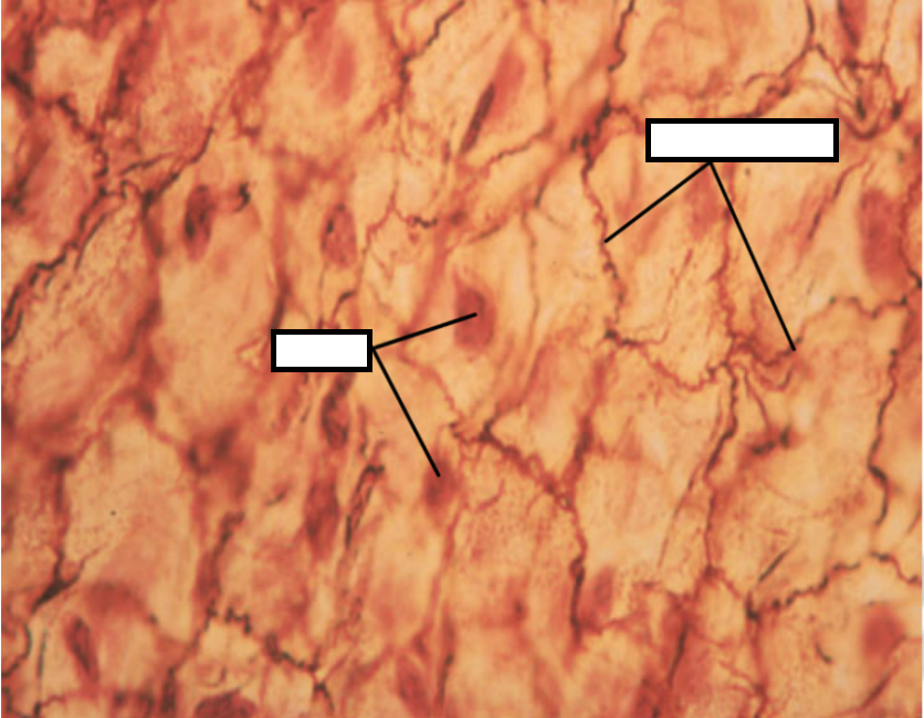
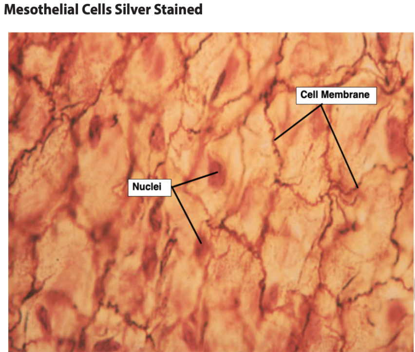
Mitochondria
Cell Membrane
Nuclei
a. Mitochondrion
b. Nucleus
c. Golgi apparatus
d. Degenerating cell
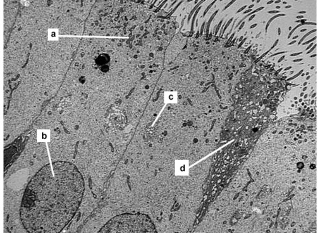
Identify structure a
Identify structure b.
Identify small round openings at c
Identify structure d.
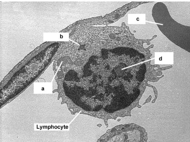
a. Erythrocyte
b. Lipid droplet
c. Nucleus pores
d. Mitochondria
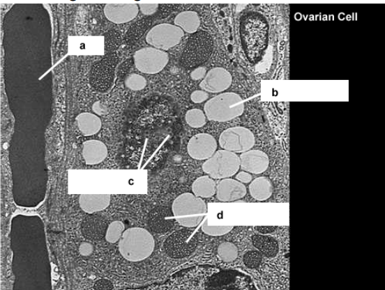
a. Nucleolus
b. Secretory granule
c. Golgi apparatus
d. Lysosomes
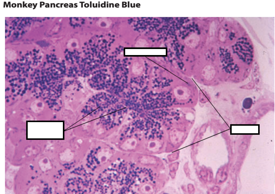
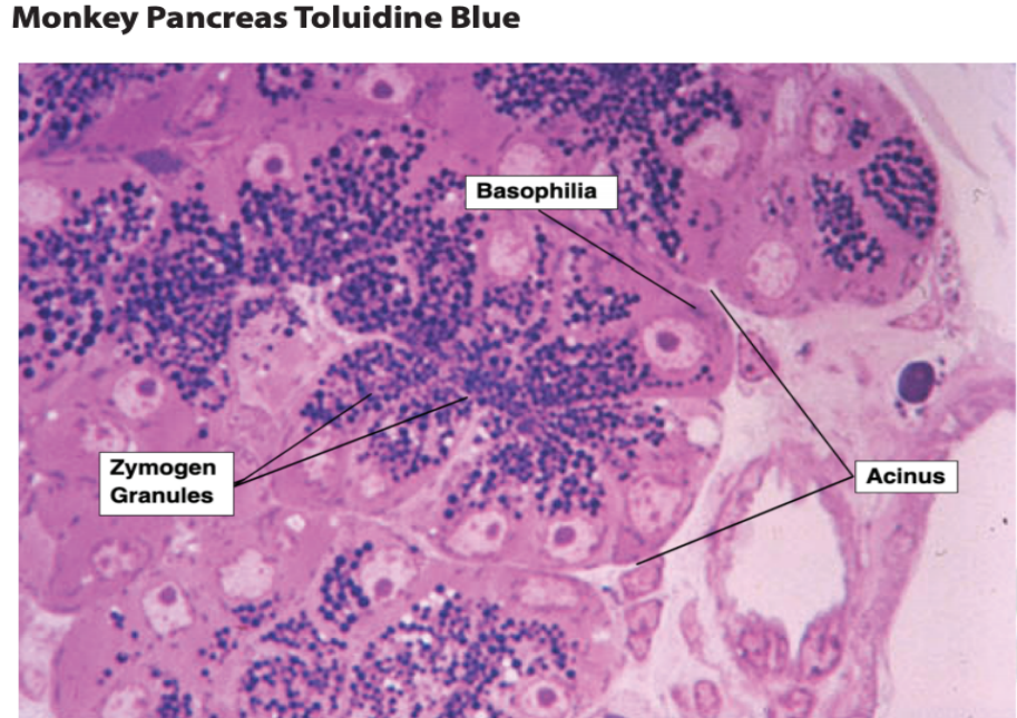
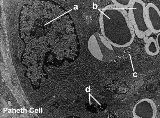
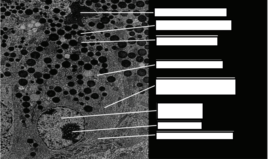
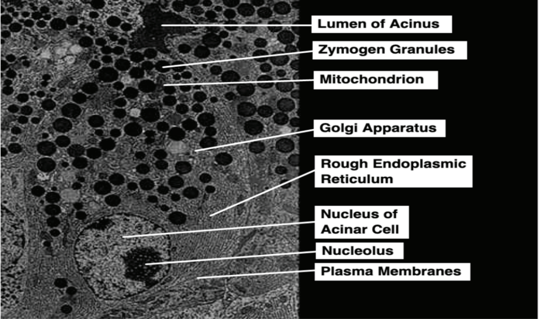
a. Lumen of Acinus
b. Zymogen granules
c. Rough endoplasmic reticulum
d. Nucleolus
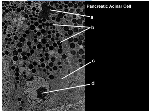
Pancreatic Acinar Cells
Lumen of Acinus
Zymogen Granules
Mitochondrion
Golgi Apparatus
Rough Endoplasmic Reticulum
Nucleus of Acinar Cell
Nucleolus
Plasma Membranes
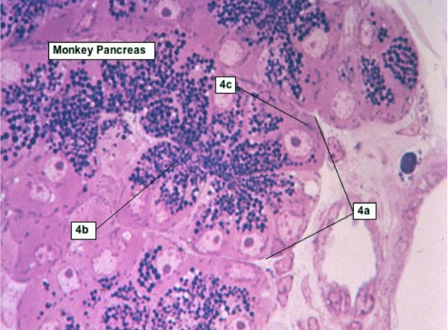
Identify secretory unit for the pancreas?
Identify small round dark-staining objects?
What is the light microscope term from the older literature for the deep purple staining region in the basal pole of the cell?
What secretory material is present in the dark staining spheres in the cells?
a. Acinus
b. Zymogen granules
c. Basophilia
d. Digestive enzymes
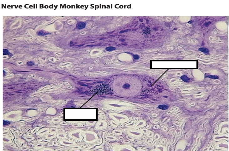
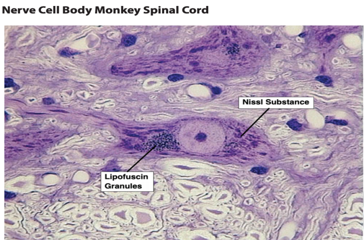
Nissl Substance
Lipofuscin Granules
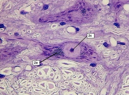
Identify blue-black cluster of granules.
What does answer 2a represent?
Identify structure. (2c)
What does euchromatic mean?
a. Lipofuscin granules
b. Lysosomes fused with substances being digested
c. Nucleolus
d. Clear (no color)
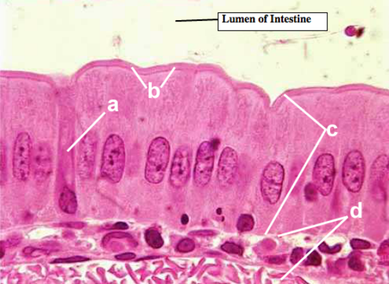
Lumine of Intestine
a. Microvilli
b. Mucous droplets
c. Rough endoplasmic reticulum
d. Nucleus-
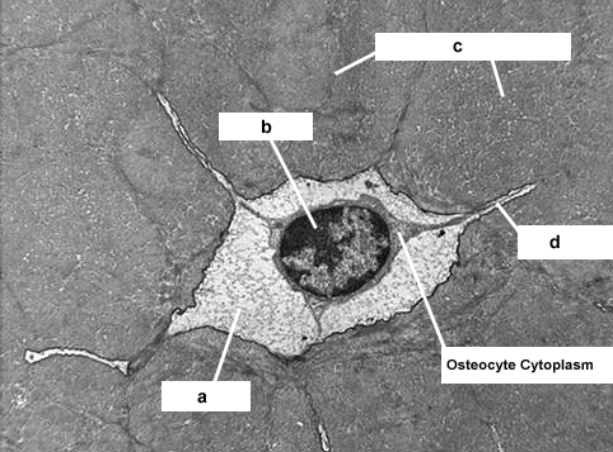
Mitochondrion
Centriole
Erythrocyte
Nucleolus
Lacuna
Nucleuos
Calcified bony matrix'
Canalicilus
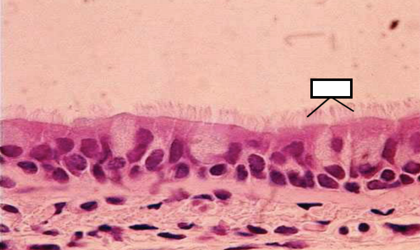
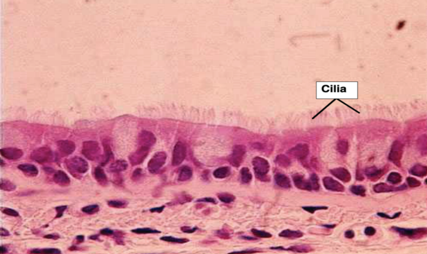
Ciliated Cells Monkey Bronchus
Cilia
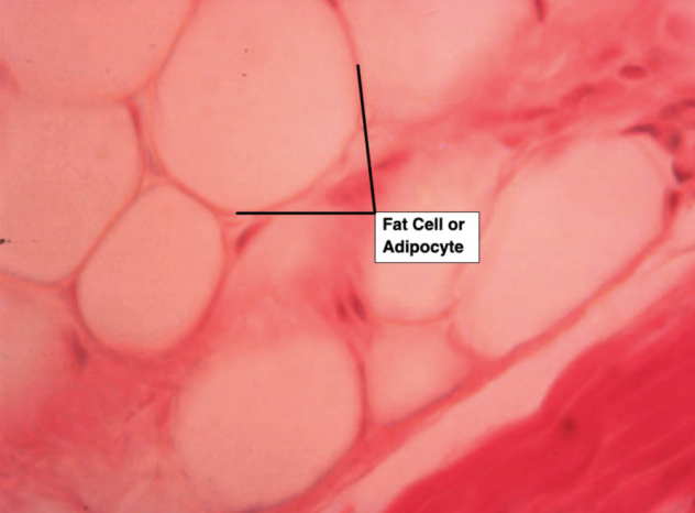
Fat Cell Human Finger Tip
a. Identify Space
b. Identify Epithelium
c. Identify blue stained cell layer
d. Identify red stained cell layer
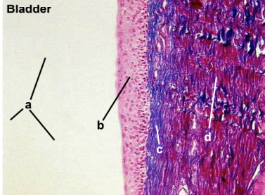
Transitional
Connective tissue
d. Muscle tissue
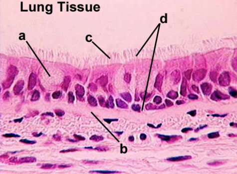
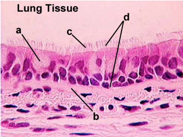
a. Goblet Cell
b. Basement Cell
c. Cilia
d. Ciliated pseudostratified columnar
Identify Cell Type
Identify cell structure
Identify type of epithelium
Identify Cell Layer
a. Goblet Cell
b. Brush Border
c. Simple Columnar
d. Lamina Proaria
Identify epithelial type
Identify epithelial layer
Identify Dark Strained Layer
Identify layer of epithelium
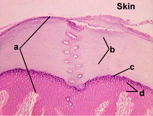
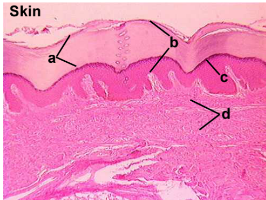
a. Stratified squamous keratinized
b. Stratum corneum
c. Stratum granulosum
d. Stratum germinativum
a. Identify substance
b. Identify entire structure
c. Identify epithelium
d. What epithelium does answer "c" look like when answer for "b" is empty?
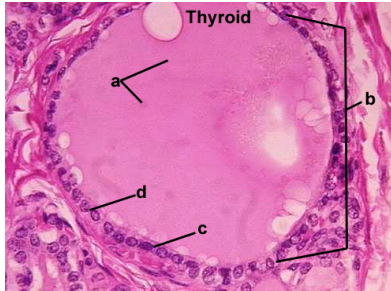
a. Thyroglobulin
b. Follicle
c. Simple cuboidal
d. Simple squamous
a. dentify space
b. Identify structures
c. Identify structure
d. Identify space
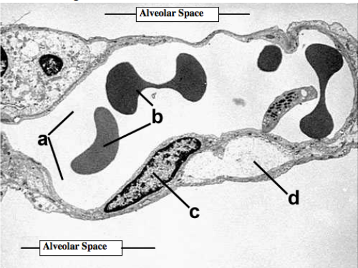
a. Capillary space
b. Erythrocytes
c. Nucleus
d. Artifact
a. Identify substance
b. Identify structure
c. Identify epithelial type
d. Identify structure
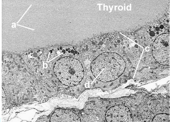
a. Thyroglobulin
b. Secretory vesicle
c. Simple cuboidal
d. Nucleus
a. Identify structure
b. Identify structure
c. Identify structure
d. Identify type of epithelium
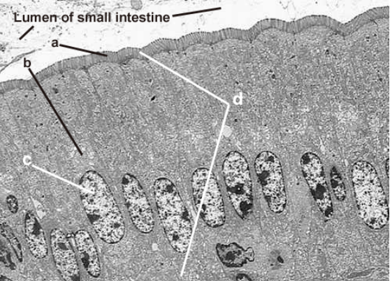
a. Microvilli
b. Golgi apparatus
c. Nucleus
d. Simple columnar
a. Identify epithelial type
b. Identify epithelial type
c. Identify entire structure
d. Identify epithelium lining tubule
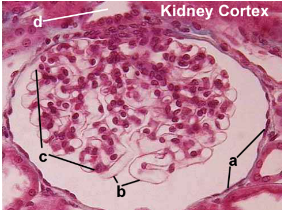
a. Simple squamous
b. Endothelium
c. Glomerulus
d. Simple cuboidal
a. Identify epithelium
b. Identify layer b
c. Identify layer c
d. What tissue type is answer c
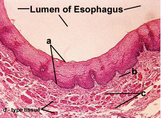
a. Stratified squamous non-keratinized
b. Lamina propria
c. Muscularis mucosae
d. Muscle
a. Identify epithelial layer
b. Identify type of epithelium
c. Identify dark stained layer of epithelium
d. Identify layer
a. Stratum corneum
b. Stratified squamous keratinized
c. Stratum granulosum
d. Dermis
a. Identify cell
b. Identify structure
c. Identify structure
d. Identify epithelium
a. Identify cell- Goblet cell
b. Basement membrane
c. Cilia
d. Ciliated pseudostratified columnar
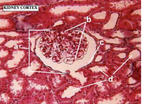
a. Identify the entire structure of A- Bowman's capsule b. Identify entire structureB- Glomerulus c. Identify type of epithelium- Simple squamous d. Identify epithelium lining tubules- Simple cuboidal
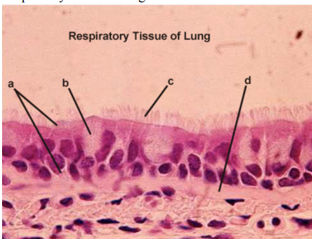
a. Ciliated pseudostratified columnar Epithelium- Identify tissue layer b. Goblet cell- Identify cell type c. Cilia- Identify structure d. Basement membrane- Identify layer
a. Smooth muscle cells
b. Fibroblasts
c. Plasma cell
d. Goblet cell
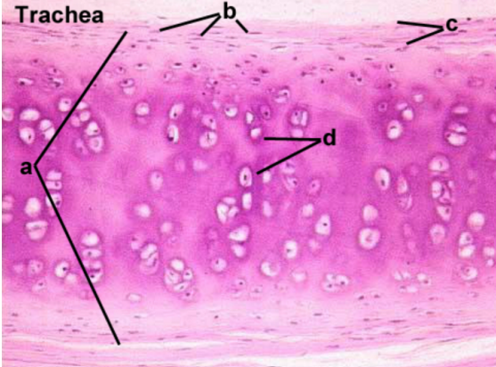
a. Hyaline
b. Chondroblasts
c. Perichondrium
d. Lacunar capsule
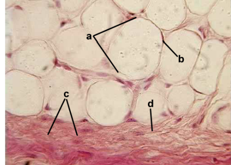
a. Fat
b. Nucleus
c. Dense irregular
d. Collagen
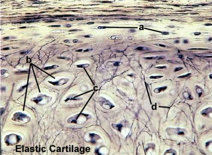
a. Chondroblasts
b. Lacunae
c. Chondrocytes
d. Elastic
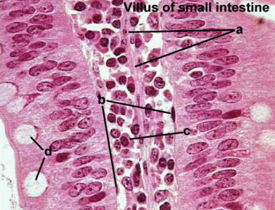
a. Simple columnar epithelium
b. Loose
c. Lamina propria
d. Goblet cell
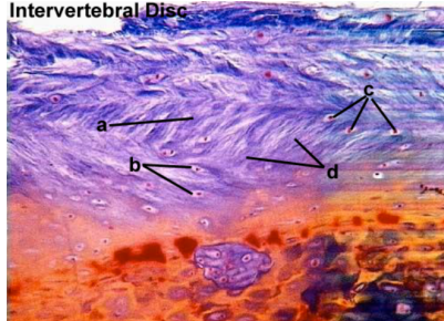
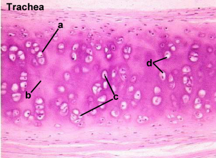
a. Fibrocartilage
b. Lacunae
c. Chondrocytes
d. Collagen
a. Lacunar capsule
b. Matrix
c. Chondrocytes
d. Lacunae
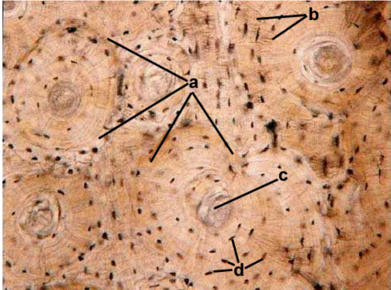
a. Osteocytes
b. Haversian canal
c. Canaliculi
d. Concentric lamellae
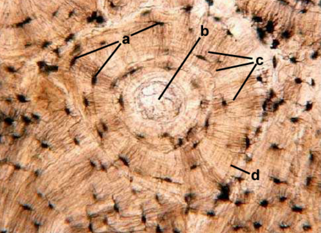
a. Haversian System
b. canaliculi
c. Haversian Canal
d. osteosytes
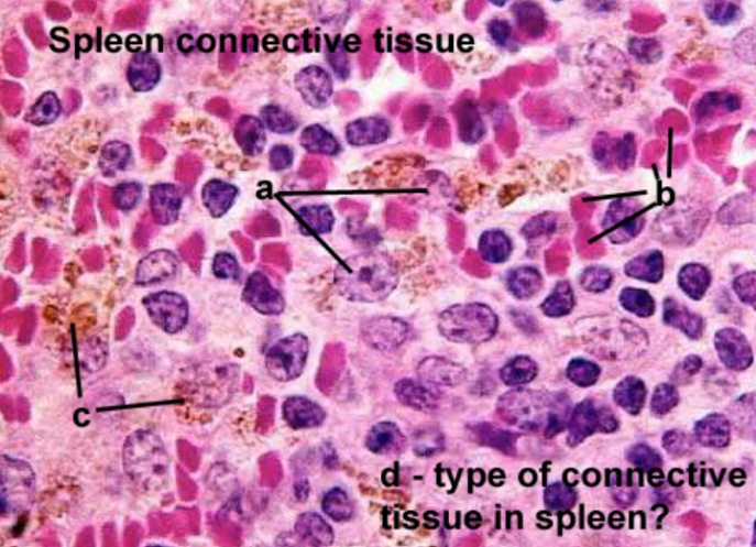
a. Macrophage
b. Red blood cells
c. Degraded hemoglobin
d. Reticula
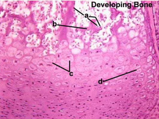
a. Osteoblasts
b. Zone of ossification
c. Zone of hypertrophy
d. Zone of proliferation
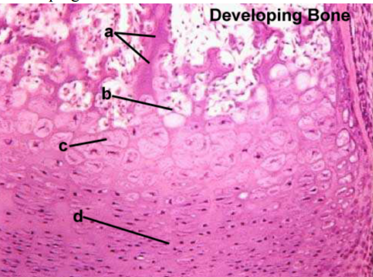
a. Calcified cartilage
b. Zone of erosion
c. Zone of hypertrophy
d. Zone of reserve cartilage