cognitive neuroscience midterm #1
1/83
There's no tags or description
Looks like no tags are added yet.
Name | Mastery | Learn | Test | Matching | Spaced |
|---|
No study sessions yet.
84 Terms
What did Ebbinghaus study with nonsense words?
He measured memory with savings and established the forgetting curve (forgetting happens quickly at first then slows down).
savings = ((initial learning rep - relearning rep)/initial learning rep) * 100
introduction of quantitative measures in psychology.
What is behaviorism?
To predict and control behavior
– No concern for what happens in between
– Rejected the use of mentalistic concepts
(e.g., the mind, imagery, thinking,
consciousness)
What are the methods of behaviorism?
Studied how stimuli in the environment
(input) influence the organism's response
(output)
Animal research:
- Classical conditioning
- Instrumental learning
What was the cognitive revolution?
a major shift in psychology during the 1950s–1970s, moving the focus away from behaviorism (which studied only observable behaviors) and toward understanding /making inferences about internal mental processes—like thinking, memory, problem-solving, and language.
ex. chomsky challenging human language/the misbehavior of organisms/human mind as a computer
How did behaviorism differ from the cognitive revolution?
behaviorism: observable behavior, mind as black box, stimulus response/conditioning experiments, learning based on reinforcement and punishments
cognitive revolution: focused on mental processes, mind as computer, lab experiments/models/RT/error analysis, based on understanding of mental rep of info
Who criticized the methods of behaviorism and why?
chomsky - language is too complex to be explained by classical conditioning alone because children make novel sentence and similar grammatical errors
the misbehavior of organisms - challenged idea that animal behavior could be shaped by operant conditioning because trained animals reverted back to instinctive behavior/stop doing trained behaviors
Describe what is meant by the information processing approach.
views the mind as a system that:
Receives input (information), Processes it, Produces an output (behavior or response)
It involves encoding, storing, and retrieving information
Input - Information from the environment (e.g., what you see or hear)
Attention - Focusing on certain information while filtering out others
Perception - Interpreting sensory input into meaningful patterns
Memory - Encoding: Getting information into the system, Storage: Keeping it over time, Retrieval: Bringing it back when needed, Decision-Making: Evaluating information and choosing a response
Output - The behavioral response (e.g., speaking, moving)
What does cognitive psychology and neuroscience contribute to each other?
cognitive psychology contributes: mental processes(e.g., memory, attention, language, thinking), experiments, models, behavior analysis, reaction times THEORIES AND MODELS FOR TESTING
neuroscience contributes: Brain structure and function (neurons, brain regions, neurotransmitters), Brain scans (fMRI, EEG), lesions, neural recordings BIO EVIDENCE FOR THEORIES
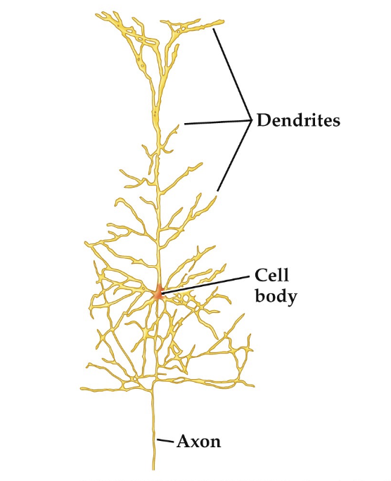
the neuron
Dendrite: Receive
signals from other
neurons
• Cell Body:
Organelles to
keep neuron alive
• Axon: Send
signals to other
neurons
• Synapse: Space
between neurons
action potential 1. Resting State (–70 mV)
The neuron is polarized.
Inside of the cell is more negative than outside.
Maintained by the sodium-potassium pump:
Pumps 3 Na⁺ out, 2 K⁺ in, using ATP.
action potential 2. Stimulus Received
A stimulus (e.g., neurotransmitter binding) causes some Na⁺ channels to open.
Na⁺ (sodium) begins to enter the neuron.
If enough Na⁺ enters and the membrane potential reaches the threshold (about –55 mV)
action potential 3. Depolarization (Rising Phase)
Many voltage-gated Na⁺ channels open.
Na⁺ rushes in, making the inside more positive.
The membrane potential spikes up to about +30 to +40 mV.
action potential 4. Repolarization (Falling Phase)
Na⁺ channels close, stopping sodium inflow.
K⁺ (potassium) channels open, and K⁺ rushes out.
The neuron becomes negative again as K⁺ leaves.
action potential 5. Hyperpolarization (Undershoot)
K⁺ channels stay open too long, so the membrane becomes more negative than resting potential (e.g., –80 mV).
This is called hyperpolarization or the refractory period.
action potential 6. Return to Resting Potential
K⁺ channels close.
Sodium-potassium pump restores balance.
Neuron returns to –70 mV and is ready for the next signal.
How do neurons communicate through the synapse? Action Potential Arrives 1
An action potential travels down the axon of the presynaptic neuron.
When it reaches the axon terminal, it triggers the process of neurotransmitter release.
How do neurons communicate through the synapse? Calcium Channels Open 2
The depolarization causes voltage-gated Ca²⁺ (calcium) channels to open.
Ca²⁺ rushes into the axon terminal.
How do neurons communicate through the synapse? Neurotransmitter Release 3
The influx of calcium causes synaptic vesicles (tiny sacs) to fuse with the presynaptic membrane.
These vesicles release neurotransmitters into the synaptic cleft by exocytosis.
How do neurons communicate through the synapse? Neurotransmitters Cross the Synaptic Cleft 4
Neurotransmitters (like dopamine, serotonin, acetylcholine, etc.) diffuse across the gap.
How do neurons communicate through the synapse? Binding to Receptors 5
Neurotransmitters bind to specific receptors on the postsynaptic membrane.
This causes ion channels in the postsynaptic neuron to open or close.
How do neurons communicate through the synapse? Postsynaptic Response 6
Depending on the type of neurotransmitter and receptor:
Excitatory signals (e.g., glutamate) → make the neuron more likely to fire (depolarization)
Inhibitory signals (e.g., GABA) → make the neuron less likely to fire (hyperpolarization)
If enough excitatory input reaches threshold, a new action potential is triggered in the postsynaptic neuron.
How do neurons communicate through the synapse? Neurotransmitter Removal 7
To stop the signal, neurotransmitters must be cleared from the synapse. This happens by:
Reuptake: Taken back into the presynaptic neuron (e.g., serotonin reuptake)
Enzymatic breakdown: Broken down by enzymes (e.g., acetylcholinesterase)
Diffusion: Drift away from the synapse
anatomical terminology
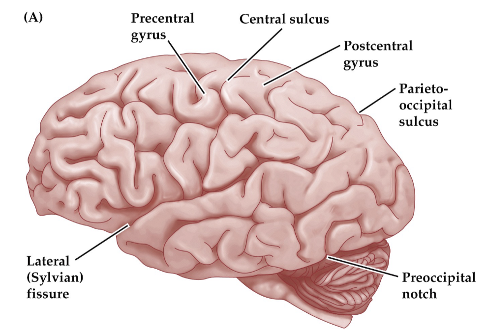
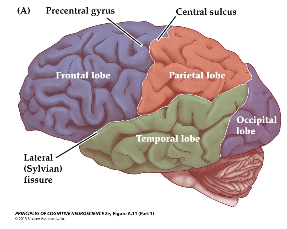
pre-central gyrus
located in front of central sulcus, containing primary motor cortex and controlling voluntary movement

central sulcus
divides the frontal and parietal lobe, separating Precentral gyrus (motor cortex) and Postcentral gyrus (somatosensory cortex)

post-central gyrus
Located posterior (behind) the central sulcus
Function: Primary Somatosensory Cortex
Processes touch, pressure, temperature, pain

parieto-occipital sulcus
Divides the parietal lobe from the occipital lobe
Visible mostly on the medial (inner) surface of the brain

lateral sulcus (sylvain fissure)
Divides: the temporal lobe from the frontal and parietal lobes
One of the deepest and most prominent sulci
calcarine sulcus
Found in the occipital lobe on the medial surface
Contains the primary visual cortex

superior temporal gyrus
Located just below the lateral sulcus
Function: involved in auditory processing and language comprehension (includes Wernicke’s area in the left hemisphere)

longitudinal fissure
Deep groove that separates the left and right hemispheres of the brain

frontal lobe
Voluntary movement (motor cortex)
Planning and decision-making
Problem-solving
Speech production (Broca’s area, usually left hemisphere)
Emotional regulation and personality
Working memory and attention

occipital lobe
Location: Back of the brain
Primary Functions:
Visual processing (primary visual cortex)
Interpreting color, shape, motion
Visual recognition and spatial processing

temporal lobe
Location: Below the lateral sulcus, sides of the brain near the ears
Primary Functions:
Auditory processing (hearing)
Language comprehension (Wernicke’s area, usually left hemisphere)
Memory formation (especially the hippocampus within this lobe)
Processing emotions and facial recognition

parietal lobe
Processing sensory information (touch, temperature, pain)
Spatial awareness and body position (proprioception)
Navigation and coordination
Understanding language and mathematics
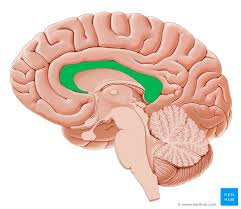
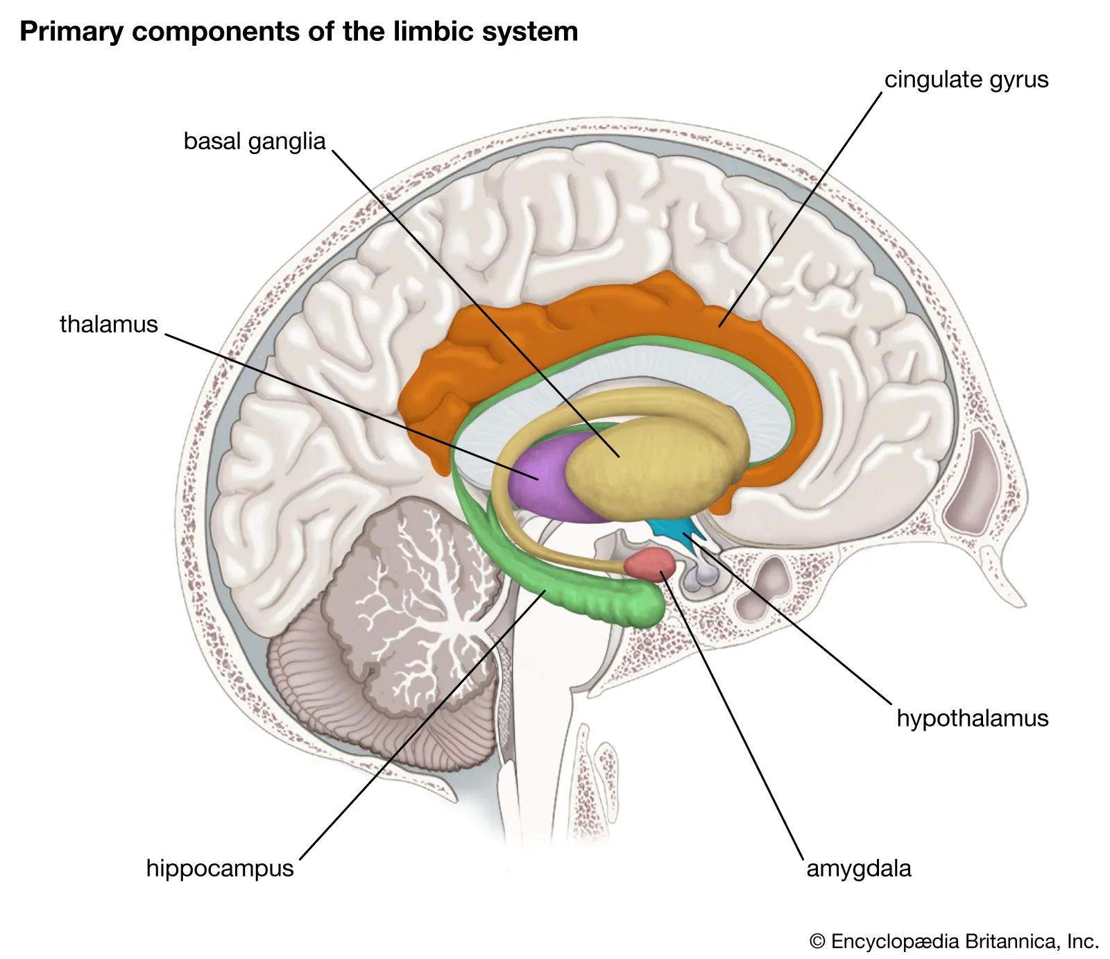
limbic system
Located deep inside the brain, including structures like the hippocampus and amygdala.
Involved in emotion, memory, motivation, and behavior.
corpus callosum
Connects the left and right hemispheres of the brain
Allows communication and information transfer between hemispheres
Enables coordinated, integrated brain function
basal ganglia
Movement regulation: initiation and control of voluntary movements
Involved in habit formation and procedural learning
Plays a role in reward processing and motivation
Dysfunction linked to disorders like Parkinson’s and Huntington’s disease

hippocampus
Crucial for forming new explicit (declarative) memories
Involved in spatial navigation and memory of locations
Plays a role in learning
Damage can cause anterograde amnesia (inability to form new memories)

amygdala
Processes emotions, especially fear and aggression
Plays a role in emotional memory formation (e.g., memories linked to strong feelings)
Involved in threat detection and emotional learning

thalamus
Acts as the brain’s relay station for sensory information (except smell)
Processes and transmits sensory and motor signals to the cerebral cortex
Involved in regulating consciousness, sleep, and alertness

hypothalamus
Regulates homeostasis (body temperature, hunger, thirst)
Controls the endocrine system via the pituitary gland
Involved in sleep-wake cycles, emotions, and sexual behavior
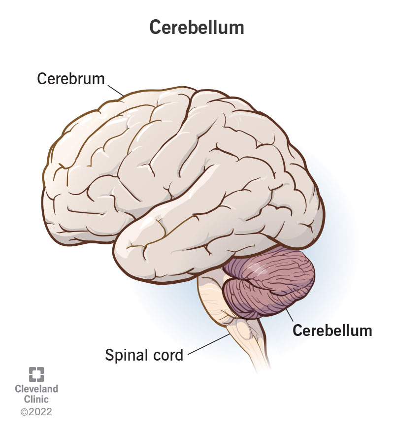
cerebellum
Coordinates balance, posture, and fine motor control
Involved in motor learning and timing of movements
Also linked to some cognitive functions like attention and language processing
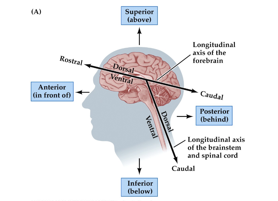
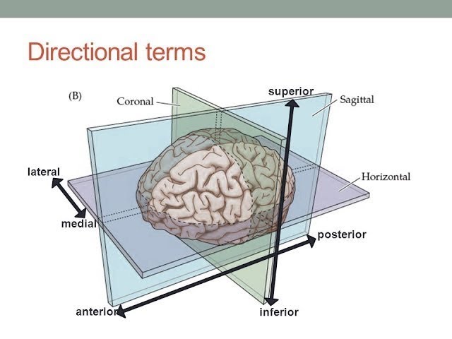
Describe the relationship between brain regions (e.g. Sylvian Fissure and Central Sulcus)
in anatomical terms
Anterior (rostral): toward the front
Posterior (caudal): toward the back
Superior (dorsal): toward the top
Inferior (ventral): toward the bottom
Medial: toward the midline
Lateral: away from the midline (toward the sides)
What did phrenology contribute to cognitive neuroscience?
Localization of Function | Phrenology introduced the idea that brain areas are associated with |
Modern Approaches:
– Try to localize brain areas associated with
cognitive processes that contribute to behavior
– Need cognitive theories that specify the
processes that underlie behaviors of interest
– Interpretation of functional imaging results is often
only as good as the theories that guided the
research
lesion
Observe or test how injuring a brain
area influences behavior then infer the
function of that areaonly on human victims of injury so it lacks anatomical precision
on animals lesions can be induced and anatomically precise
Poor spatial and temporal resolution
Disadvantages:
Heterogeneity:
– Lesion site, symptoms, subject
characteristics
• Compensatory strategies and brain
reorganization
• Downstream effects
pharmacological
Observe or test how pharmacological agents
influence behavior then infer the function of
that neurotransmitter system
can be observed in humans with chronic drug use/experimental administration/not precise
drugs for animals can be administered to specific brain region/precise
Pharmacological agents can alter
neurotransmitter functioning
Agonist: Bind to and activate receptors
Antagonists: Bind to and inactivate receptors
Poor spatial and temporal resolution
stimulation
Stimulate a brain area to alter behavior
then infer the function of that area
Positive response: Stimulation causes
behavior (e.g. stimulating primary motor cortex invokes movement)/Negative response: Stimulation inhibits
behavior
(humans) electrodes are places on the cortex before epilepsy surgery or induced from outside of the head to identify limited regions responsible for function/(animals) electrodes are placed onto cortex and used to stimulate or disrupt function
led to creation of homunculus (somatosensory/motor cortex layout)
(TMS causes electrical
current in underlying brain tissue
• Repetitive TMS can improve or impair
performance on a task
– Weak current: Positive response
– Strong current: Negative response)
Poor spatial resolution
Only affects superficial brain regions
Risk for seizure (rare)
optogenetics
Inserting light-sensitive proteins into neurons (via genes), then using light to activate or silence specific cells with high precision.
Genes for photosensitive ion channels
from algea Implanted into neurons via virus. Laser light then activates or inactivates
neurons
Invasive; not currently possible in humans.
Requires genetic modification and implanting optical fibers.
Mainly used in animal research, so translation to humans is complex.
single-cell recording
Measures correlation between stimulus/response events and electrical activity in neurons
• Can tell us about how neurons in
different brain regions process
information
• Good spatial and temporal resolution
(humans) electrodes placed on cortex pre epilepsy surgery (animals) intra(recording ap in cell)/extracellular(records electrical activity related to AP outside the cell)
Disadvantages:
Limited regions recorded in humans
Invasive and therefore mostly done in
animalsLimited to large neurons and not an
entire network
EEG
The brain’s electrical activity generates electrical field “potentials” measured on the scalp’s surface.
• EEG measures changes in electrical potentials over time through electrical activity on the scalp (best for recording neurons oriented
perpendicular to the scalp)
Poor spatial resolution — hard to tell where activity is coming from.
Only detects activity near the surface of the brain (cortex).
Prone to noise (e.g. muscle movement, blinking).
MEG
measures orthogonal magnetic activity
on the scalp (best for recording neurons oriented parallel to the scalp (in sulci))
Very expensive and rare.
Still limited in detecting deep brain activity.
Requires magnetically shielded rooms.
poor spatial resolution(sulcal activity)
PET
Measures blood flow using
radio-labeled compounds
Person given radioactive
tracer in bloodBlood travels to active
brain regionTracer decays sending
off a signal that is
detected by the machine
Disadvantages:
Do not directly measure neural activity
– Poor temporal resolution
– Safety issues
– Expensive
fMRI
Measures changes in oxygenated
blood because neural activity causes increase in blood flow
Increased neural
activity causes an
increased demand for
oxygenMagnetic properties
of oxygenated blood
is detected by the
machine
Disadvantages:
Do not directly measure neural activity
– Poor temporal resolution
– Expensive
optical imaging
Brain tissue transmits or reflects light
differently when active
Humans:
Based on underlying electrical activity
– Extracranial: Event-related optical signals
(EROS) reflection measured from outside the head
– Good temporal but poor spatial resolution
Animals:
Based on level of oxygenated vs.
deoxygenated blood
– Intracranial: Reflection measured directly from the cortical surface
– Good spatial but poor temporal resolution
Disadvantages:
– Does not directly measure neural activity
– Poor temporal resolution
– Invasive and therefore mostly done in
animals
– Poor signal to noise resolution
– Only records superficial brain regions
Define single and double dissociations? What inferences/interpretations can be better
made from double dissociations as compared to single dissociations?
Single dissociation:
– Lesion to one area of the brain and a
problem in one function while not affecting
other functions
Function A is impaired, but Function B is intact in one patient or condition.
The difference in performance could just mean Task A is harder than Task B, not that they're neurologically separate.
suggestive, but not definitive.
Double dissociation:
– Lesions in different brain areas causing
problems with different functions
Can tell us that a specific brain area is
important for a specific function
stronger evidence for functional independence
Strongly supports modularity — different parts of the brain doing different jobs
How is transcranial magnetic stimulation (TMS) like the lesion approach in some
respects but not others?
TMS is like a “virtual lesion” — it allows researchers to temporarily and safely disrupt brain function in a controlled way.
Lesion studies observe long-term consequences of real brain damage, often in clinical populations.
Therefore:
TMS is experimental, precise, and reversible.
Lesions are observational, often messy, but reflect real-world dysfunction.
Explain the relationship between neuronal activity and scalp recorded EEG
EEG measures electrical activity generated by large populations of neurons.
Specifically, it detects summed postsynaptic potentials (not action potentials) from pyramidal neurons in the cerebral cortex.
These neurons are aligned perpendicularly to the scalp, and when many fire synchronously, they generate a dipole (a separation of charge) strong enough to be picked up at the scalp.
What are the determinants of whether EEG recorded at the scalp leads to a positive vs.
negative voltage change?
Determined by the orientation of the dipole and the location of the electrode relative to the source.
In EEG, positive or negative doesn't directly map onto neural excitation or inhibition — it depends on:
Whether current is flowing toward or away from the scalp surface.
The geometry of the cortical fold (e.g., whether the source is in a sulcus or gyrus).
So, voltage polarity reflects direction of current flow, not necessarily the type of neural activity.
What is the relationship between EEG and ERP?
ERP (Event-Related Potential) is a special type of EEG analysis.
ERP = the brain's electrical response to a specific stimulus or event, extracted by averaging EEG signals across many trials.
EEG = raw, continuous brain activity.
ERP = time-locked, averaged response to specific events within the EEG.
What are the two primary pieces of information used to label ERP components?
Polarity
P = positive deflection
N = negative deflection
Latency (Timing)
Given in milliseconds (ms) after the stimulus OR as an order number.
Examples:
P300: positive peak at ~300 ms
N170: negative peak at ~170 ms
N1, P2: indicate first, second components
How are the magnetic fields measured by MEG related to the electrical dipoles measured
by EEG?
MEG and EEG both detect neural activity from electrical dipoles generated by cortical neurons.
EEG measures the voltage (electric field) created by the dipoles.
MEG measures the magnetic fields that are perpendicular to those electrical currents (via the right-hand rule).
MEG is most sensitive to tangential dipoles (in sulci), while EEG detects both radial and tangential sources.
27) How is brain activity measured with PET?
PET measures metabolic activity using a radioactive tracer (usually attached to glucose or a neurotransmitter analog).
After injection, the tracer accumulates in active brain regions.
As the tracer decays, it emits positrons, which collide with electrons, producing gamma rays.
A PET scanner detects these gamma rays and maps regions of high metabolism (indicating higher activity).
28) How is brain activity measured with fMRI?
fMRI tracks brain activity using the BOLD signal (Blood Oxygenation Level Dependent).
Active brain areas consume more oxygen; in response, the body overcompensates, increasing oxygen-rich blood to that region.
Oxygenated and deoxygenated blood have different magnetic properties, which affect the MR signal.
fMRI indirectly measures activity by detecting these blood flow changes.
Explain the basics of MRI physics including static field, gradient field, pulse sequence, and receiver coil
Component | Description |
|---|
Static Magnetic Field (B₀) | A strong, constant magnetic field aligns hydrogen protons (usually 1.5T or 3T strength). |
Gradient Fields | Small magnetic fields applied in different directions to localize signals in 3D space. Used to encode position of signals (x, y, z). |
Pulse Sequence (RF Pulse) | A radiofrequency pulse knocks hydrogen protons out of alignment. As they relax back, they emit energy. Different tissues relax at different rates. |
Receiver Coil | Detects the energy released as protons return to alignment. This signal is used to reconstruct the image |
What is the physical change within the brain that is detected by Diffusion Tensor Imaging
(DTI), and what structural characteristic of the brain is DTI used to estimate?
DTI measures the diffusion of water molecules in brain tissue.
In white matter, water diffuses along axon fibers, not across them.
DTI uses this anisotropic diffusion to estimate:
White matter tract integrity
Direction and pathways of fiber tracts (called tractography)
It helps map structural connectivity between brain regions.
What are some strengths and weaknesses of EEG, MEG, PET, and fMRI in terms of their
spatial resolution, temporal resolution, and suitability for and potential risk to normal
human subjects?
Method | Spatial Resolution | Temporal Resolution | Risk/Suitability |
|---|
EEG | Low (cm range) | High (ms) | Safe, non-invasive, suitable for children |
MEG | Medium–High (mm-cm) | High (ms) | Safe, but expensive and sensitive to magnetic interference |
PET | Low (cm) | Very Low (minutes) | Invasive (radioactive tracer), not ideal for healthy participants or children |
fMRI | High (mm) | Low (~2–6 sec) | Non-invasive, safe (no radiation), widely used in humans |
Consider an fMRI or PET study showing that activity in some brain region, X, is
associated with some behavior. Can we conclude that region X necessarily controls that
behavior? Why or why not?
No, we cannot conclude causation from fMRI or PET alone.
fMRI/PET are correlational — they show association, not control.
Region X might be involved, but not essential.
Activity could be due to:
Upstream or downstream processing
Co-activation with other regions
General cognitive effort, not task-specific processes
(To establish causation, you need intervention-based methods like lesion studies, TMS, optogenetics (in animals))
What is meta-analysis and what are the advantages over any single study?
A statistical method that combines results from multiple independent studies on the same topic.
Looks for consistent patterns across studies, increasing reliability.
Advantages:
Increased statistical power (more data = stronger conclusions)
Identifies robust effects across samples, tasks, or populations
Reduces publication bias or chance findings from individual studies
Helps detect moderators (e.g., age, method differences)
Define, compare, and contrast sensation and perception.
Term | Definition | Key Distinction |
|---|
Sensation | The raw input from sensory organs (e.g., light hitting the retina) | It’s about detecting stimuli |
Perception | The interpretation of sensory information (e.g., recognizing a face) | It’s about making meaning from sensation |
Describe the vision pathway from the eye to the brain.
Light enters the eye → focused by the cornea and lens
Hits the retina, activating photoreceptors (rods and cones)
Signal goes to bipolar cells, then ganglion cells
Ganglion cell axons form the optic nerve
At the optic chiasm, some fibers cross hemispheres
Travels via optic tract to the lateral geniculate nucleus (LGN) in the thalamus
Projects to primary visual cortex (V1) via optic radiations
Information continues along dorsal ("where") and ventral ("what") pathways
What is the difference between rods and cones?
Feature | Rods | Cones |
|---|
Light Sensitivity | Very sensitive (low light) | Less sensitive (need bright light) |
Color | No color detection | Detect color (RGB) |
Location | Periphery of retina | Concentrated in fovea |
Visual Role | Night vision, motion | Daylight, detail, color |
37) What is the difference between the magnocellular and parvocellular systems?
System | Magnocellular ("M") | Parvocellular ("P") |
|---|
Input | Rods | Cones |
Sensitivity | Motion, flicker, luminance | Color, fine detail |
Speed | Fast | Slow |
Pathway | Dorsal stream (Where/How) | Ventral stream (What) |
38) Explain the principle of retinotopic (or topographic) mapping in the visual cortex
Retinotopic mapping means that adjacent areas in the visual field are processed by adjacent neurons in V1.
Preserves the spatial layout of the visual scene.
Like a "map" of the retina on the brain's surface.
Explain the principle of cortical magnification as it applies to differences in the V1
representation of foveal vs. peripheral stimuli.
Fovea (central vision) gets disproportionately large representation in V1.
This is called cortical magnification — small central area takes up much more cortex than larger peripheral areas.
Explains why foveal vision is so detailed.
Describe how neurons in different V1 hyper columns vary in the information to which
they are responsive. What are neurons in the blobs sensitive to?
V1 hypercolumns contain:
Orientation columns: respond to specific edge orientations
Ocular dominance columns: prefer input from one eye
Blobs: sensitive to color and brightness (parvocellular input)
Each hypercolumn represents a full set of features for one small area of the visual field.
What is a receptive field?
A receptive field is the specific area of the visual field where a stimulus must appear to affect the firing of a neuron.
Receptive fields are small in early areas (like V1) and get larger in higher areas (like IT).
What is the difference between the dorsal and ventral visual pathways?
Pathway | Dorsal ("Where/How") | Ventral ("What") |
|---|
Function | Spatial location, motion, guiding action | Object recognition, identity |
Brain Areas | Parietal lobe | Temporal lobe |
Input | Magnocellular | Parvo + magno |
What are the properties of visual perception and how do they correlate with physical
reality?
Brightness | Intensity of light |
Color | Wavelength of light |
Shape | Edges, contours |
Depth | Binocular disparity, motion, cues |
Motion | Changes in position over time |
How do visual areas V1, MT, and IT differ in the types of stimuli to which they are most
sensitive?
V1 (Primary Visual Cortex) | Edges, orientation, basic features |
MT/V5 (Middle Temporal) | Motion, direction, speed |
IT (Inferotemporal Cortex) | Complex objects, faces, shapes, categories |
45) Where in the brain is category specific information stored?
Stored in high-level visual cortex, particularly ventral temporal lobe:
Fusiform face area (FFA): faces
Parahippocampal place area (PPA): places/scenes
Extrastriate body area (EBA): body parts
These areas show category-selective activation.
46) Describe the fMRI and ERP results related to face perception and perceptual expertise.
fMRI | Strong FFA activation for faces, and sometimes objects of expertise (e.g. birds for bird experts) |
ERP | N170 component larger for faces than objects; appears ~170 ms post-stimulus |
47) Compare and contrast the patterns of spared and impaired abilities shown in patients with apperceptive agnosia, associative agnosia, and prosopagnosia
Apperceptive agnosia | Cannot form a stable percept; poor copying or matching | Basic vision, light/dark, acuity |
Associative agnosia | Can perceive objects, but can’t identify them | Can copy images, describe shapes |
Prosopagnosia | Can’t recognize faces | Can recognize objects, sometimes voice or gait |
48) Describe the single-cell recording results related to object recognition.
In monkeys, neurons in inferotemporal cortex (IT) respond selectively to:
Complex shapes, faces, or specific objects
Some neurons even show invariance (e.g., respond to a face despite size, angle, position)
Shows hierarchical processing:
Early areas (V1): simple features
Higher areas (IT): complex, meaningful objects