A&P Bio-111 LAB practicle #1
 Call Kai
 Learn
Learn Practice Test
 Spaced Repetition
 Match
Match Flashcards
 Knowt Play
1/89

Earn XP
Description and Tags
By Angela22303 Angela B. Brookdale Commuity College Originally on Quizlet
Name | Mastery | Learn | Test | Matching | Spaced | Call with Kai | Chat |
|---|
No analytics yet
Send a link to your students to track their progress
90 Terms
Anatomical Position
erect, head level, eyes forward, feet flat on the floor, arms at sides, palms facing forward
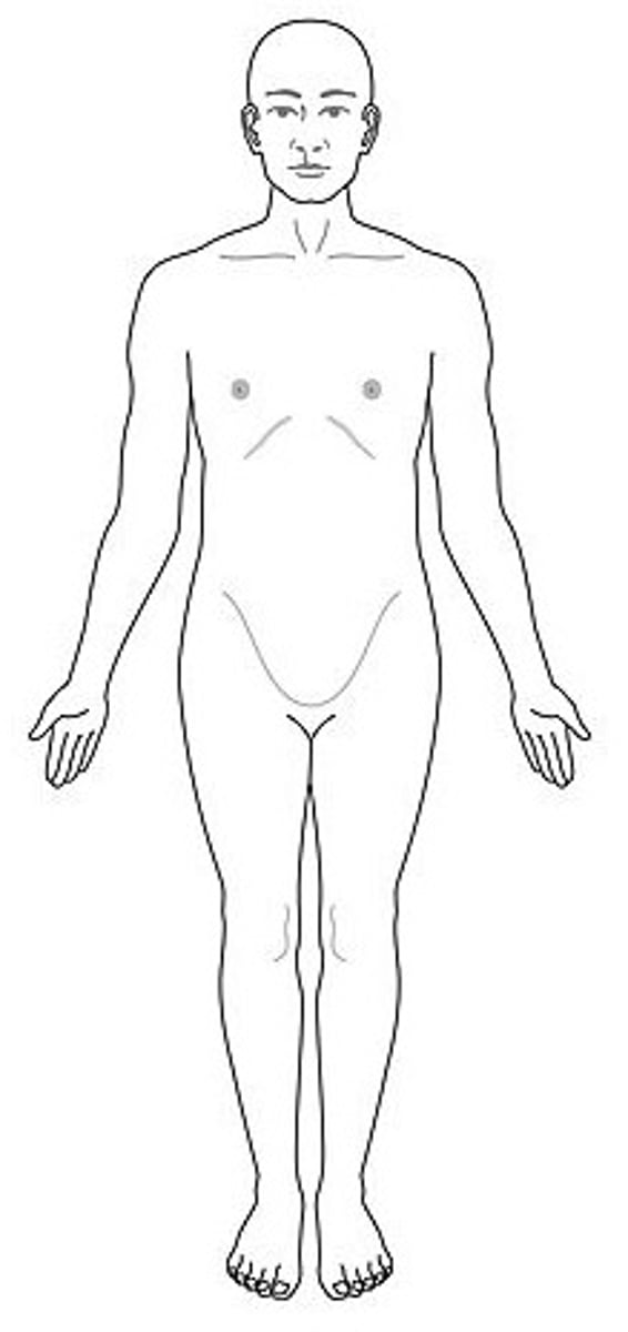
Superior
above
Inferior
below
Anterior (ventral)
front
Posterior (dorsal)
back
Medial
near the midline
Lateral
away from the midline
Proximal
closer to the point of attachment
Distal
Father from the point of attachment
Superfical
towards the surface
Deep
Away from the body surface; more internal
Ipsilateral
on the same side
Contralateral
on the opposite side
Sagittal plane
divides body into left and right
Mid-Sagittal plane
divides the body directly down the middle
Parasagittal plane
divides body into unequal parts
Frontal plane (Coronal plane)
divides the body into anterior and posterior
Transvers palne
horizontal; divdes the body into superior and inferior portions
Oblique plane
passes through the body at an angle
Craninal Cavity
encases the brain
vertebral (spinal) cavity
runs within the bony vertebral column, encloses the spinal cord
Thoracic cavity
formed between the ribs, muscles of the chest, the breastbone and chest portion of the spine; houses the heart and lungs
What separates the thoracic cavity from the abdominal cavity?
the diaphragm
Pericardial Cavity
surrounds the heart
Pleural Cavity
surrounds each lung
Mediastinum
thoracic - lungs
Abdominopelvic cavity
exdtends from the diaphragm to the groin and is encircled by the abdominal wall of the pelvis
What does the abdominal cavity hold
most of the large intestine, small intestine, stomach, liver, spleen, and the gallbladder
What does the pelvic cavity hold
part of the large intestine, urinary bladder, reproductive organs
Retroperitoneal
organs that are external to the lining of the abdominopelvic cavity
What organs are considered to be Retroperitoneal
kidneys and pancreas
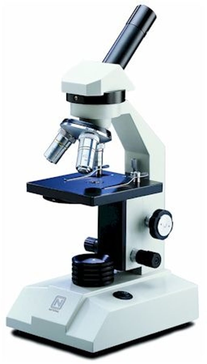
Microscope
magnifies objects
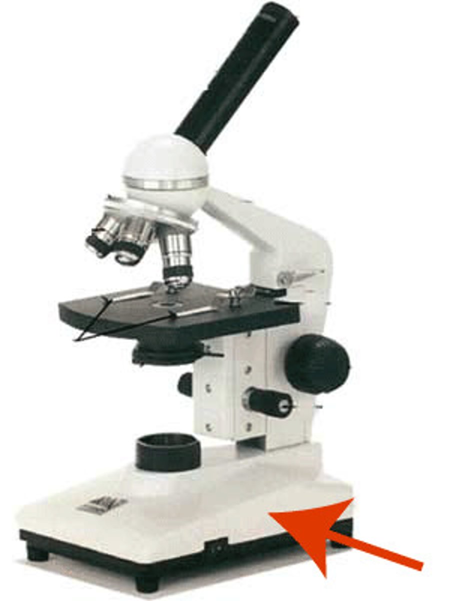
Base
supports instrument
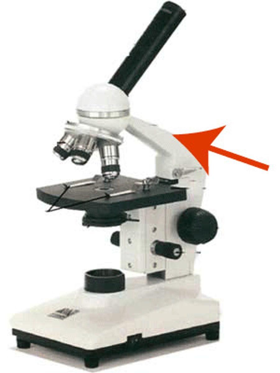
Arm
connects the base to the body
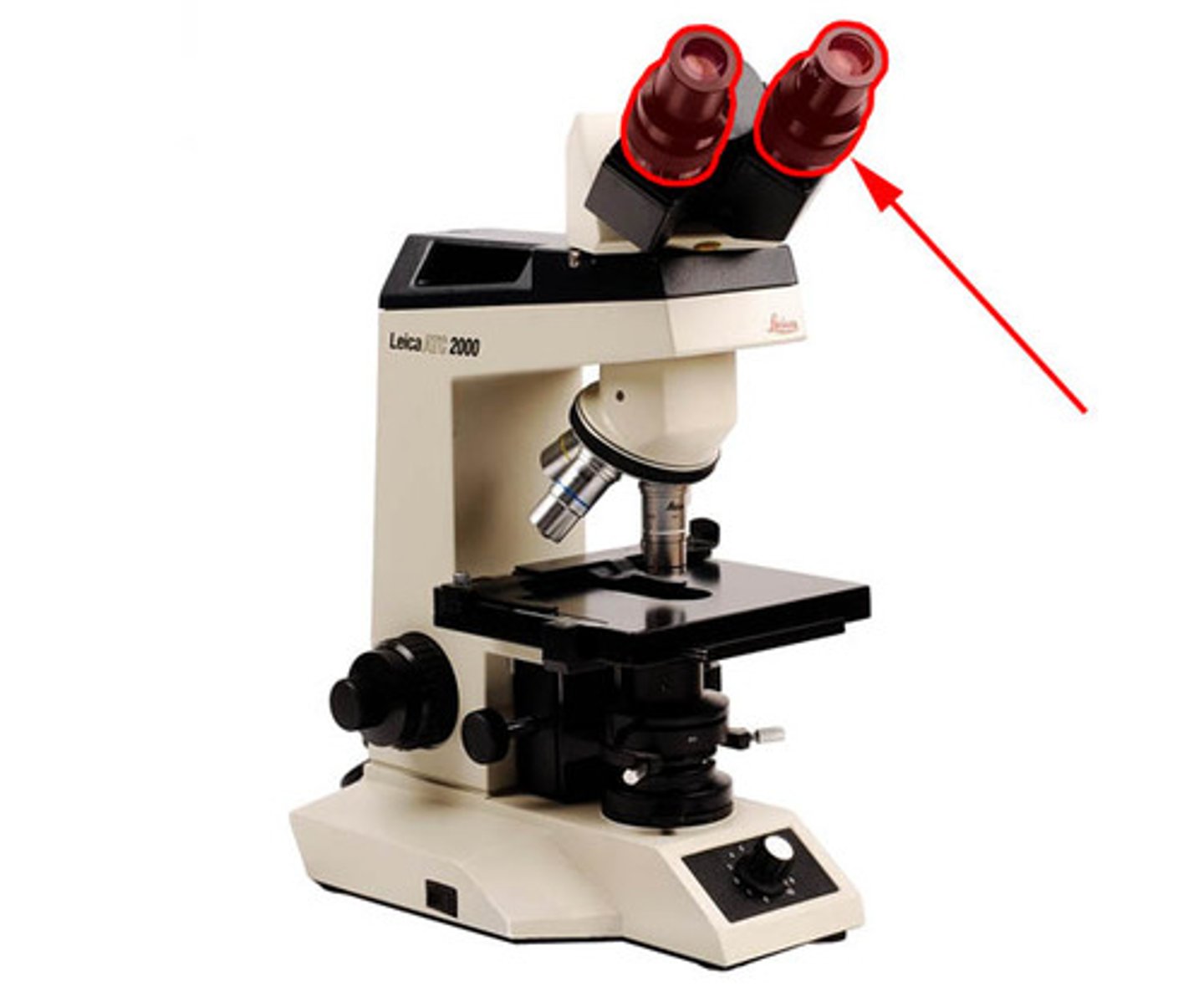
Oculars (eyepieces)
series of lenses to look through to view the specimen
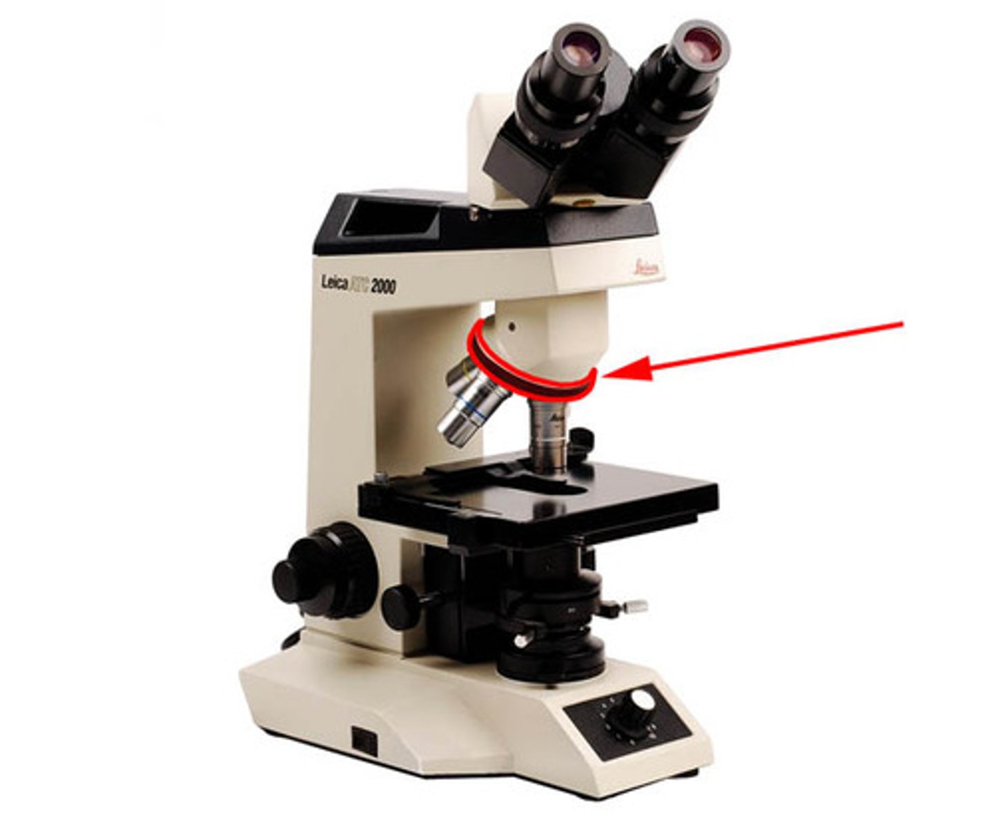
Revoling Nosepiece
rotates to change objective lenses
Objective Lenses
scan the specimen
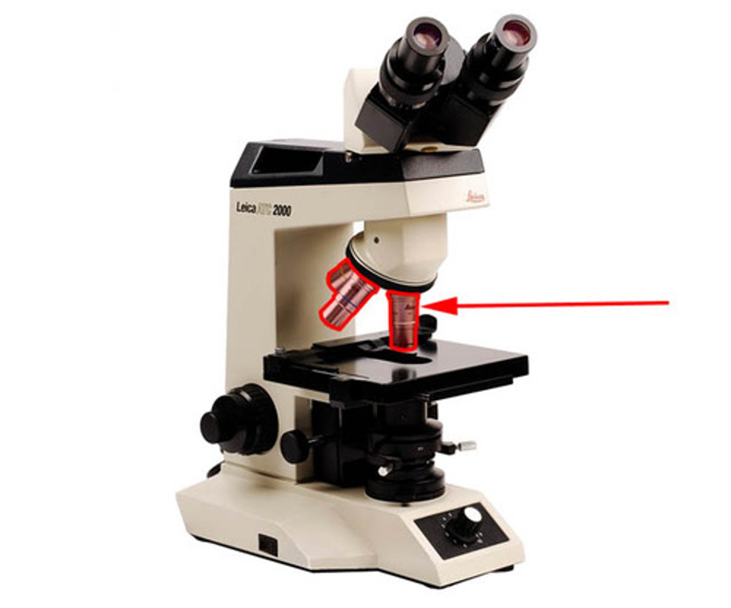
Scanning Power Objective
4X lens (red)
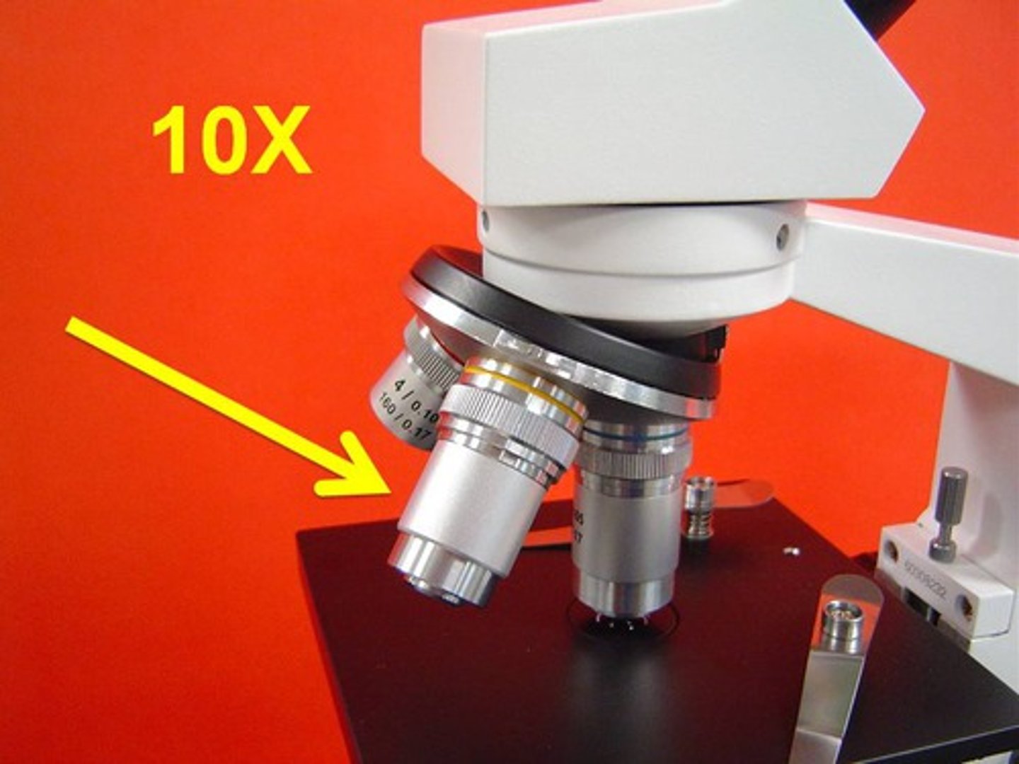
Low Power Objective
10X lens (yelllow)
High (dry) power objective
magnifies 40x (blue)
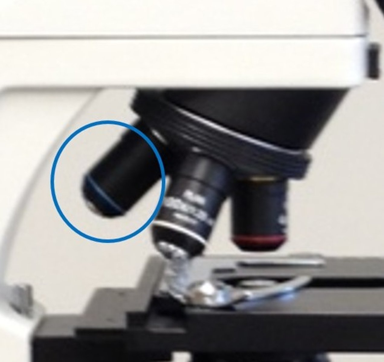
Stage
holds and supports the slide/ specimen
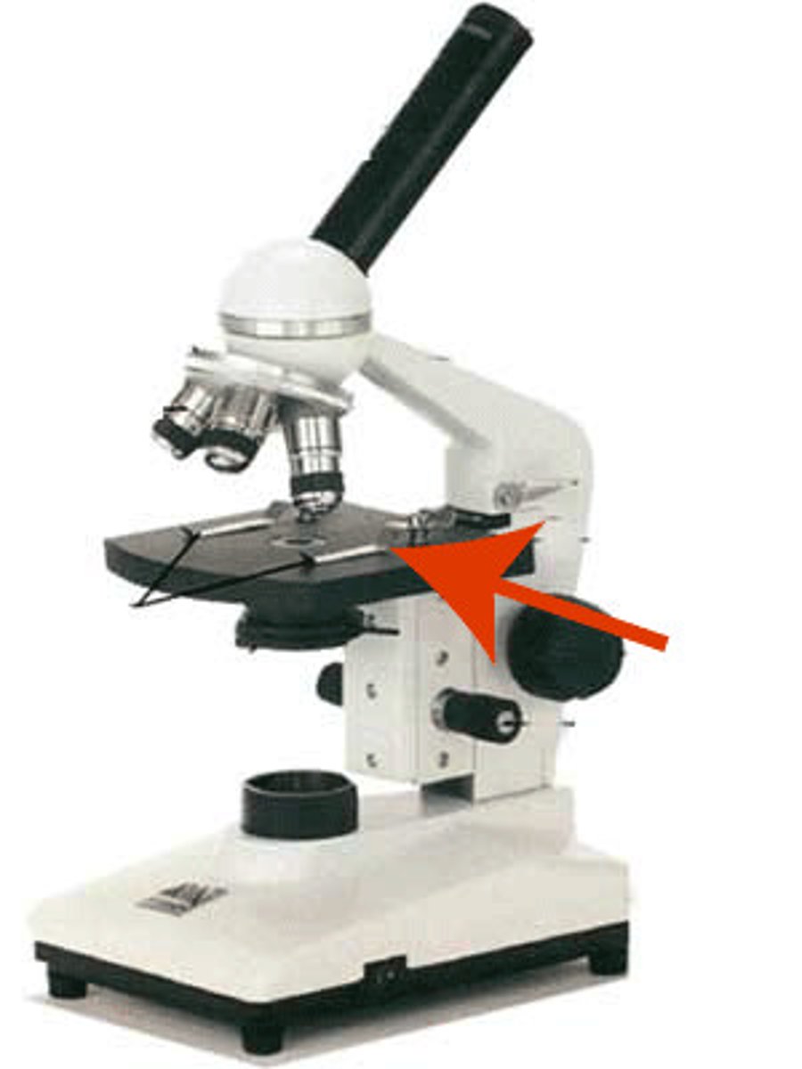
Diaphragm
Regulates the amount of light on the specimen
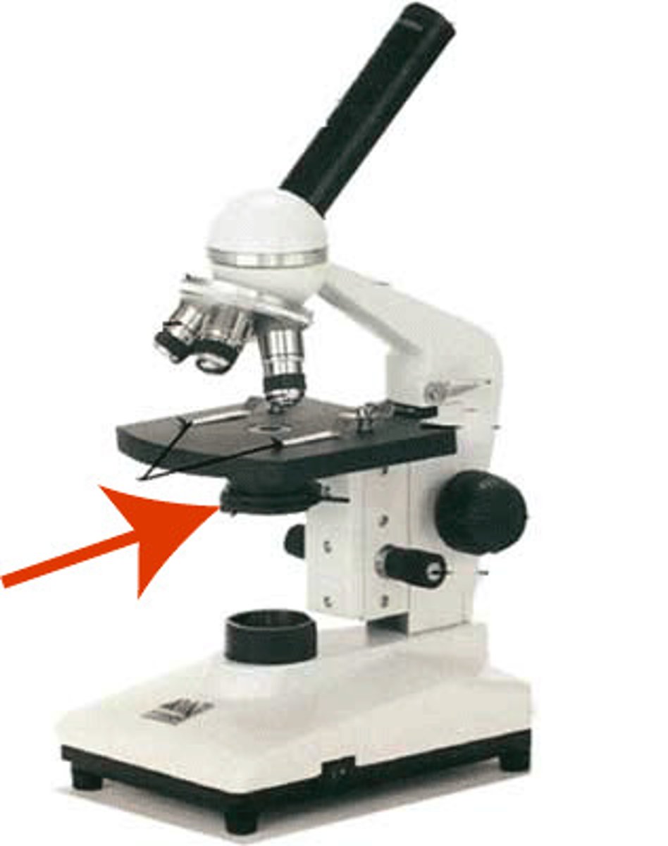
Light Source (illuminator)
contains light
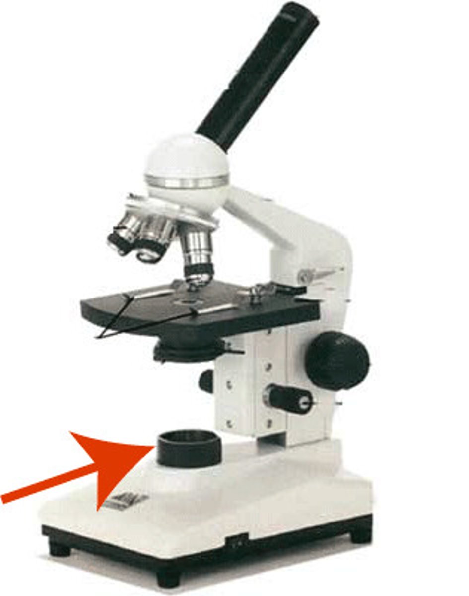
Slide Position Adjustment Knob
adjusts the position of the slide on the stage
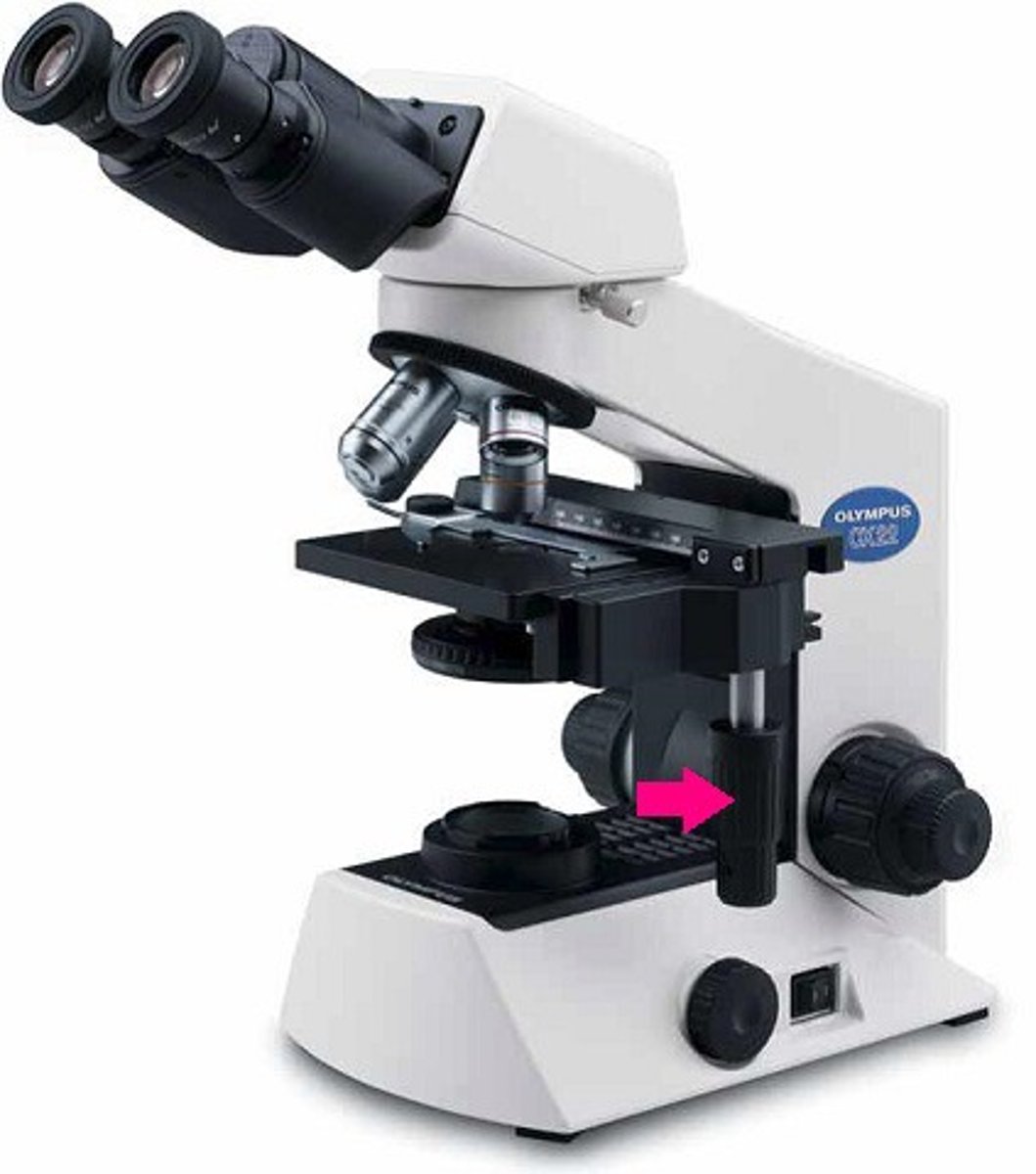
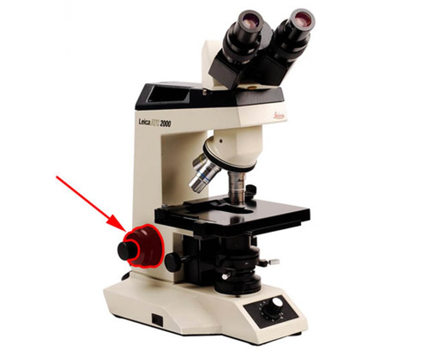
Coarse Adjustment Knob
used for inital focusing
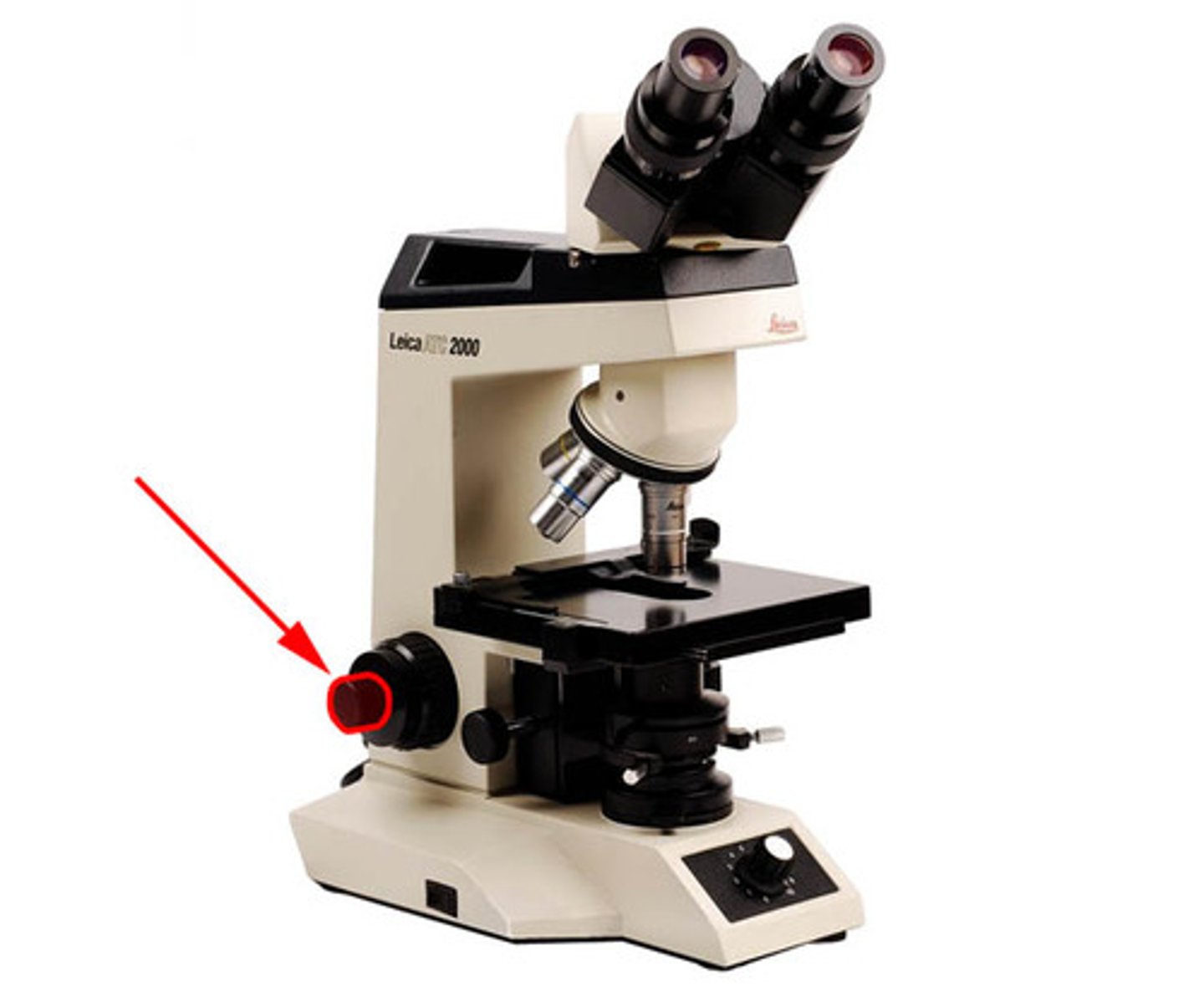
Fine Adjustment Knob
used for final focusing
Cell Theory
The cell is the basic unit of life
All organismis consits of cells
All cells come from pre-existing cells
What 3 componets make up a cell
Plasma membrane, Nucleus, Cystoplasm
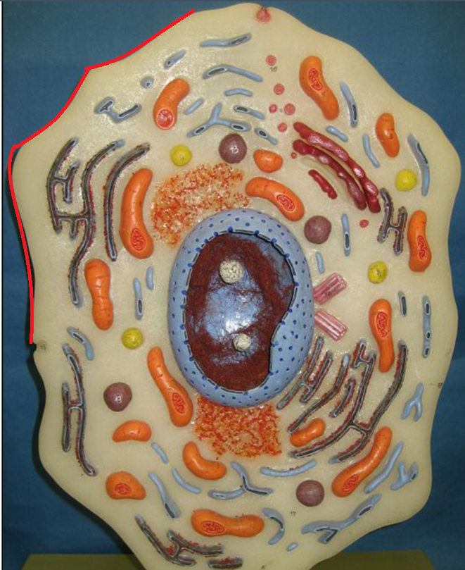
Cell Membrane
outter barrier of the cell; controls what enters & exits the cell
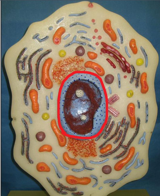
Nucleus
house DNA; controls cell structure & functions
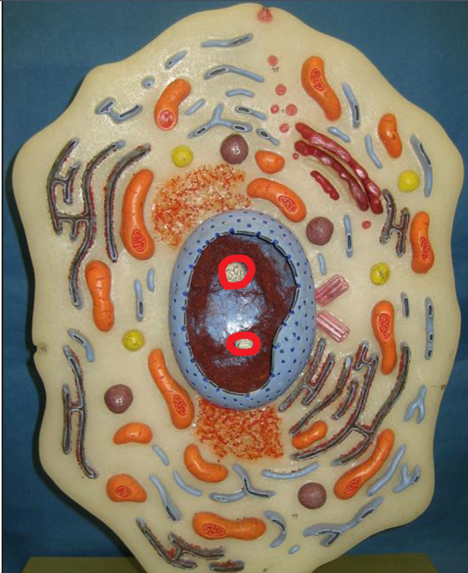
Nucleolus
produces ribosomes
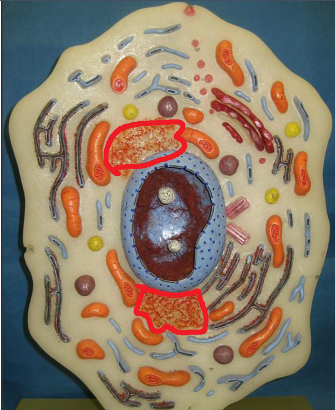
Ribosomes
Makes proteins
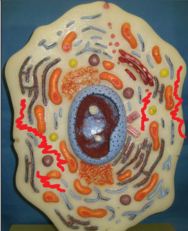
Cytosol
Fluid portion of cytoplasm that surrounds organelles; keeps out bad
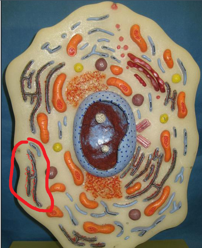
Rough Endoplasmic Reticulum
nettwork of flattened sacs that make glycoproteins & phosphlipds
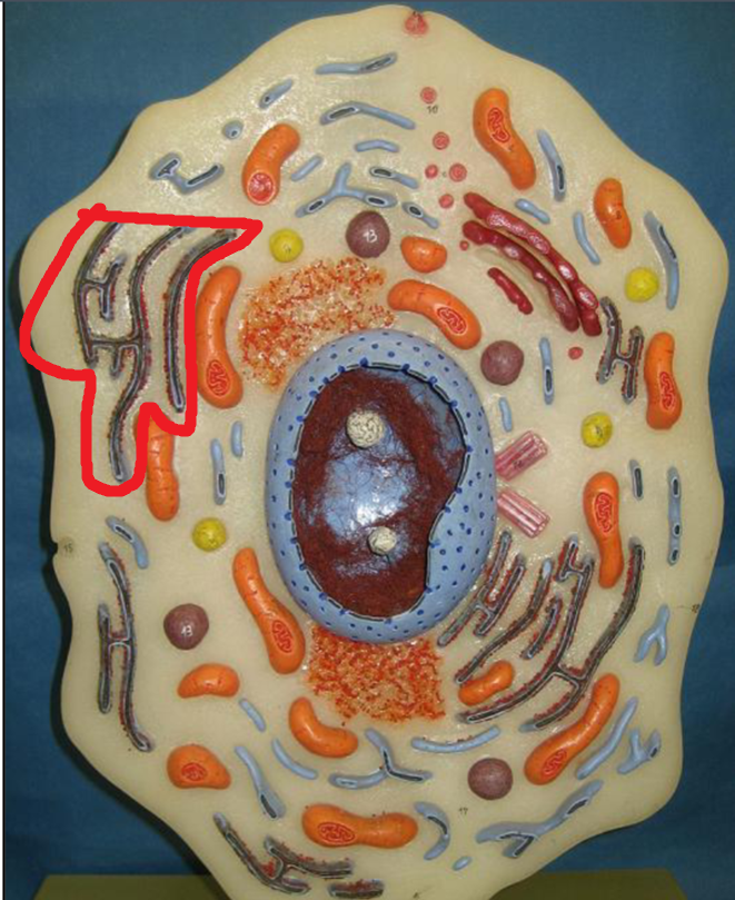
Smooth Endoplasmic Reticulum
network of tubules that make (synthesize) lipds (fatty acids) & steroids
Golgi Apparatus
modifes, sorts, packages, & transports proteins from rough endoplasmic reticulum
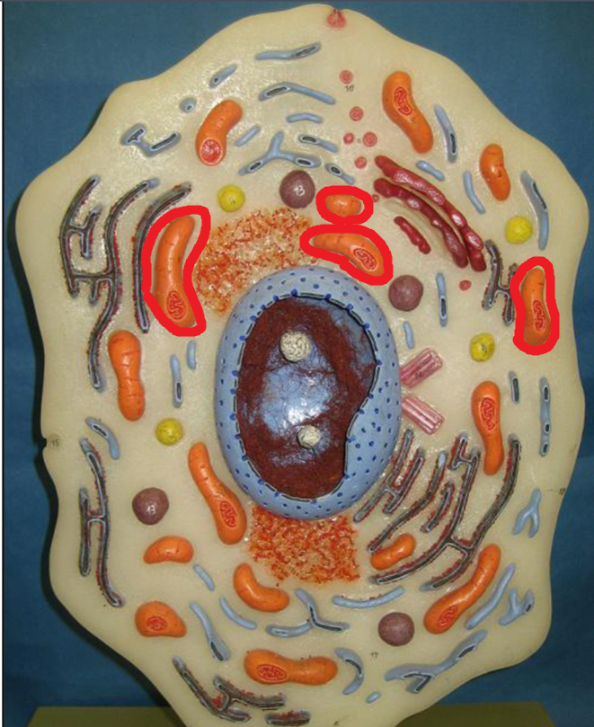
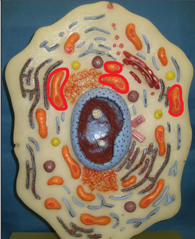
Mitochondria
"Powerhouse" of the cell; generates energy or ATP
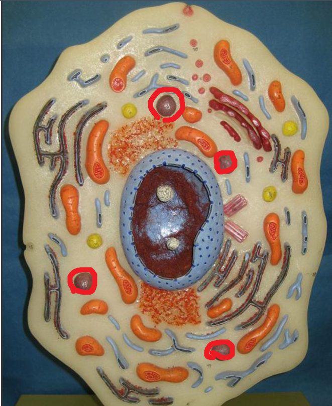
Lysosome
contain digestive enzymes that breakedown molecules
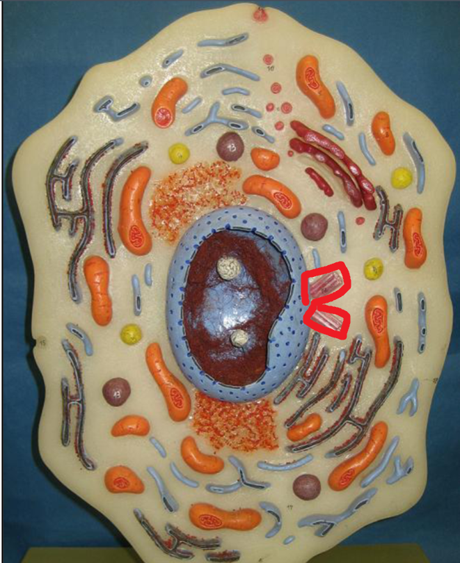
Centrioles
organizing center for growth of mitotic spindles needed for cell division
Cytoskeleton (not seen on model)
network of protein filaments that extend through the cytosol
Histology
study of tissues
What are the 4 types of tissues?
epithelial, connective, muscle, nervous
Epithelial Tissue
covers the surface of the body, lines hollow organs, and acts as glandular tissues
Connective Tissue
act to support and protect other tissues
Muscle Tissue
produces movemnt
Nervous Tissue
communicates with and controls other parts of the body
Characteristics of Epithelial tissues
make excellent barriers
have polarity- basal surface (attached to deeper tissues) & an apical surface (exposed to environment)
Avascular- contain no blood vessels
Innervated- connected to the nervous system
Regenerative- worn out & dead cells continually replaced
How are Epithelial tissues classified?
shape and number of layers
What are the 3 types of layers Epithelial tissues can be classified by?
Simple
Pseudostratified
Stratified
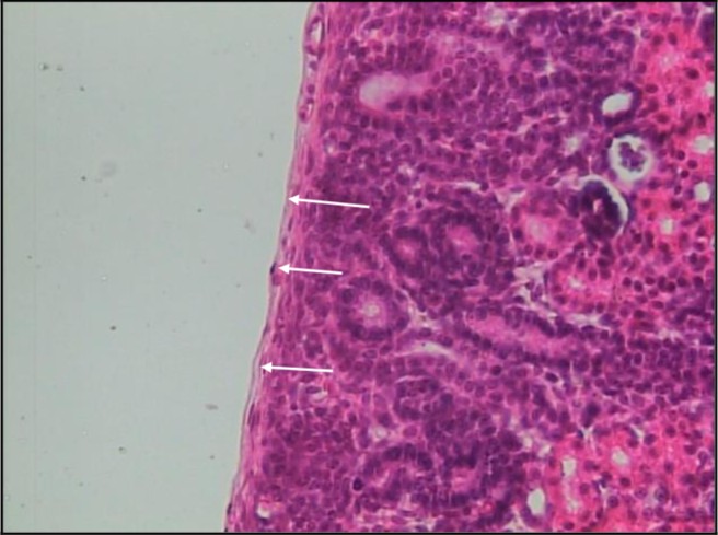
Simple Squamous epithelium
epithelial tissue with 1 layer
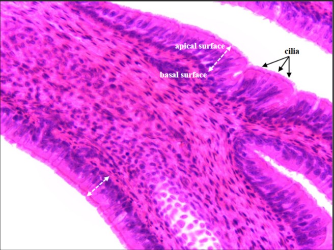
Pseudostratified Columnar epithelium
appears to have multiple layers, but all its cells lie on the basement membrane, making it simple epithelium
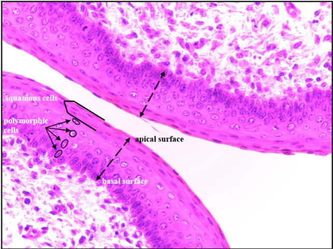
Stratified Squamous epithelium
epithelium: multiple layers
What are the 3 cell shapes of Epithelial tissues
squamous, cuboidal, columnar
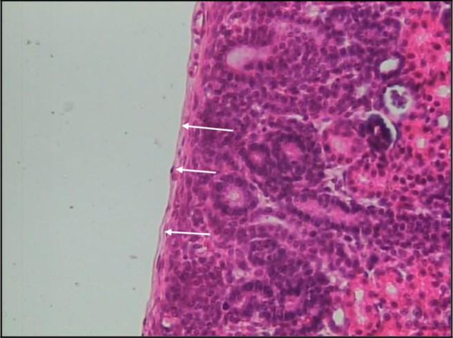
Simple Squamous epithelium
tile like
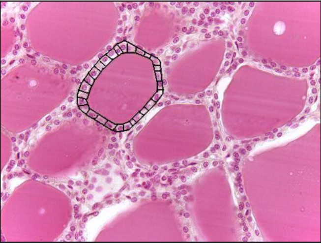
Simple Cuboidal epithelium
cube like
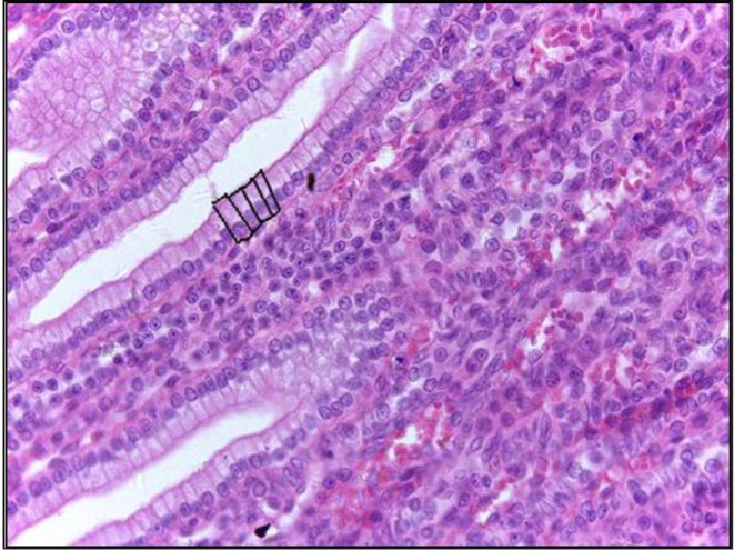
Simple Columnar epithelium
column-like
What is the function (s) of Simple Squamous epithelium
filtration, diffusion, rapid exchanges of materials
Where is the location of Simple Squamous epithelium tissues
air sacs of lungs; kidney filtation; lines of blood vessels; serous membrane
What is the function of Simple Cuboidal tissues
secretion
Where is the location of Simple Cuboidal epithelium tissues
Kidney tubulus & Glands
What is the function of Simple Columnar tissues
Secreation & Absorption
Where is the location of Simple Columnar epithelium tissues
Kidney ducts, lines of the GI tract, lining of spinal cord
What is the function of Pseudostratified Columnar epithelium tissue
secreation; may be ciliated (hair like projection)
Where is the location of Pseudostratified Columnar epithelium tissue
Respiratory Tract
What is the function of Stratified Squsamous epithelium tissue
protection (abrasion, water loss, UV lights) & its the 1st line of defense
Where is the location of Stratified Squsamous epithelium tissue
Keratinized skin; mouth; esophagus
What is the function of Transitional epithelium tissue
streches to accommodate changes in volume
Where is the location of Transitional epithelium tissue
lines portions of the urinary system (bladder)
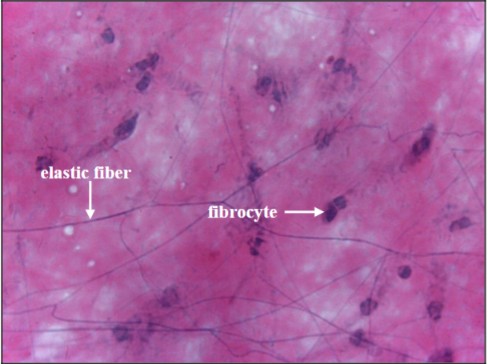
Function & Location of: Areolar (loose) CT
Function: strength, elasticity, & support
Location: skin, mucous membrane, around blood vessels, nerves, organs