KIN 223 EXAM 4 DIAGRAMS
1/27
There's no tags or description
Looks like no tags are added yet.
Name | Mastery | Learn | Test | Matching | Spaced |
|---|
No study sessions yet.
28 Terms
Check all of the statements that are true regarding the changes that occur in the muscular system as a result of aging.

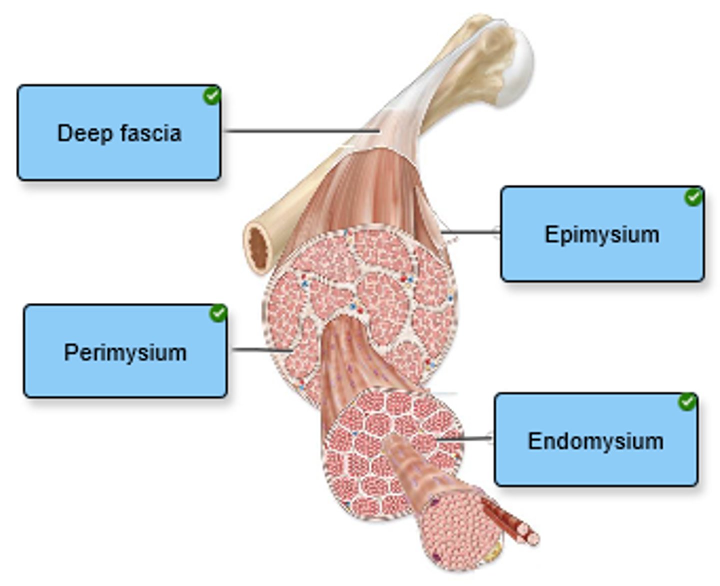
Label the connective tissue in the figure.
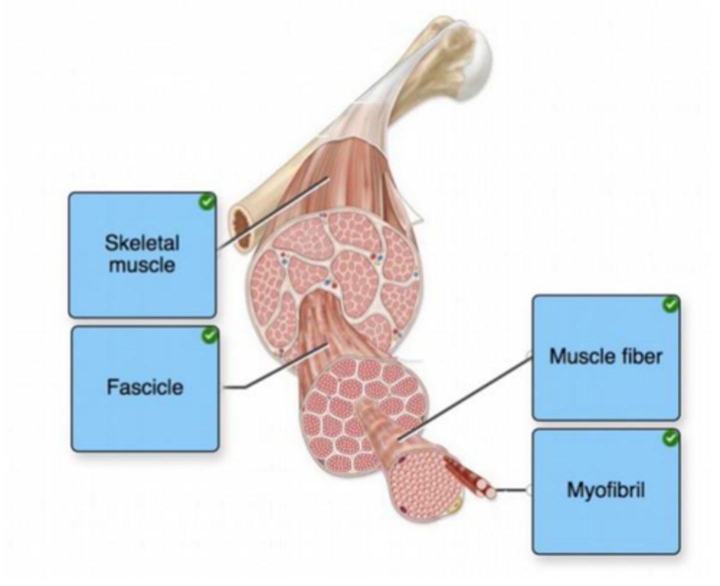
Label the components of skeletal muscle.
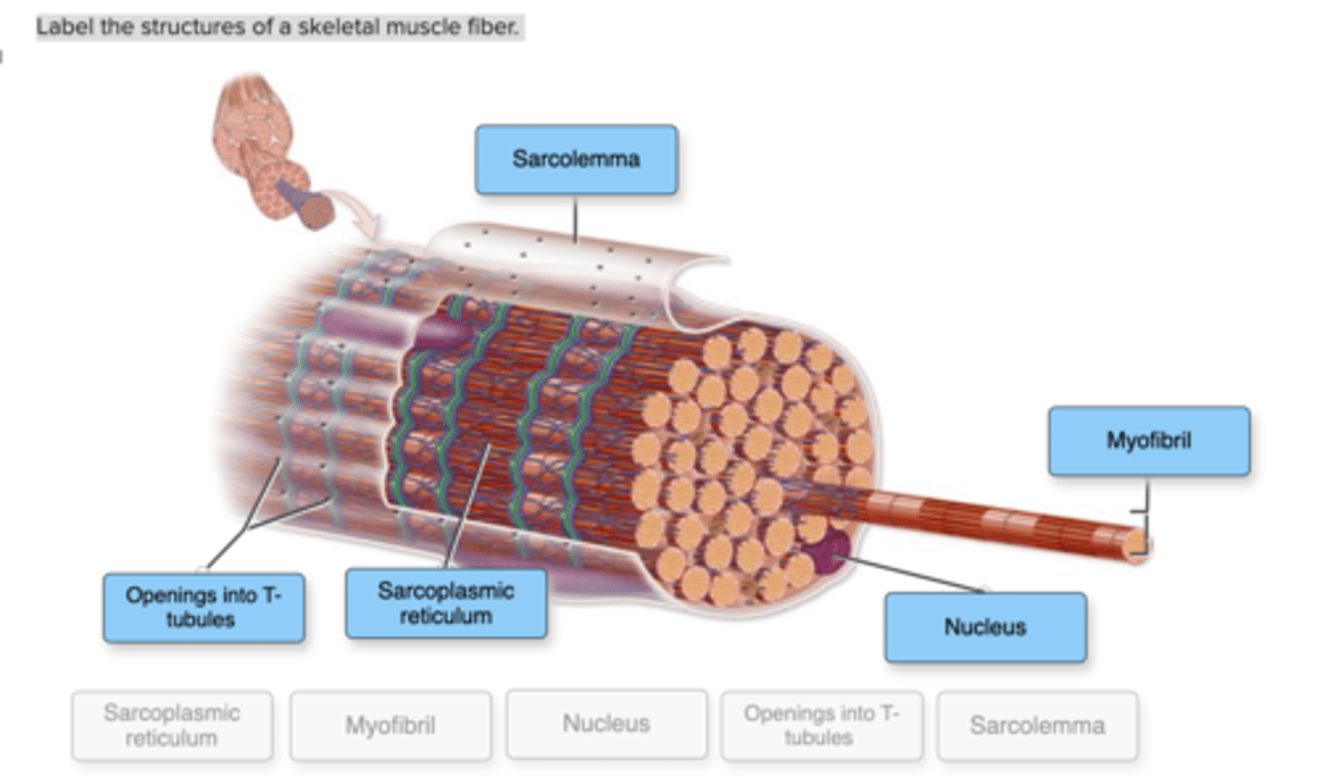
Label the structures of a skeletal muscle fiber.
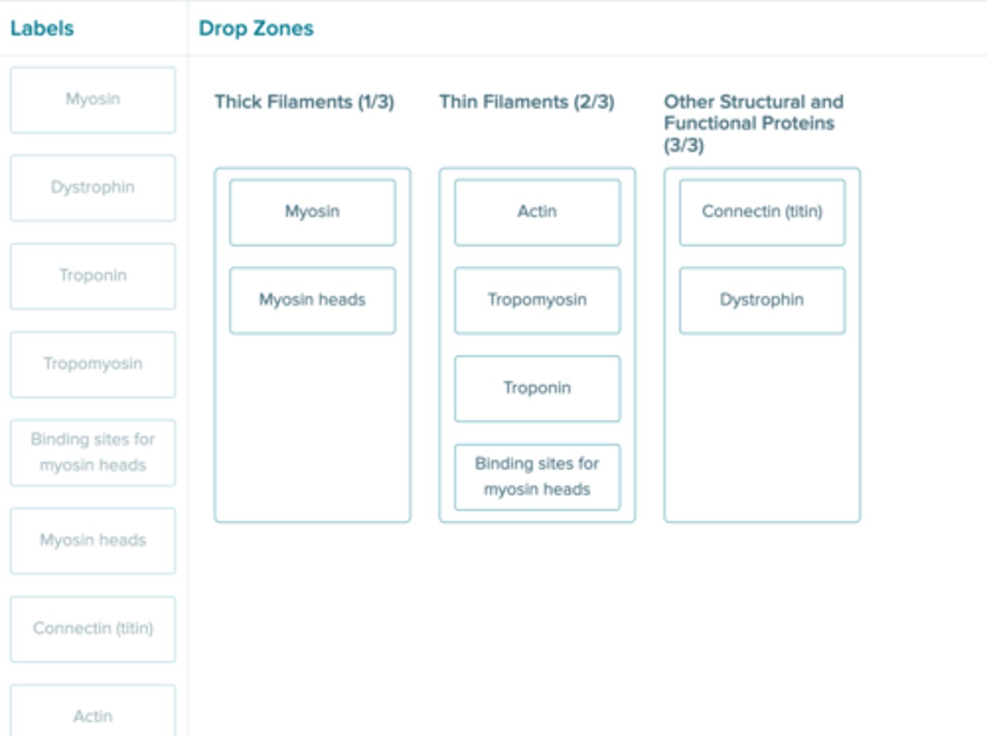
Determine whether each structure is part of a thick filament, part of a thin filament, or is a different structural/functional protein.
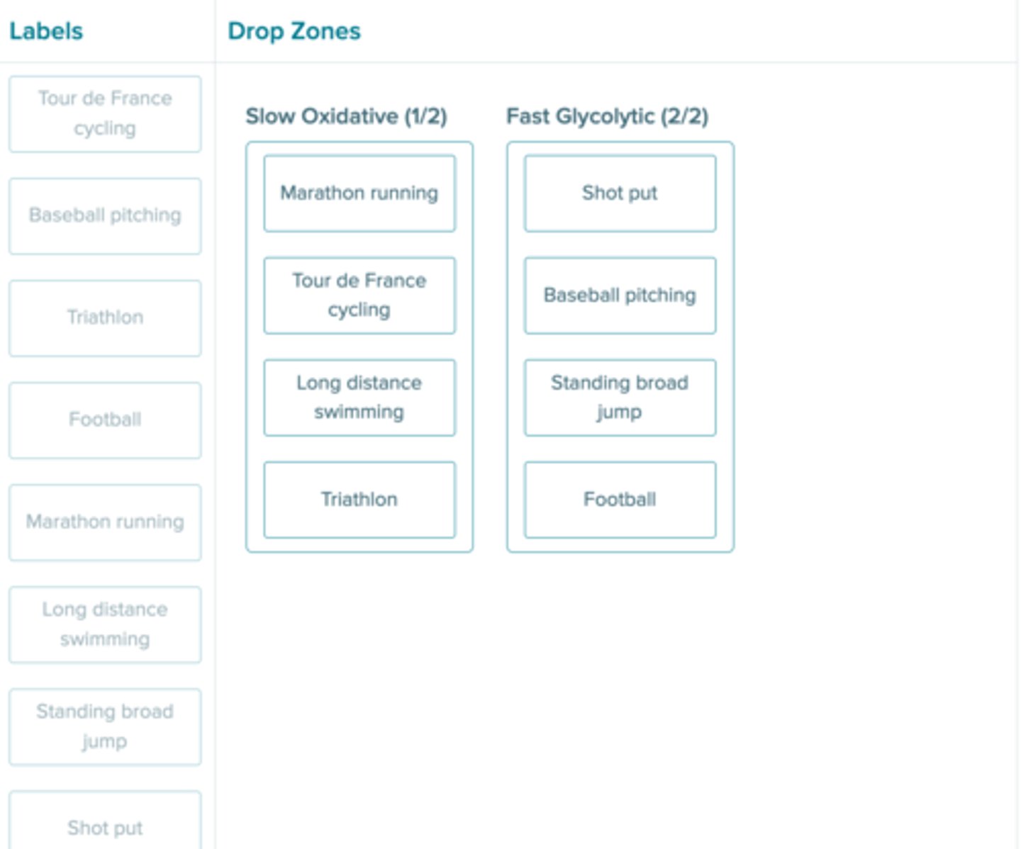
Sort each movement by the type of motor unit used to achieve the movement.

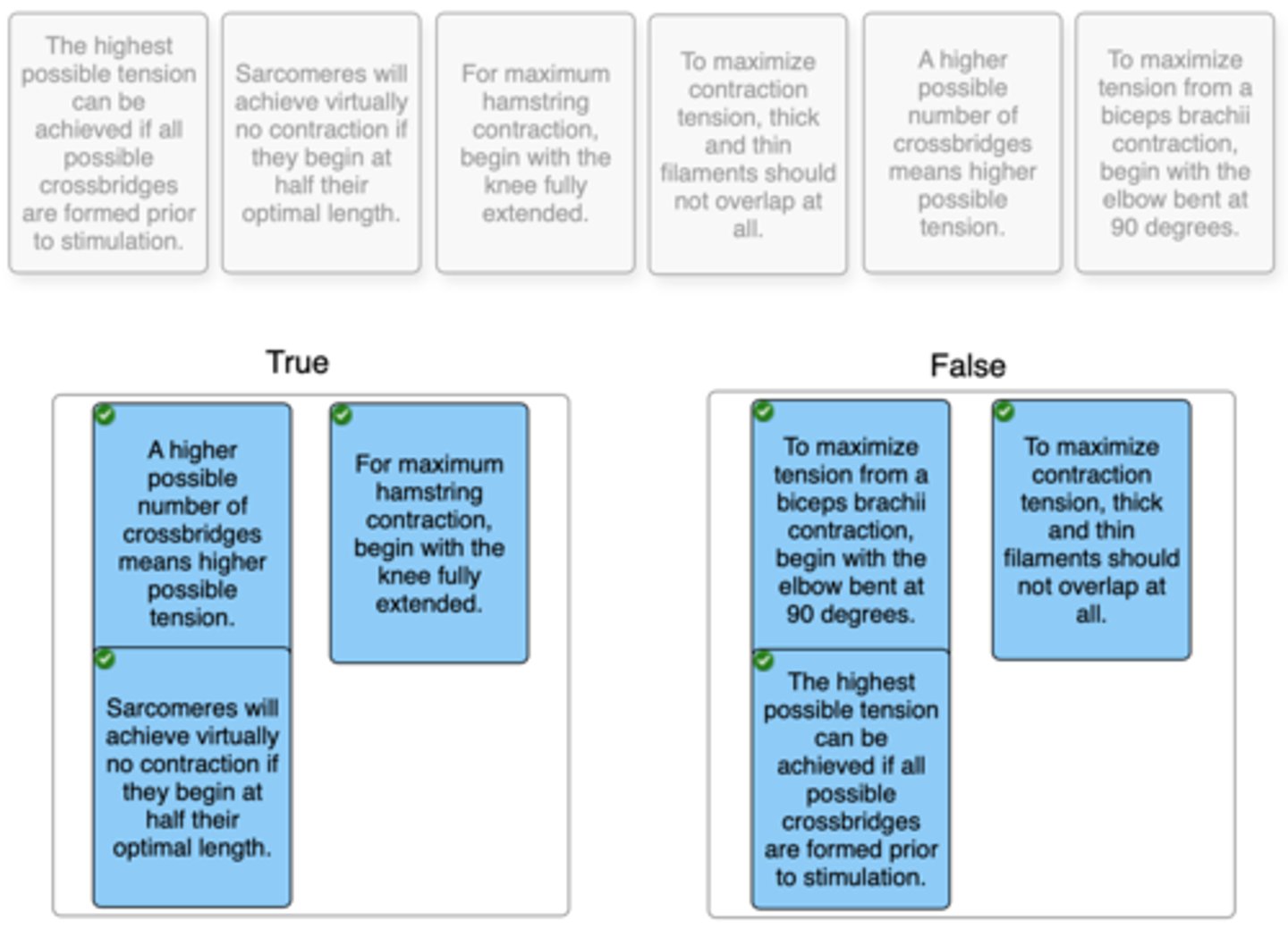
Click and drag each statement into the correct box to identify whether it is true or false regarding the length-tension relationship.
If gene therapy in the future would allow one to artificially alter the expression of muscle fibers for athletic success, which increase in fiber types would make the most sense for the following activities?
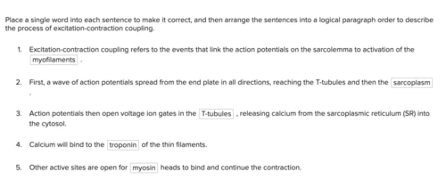
Place a single word into each sentence to make it correct, and then arrange the sentences into a logical paragraph order to describe the process of excitation-contraction coupling.
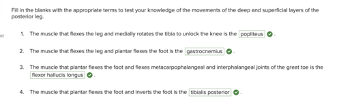
Fill in the blanks with the appropriate terms to test your knowledge of the movements of the deep and superficial layers of the posterior leg.
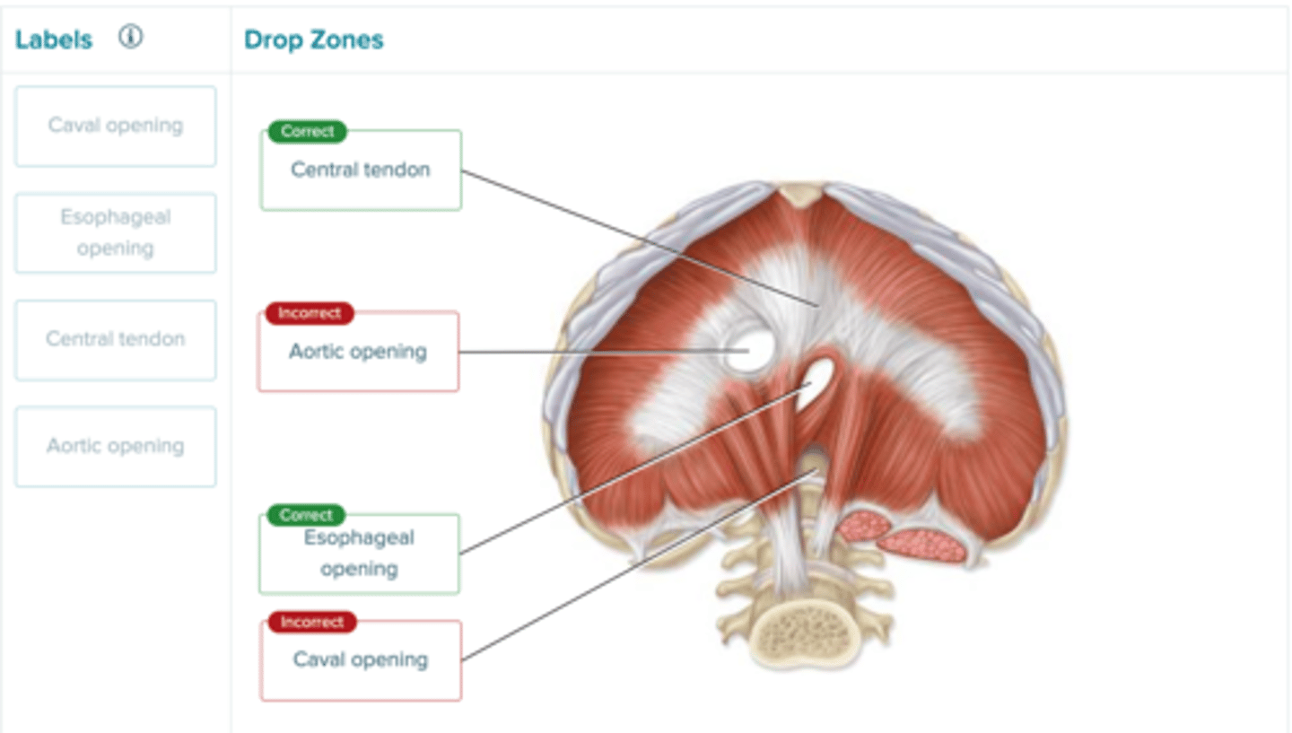
Label the parts of the diaphragm in the image.
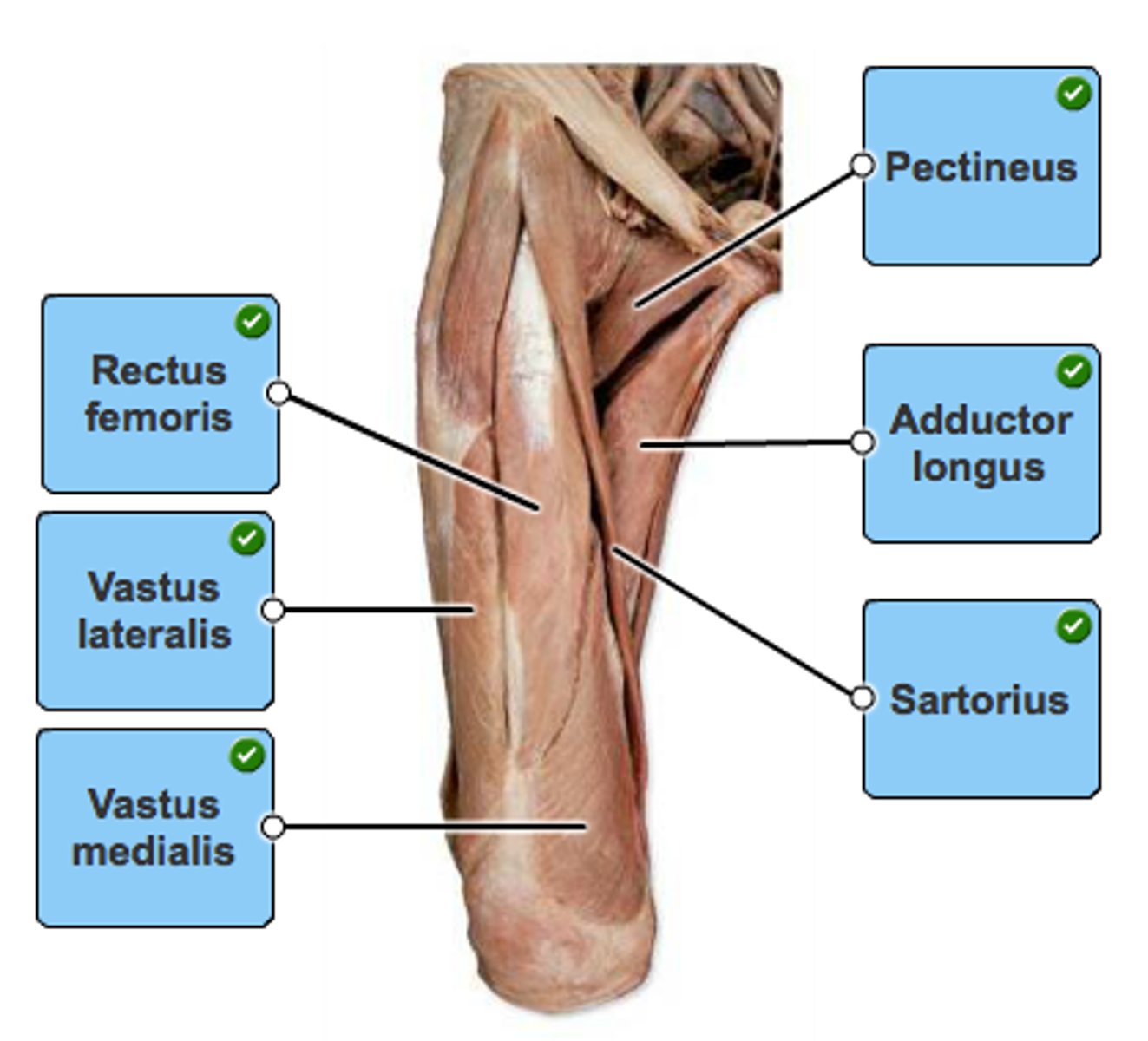
Label the muscles of the anterior thigh.
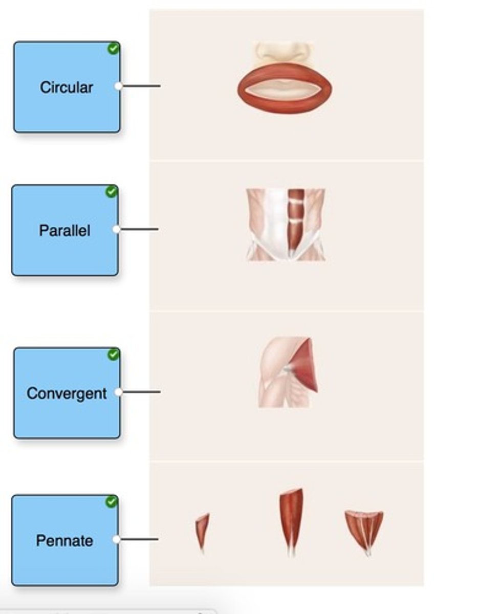
Identify the organizational pattern of the fascicles in each muscle.
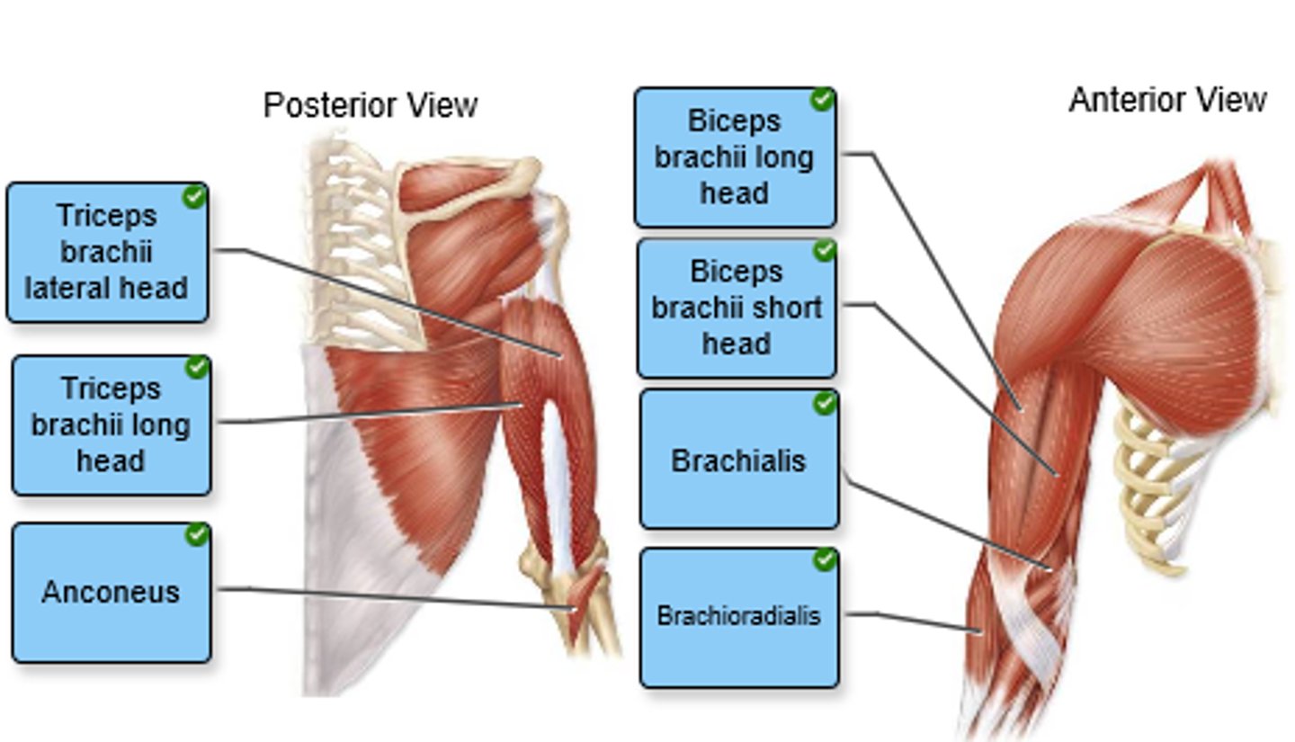
Label the anterior and posterior arm muscles in the figure.
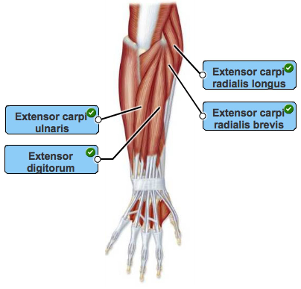
Label the muscles of the right posterior forearm in the figure. Not all labels will be used.
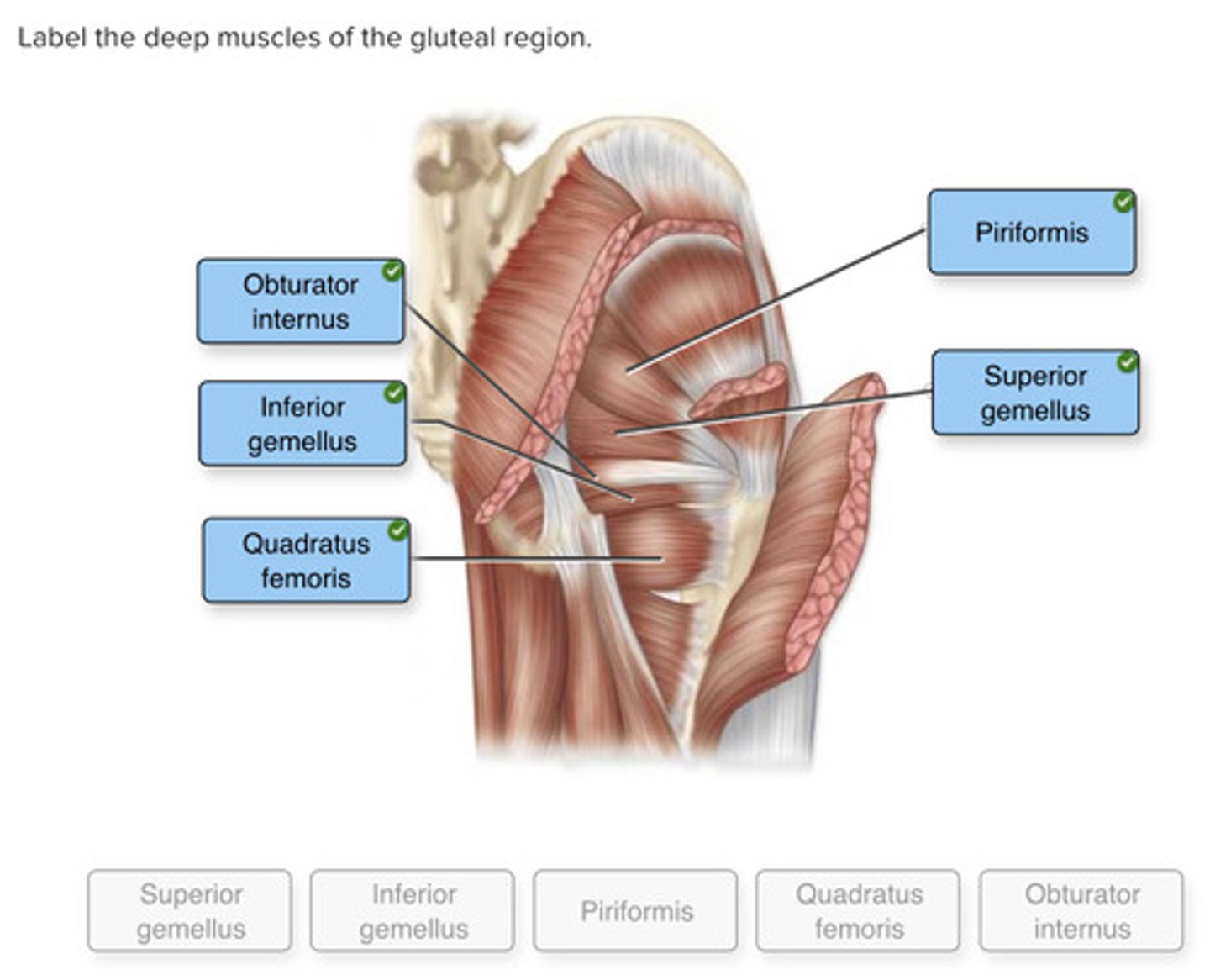
Label the deep muscles of the gluteal region.
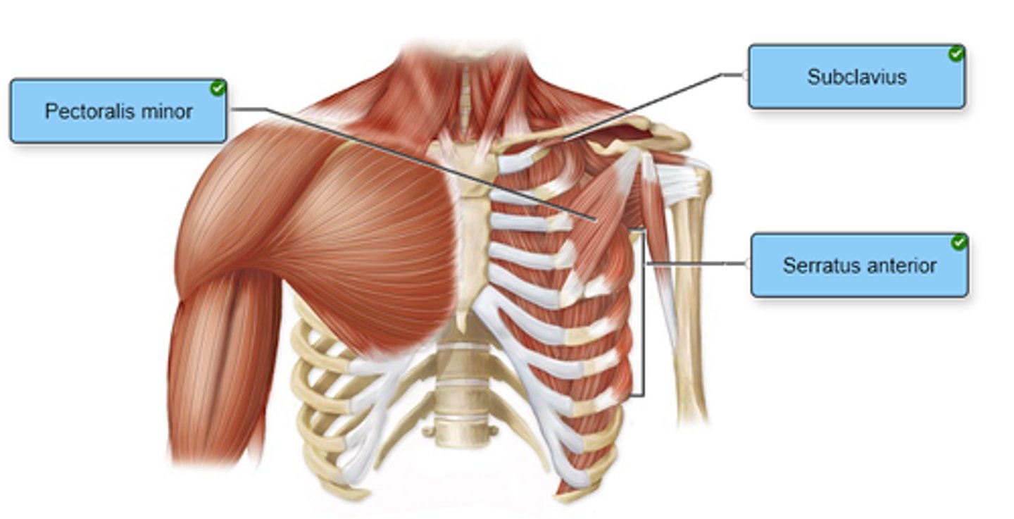
Label the anterior thoracic muscles in the figure.
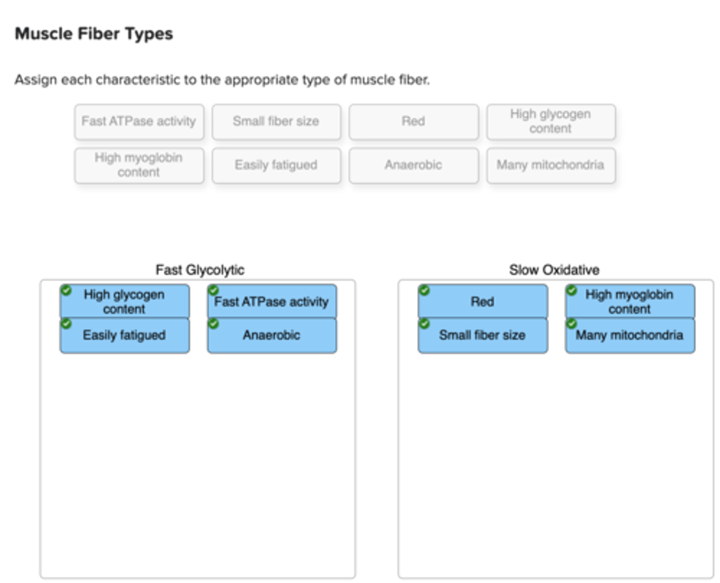
Assign each characteristic to the appropriate type of muscle fiber.
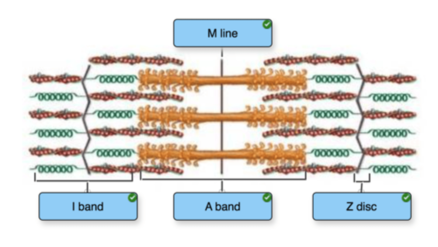
Label the structures of a sarcomere.
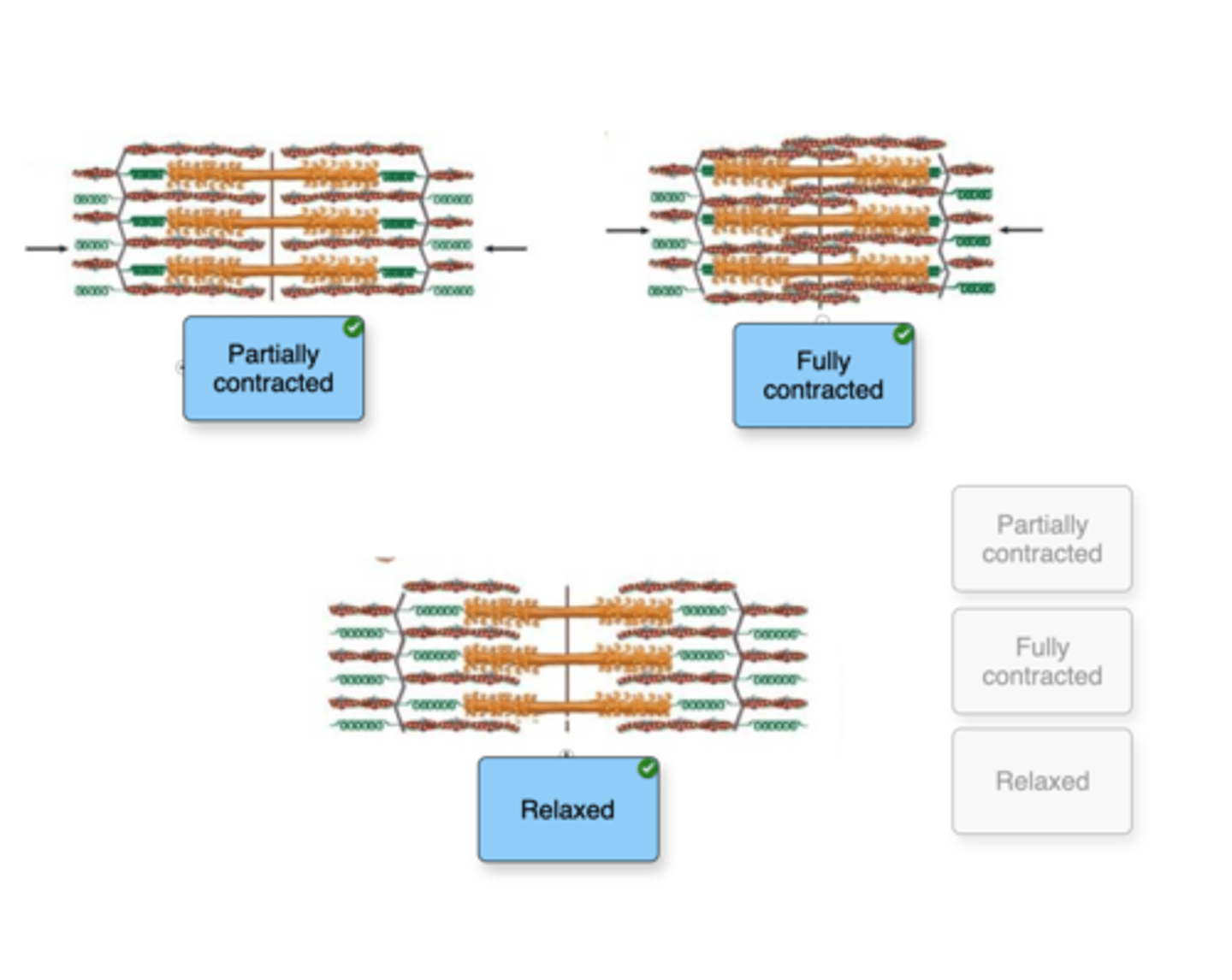
Based on your understanding of the process of skeletal muscle contraction, indicate whether each figure represents a relaxed, partially contracted, or fully contracted skeletal muscle.
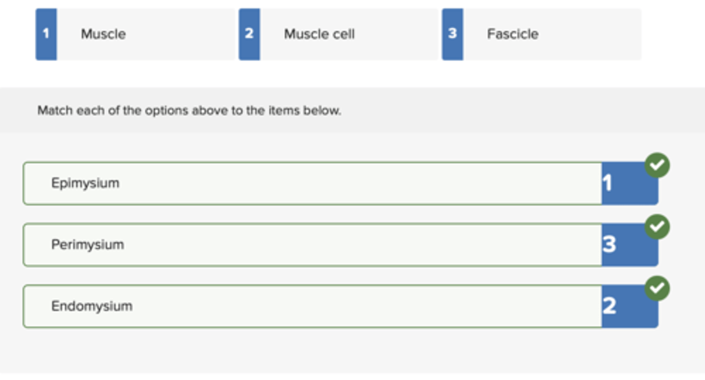
Match each structure with the connective tissue layer that surrounds it.
Epi
Peri
Endo
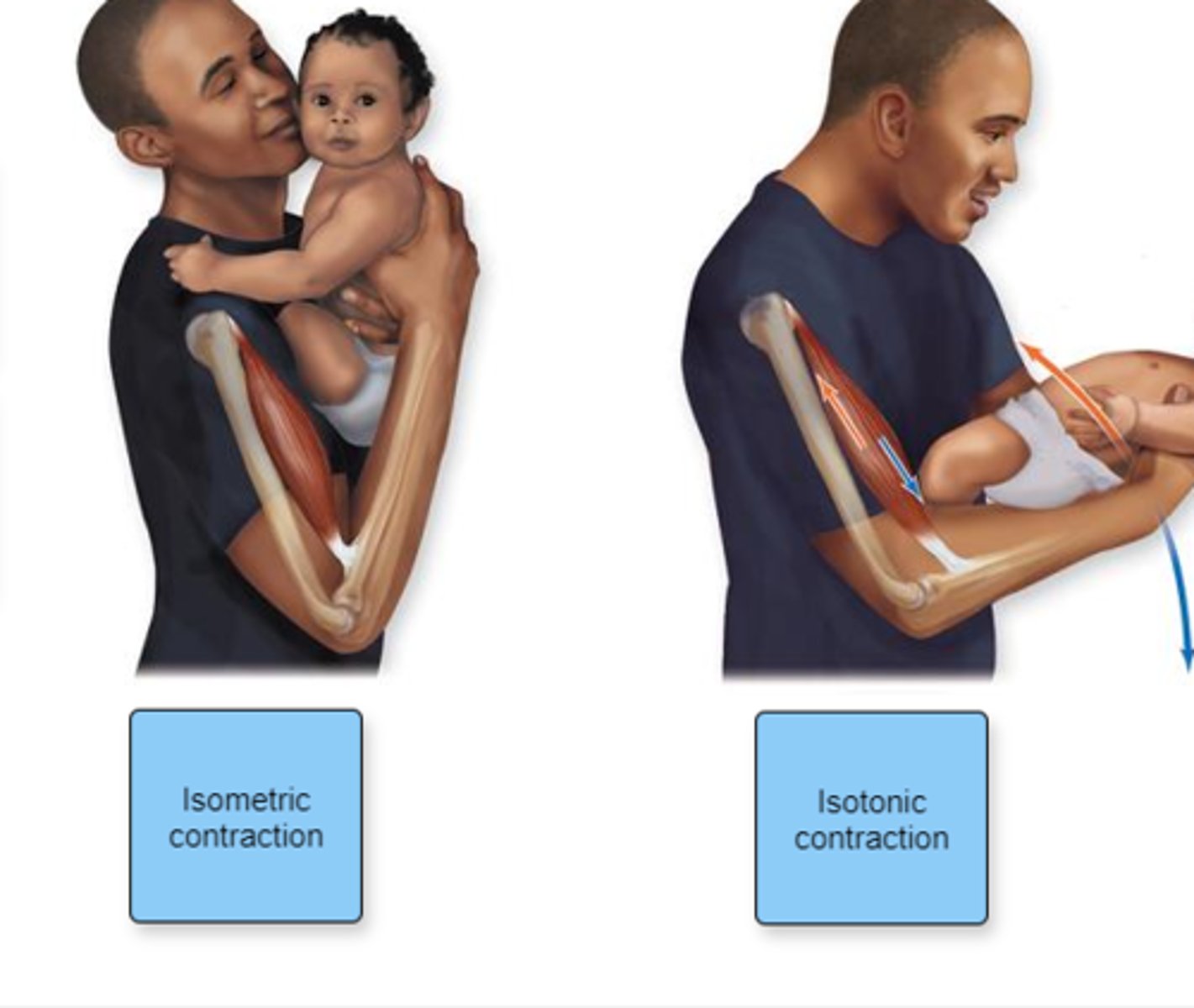
Indicate which type of contraction each figure represents.
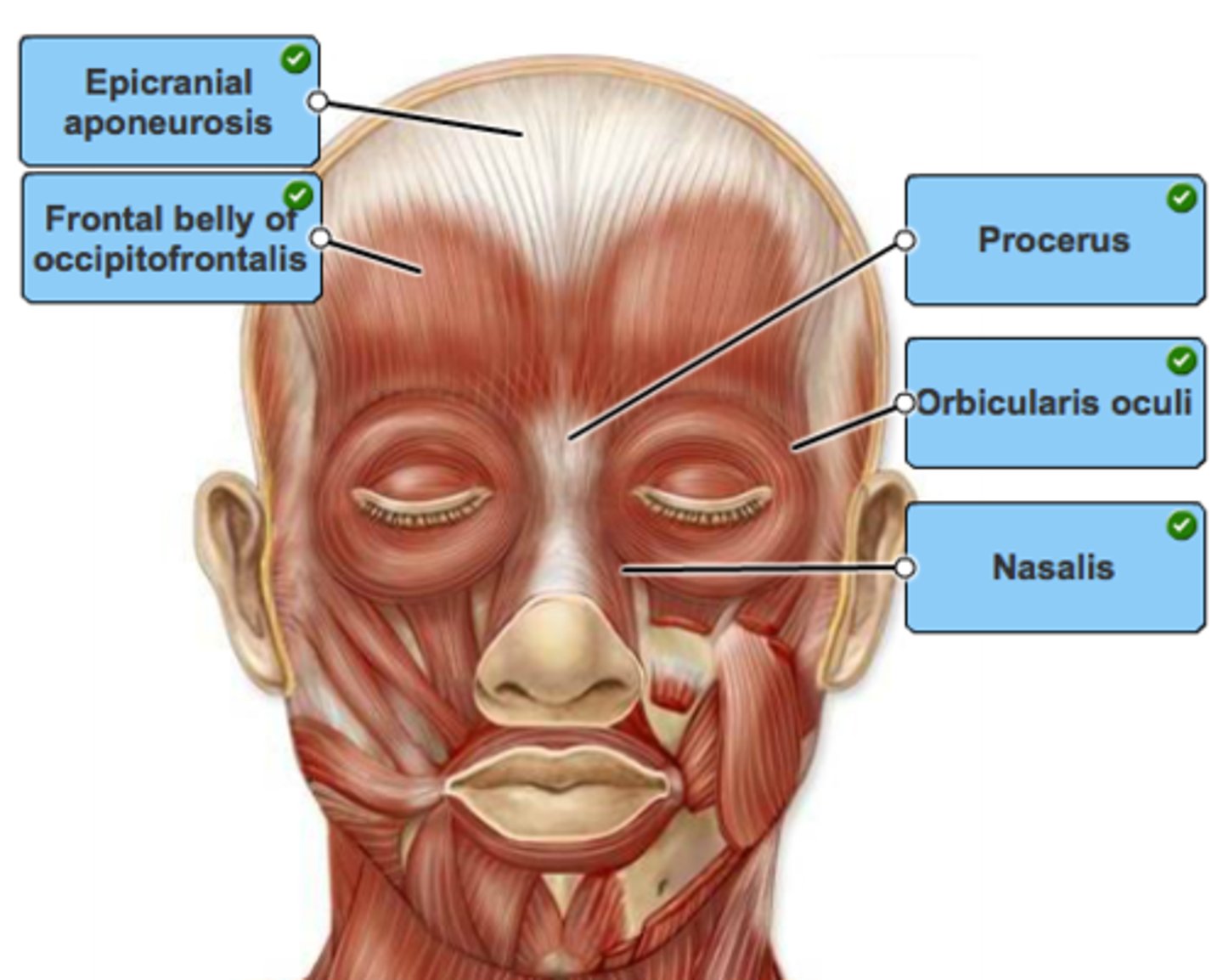
Label the muscles of the forehead and muscles around the eyes in the figure.
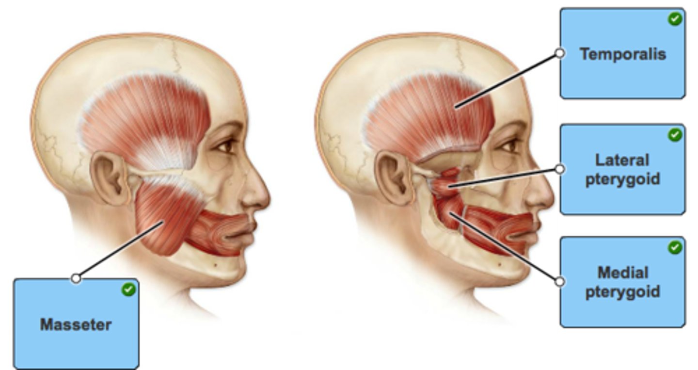
Label the muscles of mastication in the figure.
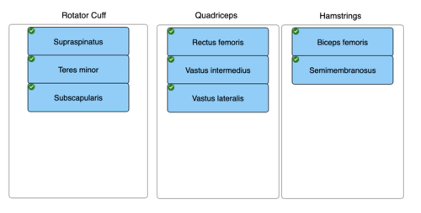
Drag each muscle to the muscle group in which it belongs.
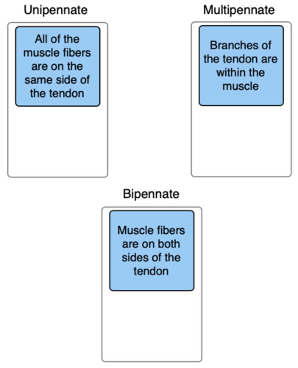
Classify the type of pennate muscle with its description. Not all labels will be used.
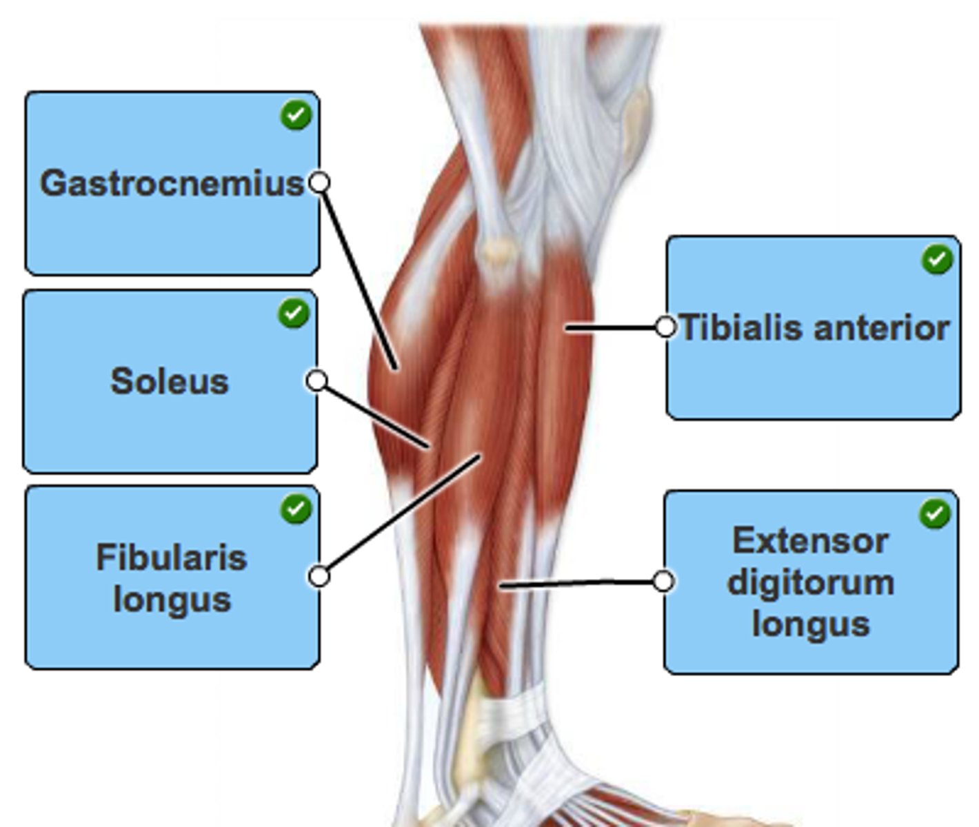
Label the muscles in a lateral view of the leg.
Label the anterior and posterior arm muscles in the figure.
