Chapter 4
 Call Kai
 Learn
Learn Practice Test
 Spaced Repetition
 Match
Match Flashcards
 Knowt Play
1/254

There's no tags or description
Looks like no tags are added yet.
Name | Mastery | Learn | Test | Matching | Spaced | Call with Kai | Chat |
|---|
No analytics yet
Send a link to your students to track their progress
255 Terms
What are tissues?
Group of cells of similar structure that perform a common function
What is histology?
the microscopic study of tissues and organs through sectioning, staining, and examining those sections under a microscope
can observe and evaluate
What are intracellular attachments or cell junctions?
Specialized structures that connect neighboring cells, facilitating communication and strength among tissues
3 types
What are the three types of cell junctions?
tight
gap
desmosomes

What are cell adhesion molecules?
CAMs: transmembrane proteins responsible for cell junctions
Tight junctions
closed; prevent movement between cells
impermeable junctions
form continuous seals around the cell
prevent molecules from passing between cells

Where are tight junctions (epithelial)?
stomach: enterocyte cells
brain (areas that need to be sealed)
Desmosomes
small openings/spaces; anchor cells to each other
anchoring junctions
bind adjacent cells together like molecular Velcro
help keep cells from tearing apart
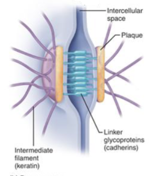
Where are desmosomes?
used as anchors; found in basal and lateral side
Gap junctions
form passageways
communicating junctions
allow ions and small molecules to pass from cell to cell
particularly important in heart cells and embryonic cells

Where are gap junctions (epithelial)?
heart (allow electrical signals to pass)
What is the behavior of desmosomes as cell juncitons?
resist damage for stretching and twisting (blister)
connection between epidermis and dermis
What is the importance of hemidesmosomes?
attached to another tissue; maintain cell structure together
Where are desmosomes found?
connection between epidermis and dermis
How is the connection between epidermis and dermis created?
hemidesmosomes and basal lamina
What are hemidesmosomes?
tie epidermal cells to fibers of the basal lamina
What is the basal lamina?
basement membrane; acellular layer between epidermis and dermis
What is an autoimmune disease associated with cell junctions?
Pemphigus
blisters in mouth, esophagus, eyes, nose, vagina, anus
How are cell junctions important in resistance to damage?
pressure from skin rubbing on a new shoe, for example, anchoring junctions like hemidesmosomes and desmosomes provide strength and stability to epithelial tissues; deal with the friction
blister forms (ECF fills from movement)
What are the four primary tissue types?
epithelial
connective
muscle
nervous
What is the specialized function of epithelial tissue?
covers exposed surfaces
lines internal pasageways and chambers
produces glandular secretions
forms GLANDS
What is the specialized function of connective tissue?
fills internal spaces
provides structural support (soft organs)
stores energy
ALMOST EVERYWHERE
What is the specialized function of muscle tissue?
contracts to produce movement (and force!)
includes skeletal muscle, cardiac muscle, and smooth muscle
3 types: skeletal, cardiac, smooth
What is the specialized function of nervous tissue?
conducts electrical impulses
carries information
sends electrical signals
What makes up epithelial tissue?
sheets of cells that cover: exposed surfaces, line internal cavities and passageways
Where can epithelial tissue be found?
integumentary system
pleural cavity (resp. sys)
digestive system (intestine)
urinary and reproductive systems
Characteristics of epithelial tissue:
cellularity
specialized cell junctions
supported by connective tissue
polarity
avascularity
high regeneration (sheets or layers)
What is cellularity?
cells bound tightly together by specialized junctions
property of epithelial tissue: no space in between tissue, just the cells themselves
Ie Lumen of intestine of kidney: cellularity and specialized junctions → apical membrane
faces the lumen
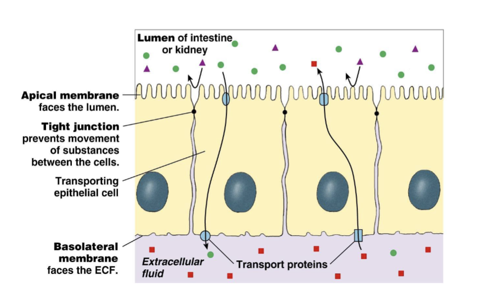
Ie Lumen of intestine of kidney: cellularity and specialized junctions → tight junction
prevents movement of substances between the cells
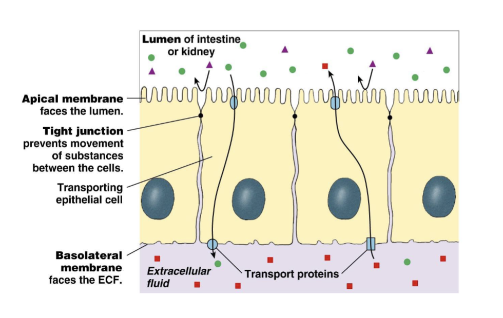
Ie Lumen of intestine of kidney: cellularity and specialized junctions → basolateral membrane
faces the ECF

Epithelial cells are usually _____ and interconnected by ____.
Epithelial cells are usually packed together and interconnected by intercellular attachments.
At their basal surfaces, epithelia are attached to what?
a basement membrane that forms the boundary between the epithelial cells and the underlying connective tissue
What are epithelial cells supported by?
connective tissue
How does epithelial tissue demonstrate cellularity?
hemidesmosomes and desmosomes create high density of cells (layering) of fibers → basement membrane
What does the basement membrane look like, in relation to supporting epithelial cells?

What is polarity?
tight junctions polarized the cell membrane in two regions
distribution of the organelles is different (cell function)
What are the two sides in a cell (in relation to polarity)?
apical (lumen: space in tubular structures; ie: intestine)
basolateral (ECF)
Where are tight junctions found (which side)?
apical side of the cell
Where are organelles found (which side)?
towards the ECF
The nucleus is ___ to the tight junctions.
inferior
The nucleus is ___ to the ECF.
proximal
What membrane faces the lumen?
apical
What membrane faces the ECF?
basolateral
What is avascularity in relation to epithelial tissue?
Epithelial tissue does not contain blood vessels
thus → can’t bleed if damaged
How are epithelial cells important: composed of layers and high regeneration?
replacement of cells
Why is regeneration important to epithelial cells?
composed of layers → stem cells are the most inferior, so they grow upward to replace dead or damaged cells, ensuring tissue integrity and function.
What are the major functions of epithelial tissue?
protection
control of permeability
provide sensation
produce specialized secretion
Example how epithelial tissue provide sensation:
neuroepithelia: epithelia found in special sense organs
Example of how epithelial tissue produces specialized secretion
unicellular glands
glandular epithelia
What are the specializations of epithelial cells on the apical side?
microvilli, cilia, sterocilia
What are microvilli?
absorption and secretion
extensions of cytoplasm
What are cilia?
movement
extension of cytoplasm
in areas with mucous
What are stereocilia?
sensory organs (inner ear)
one important cilia → kinocilium and smaller steriocilia in the inner ear
What is long cilium?
flagellum
sperm: unique cell with the same function of movement
How are epithelial cells classified?
number of cell layers
simple and stratified
cell shapes
What are the epithelial cell shapes?
squamous (plate or scale)
cuboidal (cube)
columnar (rectangle)
What is the classification of epithelium with one layer?
simple
What is the classification of epithelium with more than one layer?
stratified

simple squamous epithelium
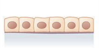
simple cuboidal epithelium

simple columnar epithelium
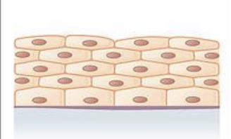
stratified squamous epithelium
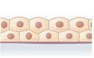
stratified cuboidal epithelium
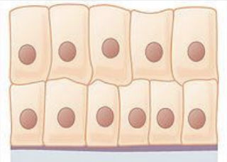
stratified columnar epithelium

pseudostratified columnar epithelium
What is simple squamous epithelium?
reduces friction; controls vessel permeability; performs absorption and secretion
single layers help to decrease diffusion distance
cells are thin, flat, and irregular
Location of simple squamous epithelium
respiratory exchange surface (CO2 and O2)
circulatory system (blood vessels: endothelium and lymphatic vessels), and lining of body cavities (mesothelium).
What is cuboidal epithelium?
cells have hexagonal box shape
perfectly round nucleus
Location of cuboidal epithelium
regions where secretion or absorption take place
What are regions where secretion or absorption take place?
stomach
intestinal tract
uterine tubes
excretory ducts
Where can you find cuboidal epithelium?
Regions where
secretion or
absorption take place:
Stomach
Intestinal tract
Uterine tubes
Excretory ducts
What is simple columnar epithelium?
protection; secretion; absorption
height of the cell is greater than the width
cells with/wo microvilli
Location of simple columnar epithelium?
areas where absorption and secretion occur
stomach
intestine
uterine tubes
gallbladder
What is stratified squamous epithelium?
provides physical protection against abrasion, pathogens, and chemical attack
keratinized
non-keratinized or mucosal
cells at the surface layer are flat
Found in areas where mechanical or chemical stress are severe
What is keratinized (stratified squamous epithelium)?
where dehydration occurs
palmar skin
keratin prevents water loss and protects from mechanical stress
What is non-keratinized or mucosal (stratified squamous epithelium)?
keep moist
oral cavity, pharynx, esophagus, rectum, anus, vagina
protects from abrasion and chemicals due to layers
Location of stratified squamous epithelium
found in areas where mechanical or chemical stress are severe
What is stratified cuboidal epithelium?
protection and secretion
found with lots of ducts and glands
circular nuclei
Location of stratified cuboidal epithelium
lining the ducts of sweat and mammary glands
What is stratified columnar epithelium?
rare; protection
two or more layers (first is columnar)
Location of stratified columnar epithelium (rare)
small areas of the pharynx, salivary ducts, urethra, and anus
superficial side has columnar shape
What is pseudostratified columnar epithelium?
cell nuclei are located at varying distance from the surface (close to basal area)
usually ciliated cells
Location of pseudostratified columnar epithelium
Nasal cavities
Trachea
Bronchi
portions of the reproductive male tract.
What is transitional epithelium?
dome-shaped cells
stratified epithelium that can stretch without damaging epithelial cells
relaxed: bubble shape
stretched: look squamous
Location of transitional epithelium
Urinary system structures such as:
Urinary bladder, ureters, lining the renal pelvis
What do epithelial cells form?
our glands; produce specialized secretions
What are the types of secretions (epithelial cells which form our glands)?
exocrine
endocrine
What are exocrine glands?
release their secretions onto epithelial surface through epithelial ducts (or surface)
Examples of exocrine glands
serous, mucosal, mixed glands
Serous glands (exocrine)
water and proteins— enzymes
Mucous glands (exocrine)
glycoproteins (mucins)
mucus
Mixed glands (exocrine)
serous and mucus secretions (water, proteins, enzymes)
What are endocrine glands?
release their secretions into the interstitial fluid (ductless glands)
What are examples of endocrine glands?
hormones
associated with the endocrine system
Hormones (endocrine gland)
chemical substances that diffuse into the blood to different tissues, organs
travels through blood
What is the structure of exocrine glands?
multicellular
unicellular
Multicellular exocrine glands
ducts: simple
secretory part: tubular and alveolar
Unicellular exocrine glands
goblet cells
secrete mucins

Where simple tubular ducts?
intestinal glands
