(1) Digestive system (without stomach)
1/69
Earn XP
Description and Tags
Digestive system incl: apparatus digetorius, teeth/glands of oral cavity, contains intestine, large intestine, liver,
Name | Mastery | Learn | Test | Matching | Spaced |
|---|
No study sessions yet.
70 Terms
the oral cavity is divided by closed jaws into?
cavum oris proprium (the proper oral cavity)
vestibulum oris (the one in the mouth)
consist of the vestibulum buccale and labiale
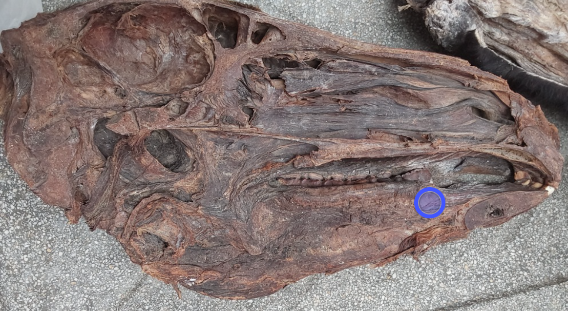
what is this? Seen when removing the tongue, seen between molar teeth and cheeks.
vestibulum buccale
In ru: papilla conicae are seen here in the buccae.
Part of vestibulum oris.
Buccae (the cheeks) form the lateral walls of vestibulum buccale and is without a bony base. The musculus buccinator is right under the skin in this area.
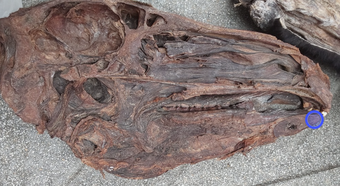
what is this?
Labium inferius - part of rima oris which the cavum oris is bounded by.
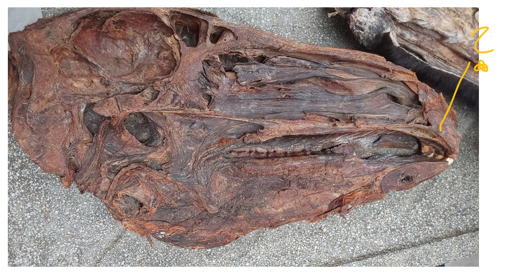
what is this?
labium superius - part of rima oris, which the cavum oris is bounded by.
space between incisor teeth and lips is?
Vestibulum labiale - between incisior teeth and lips, part of vestibulum oris.
which part makes up the cavum oris with vestibulum oris?
cavum proprium oris - This together with vestibulum oris, makes up the cavum oris, and they are divided by closed jaws. The cavum oris proprium → palatum durum and palatum molle.
The roof of oral cavity consist of?
Palatum durum - It has a bony base, and have the structures
rhaphe palatini (the streak in the middle)
rugae palatinae (ridges)
palatum molle/velum palatinum - caudal continuation of palatum durum. This is of mainly muscles.
m. palatinus - responsible for active movement, m. tensor veli palatini tenses and the m. levator veli palatini raises the palate.
uvula - in some animals (“drøvel”)
arcus palatoglossus (on sides of uvula, down)
arcus palatopharyngeus (on side of uvula, upper)
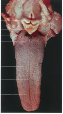
which structure forms the bottom of cavum oris? What are the muscles involved?
Lingua - Tongue forms the bottom of cavum oris. (Bounds the cavum oris with palatum durum, molle and rima oris).
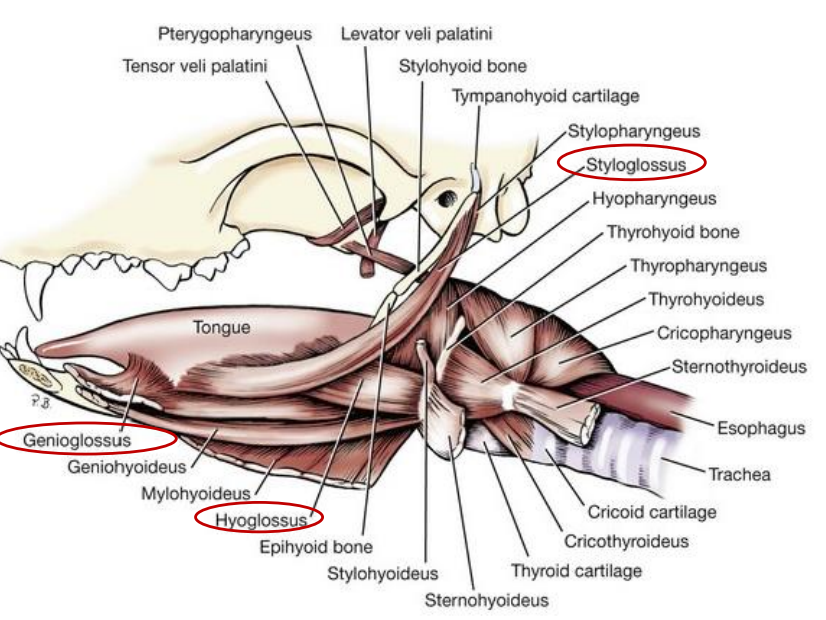
Includes extrinsic muscles of tongue:
m. genioglossus - near septum linguae, moving tongue rostrally
m. hyoglossus - moves tongue caudally
m. styloglossus - shortens tongue, elevates apex
hyoid muscles also belong here, like m. mylohyoideus which suspends the tongue.
Intrinsic muscles:
m. lingualis proprius → fibrae longitudinales superficiales et profundae, fibrae transversae, fibrae perpendiculares.
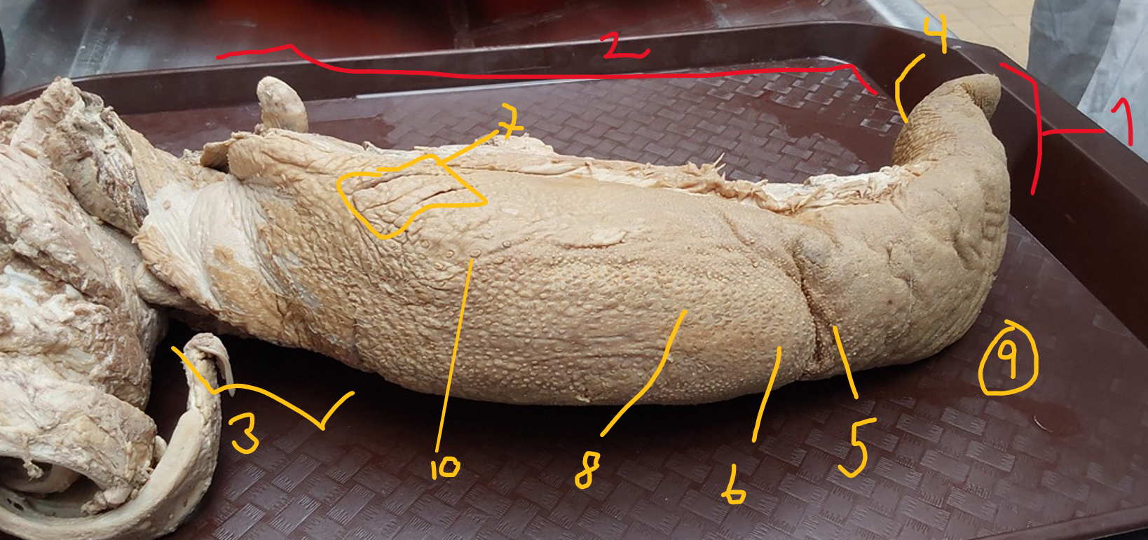
describe the structures of lingua.
apex lingua
corpus lingua
radix linguae
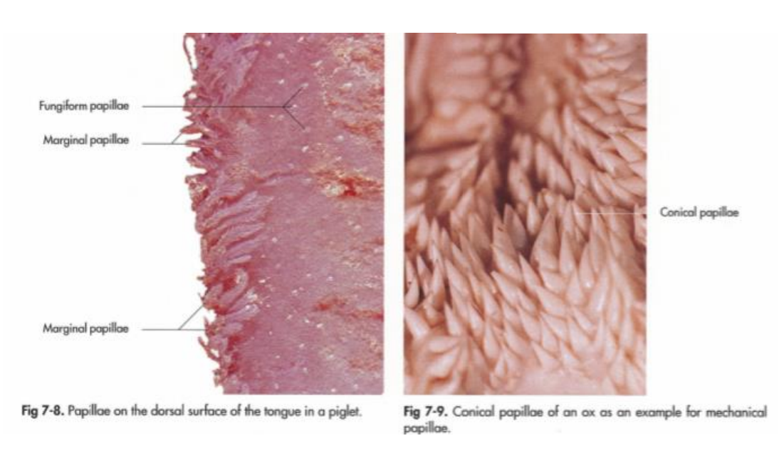
Papillae fungiformes (larger than filiformes, on borders of linguae and sometimes on the apex ventral surface (under)
fossa linguae (seen in ru)
torus linguae (seen in ru)
papillae foliatae (on borders of radix, consisting of parallel leaves of CT which are separated by small furrows, absent in ru)
papillae filiformes (on dorsum of tongue, soft in eq, ca + su or horny in ru + fe threads directed caudally)
dorsum linguae (surface of tongue, opposite of hard palate)
facies ventralis linguae is the other, where frenulum linguae (the string) arises.
margo linguae (ventral surface + dorsum meets here)
papilla vallatae (on dorsum rostrally to radix, surrounded by circular cleft, larger than fungiformis)
in car: 2-3, bo: 8-18, small ru: 12-24 on radix.
in eq + su → present one pair of big papilla vallatae.
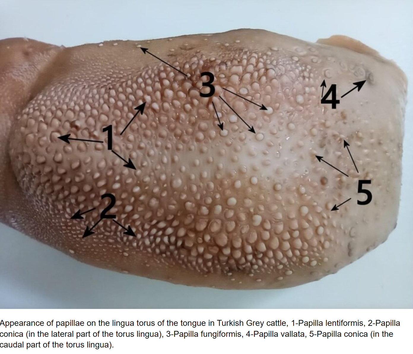
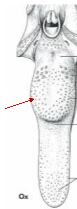
Which papilla type can we see here?
papillae conicae - larger than filiformes, present in ru, on torus linguae especially.
In horse tongue contains a slender bar of —- which lies in the median plane below the mucous membrane
cartilago dorsi linguae
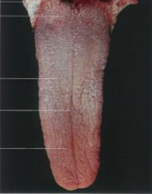
the midline of tongue in dogs, is called? What structure is seen on ventral surface in car?
sulcus medianus linguae - dividing the tongue in carnivores.
ventral surface of car tongue has lyssa (rod shaped, fibrous body lying in the median plane, going from apex linguae to radix linguae, in fe - forms a fatty bar)
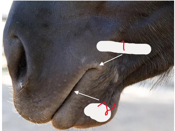
externally on the angle of mouth we see?
angulus oris - The angle of mouth is seen externally. Part of rima oris together with the lips (labium). This is where the labium inferius et superius meet.
rima oris
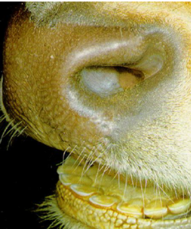
What are types of areas, the upper lip together with septum of nostrils forms externally?
planum nasolabiale (bo)
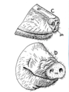
upper lips forms in su and in car, ov and cap?
Planum rostrale (su)
planum nasale (car + small ru)
The planes are divided by sulci - into small areae, moistened by glands. In car + small ru: gl. nasalis lateralis - keeps the nasal planes moist. This is absent in bo, in others it moistens the air.
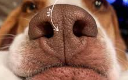
divides the planum nasale of carnivores and small ruminants?
Philtrum
What is the folds between labial mucosa of lips and gums called?
frenulum labii superioris (of upper lip)
frenulum labii inferioris (of lower lip)
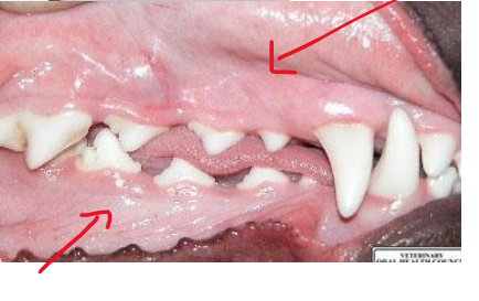
what is this?
Gingivae - gums
A part of the oral mucosa, fused with periosteum. It covers the processus alveolares of bones and palatum durum.
Ru: pulvinus dentalis, which replaces the upper incisor teeth.
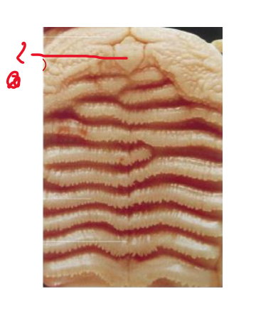
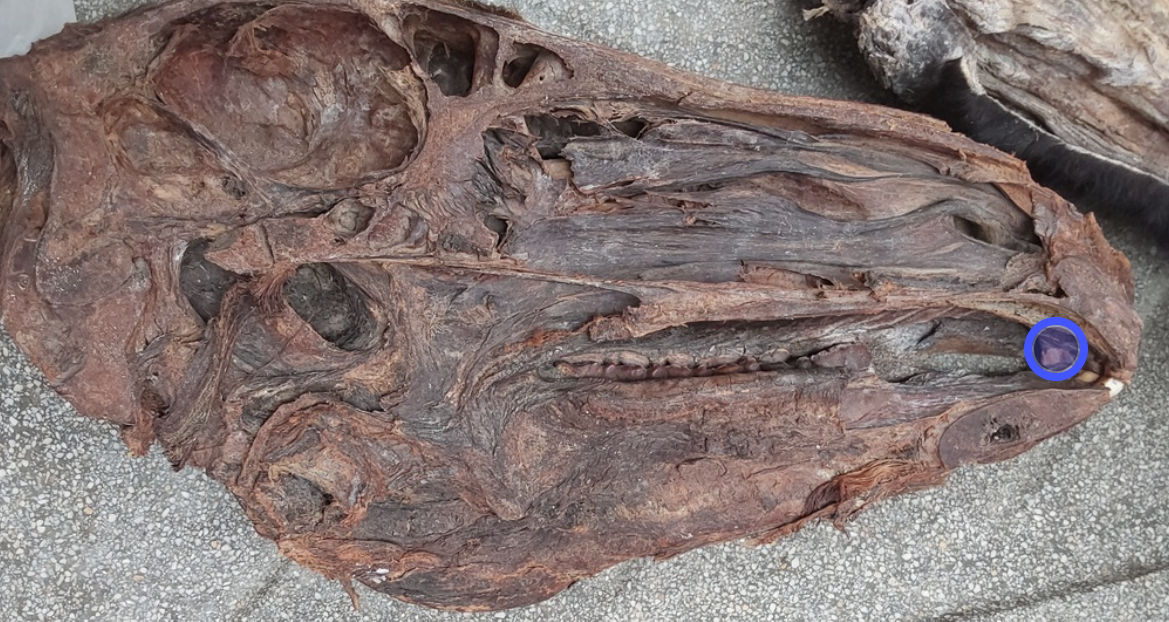
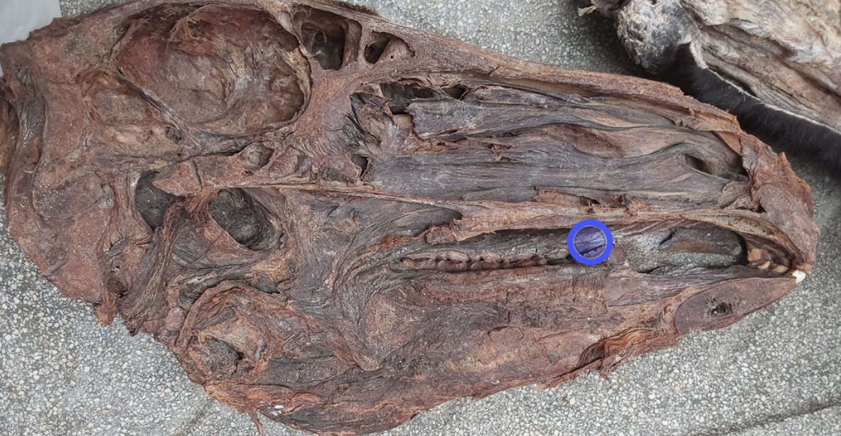
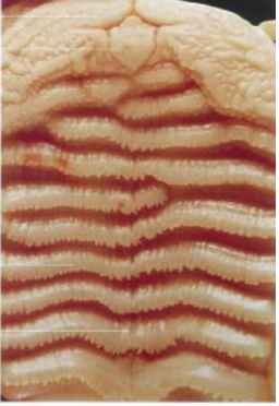
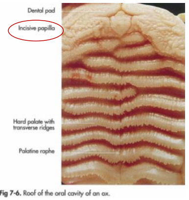
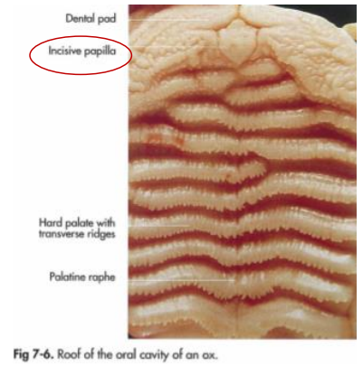
Structure in the gingiva, seen on the roof of oral cavity of an ox.
papilla incisiva - located behind to the upper incisor teeth or dental pad and is flanked on each side by the openings to ductus invisivus which goes through the hard palate.
gll. salivariae are divided acc. to the size to the?
gll. salivariae minores
gll. salivariae majores
Both produces saliva which is mixed with food during mastication, acting as a lubricant.
The glandulae salivariae minores consist of?
Located in the tunica mucosa of oral cavity, to these belong:
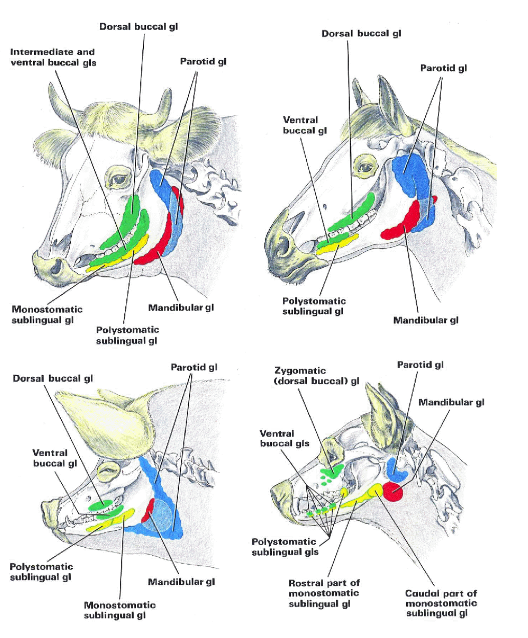
Glandulae buccales
gll. buccales dorsales → in car, these lie medially to arcus zygomaticus, called gl. zygomatica.
ventrales
ru: also intermediae
Ducts of these glands open into the vestibulum buccale, duct of gl. zygomatica opens on the papilla zygomatica
glandulae linguales
glandulae palatinae
glandulae labiales
The glandulae salivaries majores consist of?
gl. parotis
located caudally to ramus mandibulae, ventrally to ala atlantis, saliva is sent through the ductus parotideus
ru + car: ductus parotideus crosses lateral surface of m. masseter.
eq, bo + su: ductus runs firstly on medially of mandible, then at level of incisura vasorum facialium, opening at papilla parotidea (in vestibulum buccale)
gl. mandibularis
bw. basihyoideum + ala atlantis, saliva is sent by ductus mandibularis, opening is on caruncula sublingualis.
gl. sublingualis monostomatica
with a single ductus sublingualis major opening on the caruncula sublingualis.
gl. sublingualis polystomatica
with numerous ductus sublinguales minores, opening in the recessus sublingualis lateralis space on both sides of frenulum of tongue.
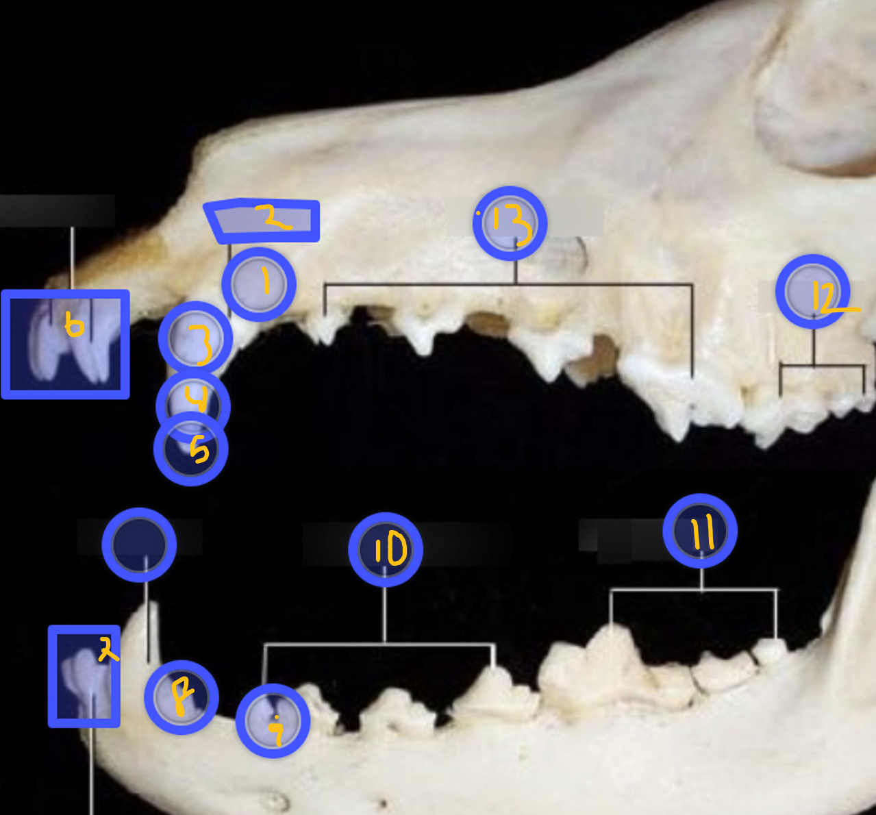
Identify these structures of teeth, how is the teeth arranged?
The teeth are aranged in the arcus dentalis superior and the arcus dentalis inferior, the small space between the teeth is called diastema (typical in car)
apex radicis dentis - foramen apicis dentis leads into the cavum dentis and pulpa dentis inside the tooth
dentes canini - deciduous (dentes decidui), meaning its one of the non-permanent teeth.
radix dentis
cervix dentis
corona dentis
dentes incisivi - incisor teeth - arcus dentalis superior. the upper ones. A type of dentes decidui, meaning it is non-permanent.
dentes incisivi - incisor teeth - arcus dentales inferius.
margo interalveolares (bigger space bw. teeth)
diastema - space in bw. teeth
dentes premolares
dentes molares
dentes molares - dentes permantes, type of permanent teeth
dentes premolares - dentes decidui, meaning its one of the non-permanent teeth.
what are the surfaces of the crown of tooth (corona dentis)?
facies vestibulartis - toward oral vestible
facies lingualis - in contact with tongue
facies contactus - surface which faces the other teeth
facies mesialis - surface toward next rostral tooth in the arcus dentalis, nearer the medial line.
facies distalis - surface directed to next caudal tooth.
facies occlusalis - surface facing its antagonist in the opposite jaw (on top)
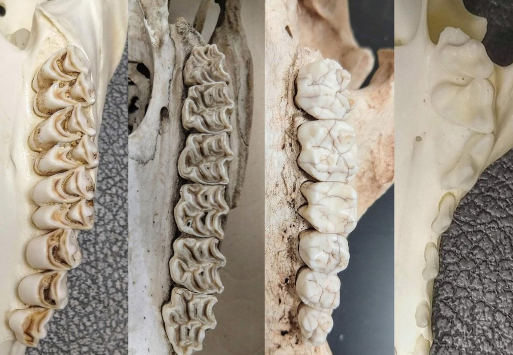
On basis of shape and formation of the facies occlusalis of corona dentis, we can describe 3 types of tooth:
bunodont type (byonos - tubercle, low hill) - in car + su, like a multitubercular tooth, flatter occlusal surface, intended for crushing the food.
selenodont type (selenos - moon) - present in ru, dentine surrounds semilunar invaginations
lophodont type (lophos, ridge) - present in eq, surface forms folds + ridges)
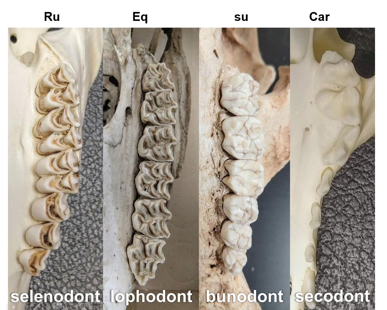
The 3 substances of tooth?
1. dentinum (hard osseus tissue) - (periodontium, made of fibrous CT that links the cementum with alveolar wall, anchoring the tooth into the jaw)
2. enamelum (hardest, covers the corona dentis)
3. cementum (on radix dentis)
what means by that teeth are diphyodont?
first erpted teeth are replaced by a single set of teeth in older animals - dentition of domestic mammals.
first set of teeth: deciduous teeth (dentes decidui) - milk teeth
permanent teeth (dentes permanentes) - will replace the milk teeth
Polyphyodont - some animals can have sets of teeth that will erupt throughout the animal`s life.
Classification of teeth is based on growth ?
brachyodont type - in car, su + humans, stopped growing.
hypsodont type - never stops growing, only corpus dentis + rooth, in rodents.
semihypsodont type - similar to hypsoondt but when the tooth begins to wear out, the growth stops - like incisor teeth of eq.
Amount of teeth each species has?
stallion
mare
su
ru
ca
fe
Stallion: around 40
deciduous: 28
mare: 36-38
deciduous: 24
su: 44
deciduous: 28
ru: 32
deciduous: 20
ca: 42
deciduous: 28
fe: 30
deciduous 26
eq: each incisor teeth has a central infundibulum dentis, lined with layer of cementum and together with food → black teeth appearance. The tooth will become shallower by age and bottom of infundibulum will form stella dentaria. Dens lupinus - woolf tooth.
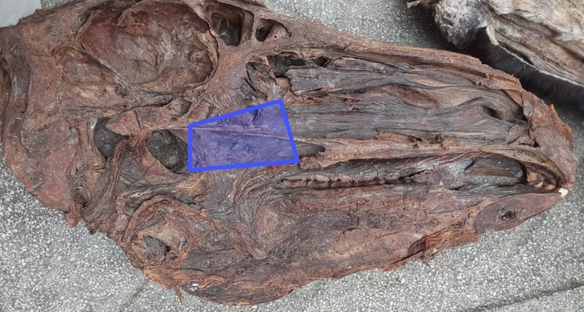
this area is of pharynx?
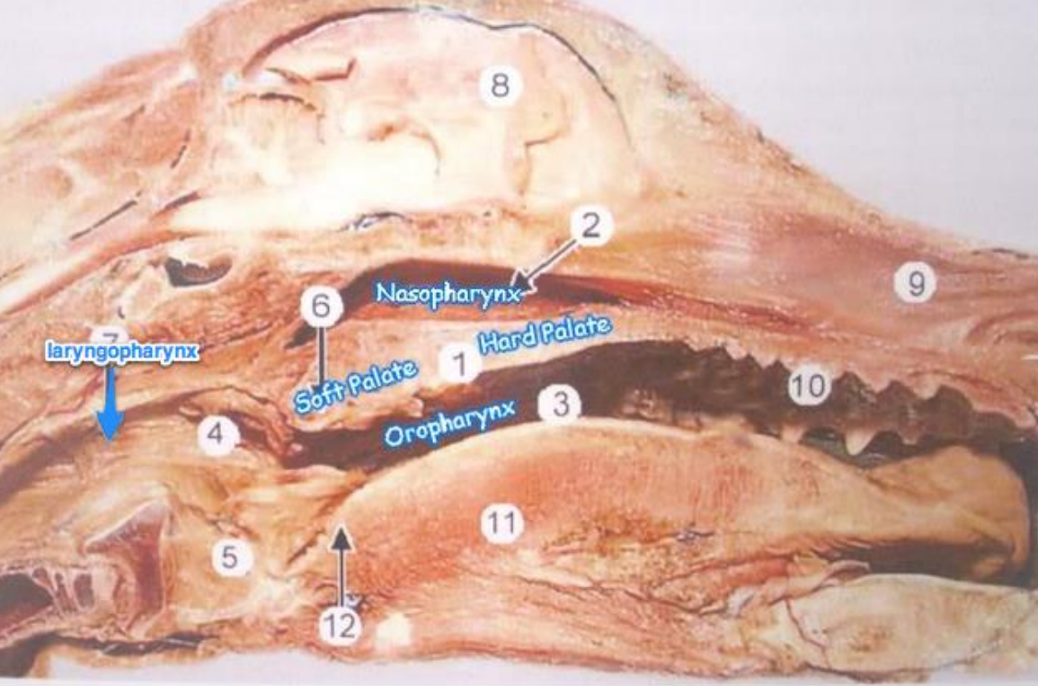
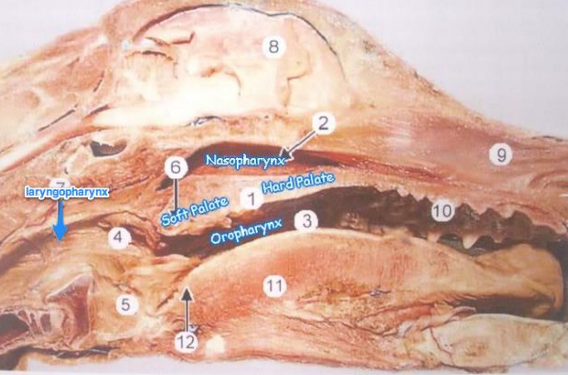
Pars nasalis pharyngis: dorsally to palatum molle. Ostium tubae auditivae is here, which connects the pars nasalis and middle ear cavity. (1 of the 3 main parts of pharynx)
this is what main part of pharynx?
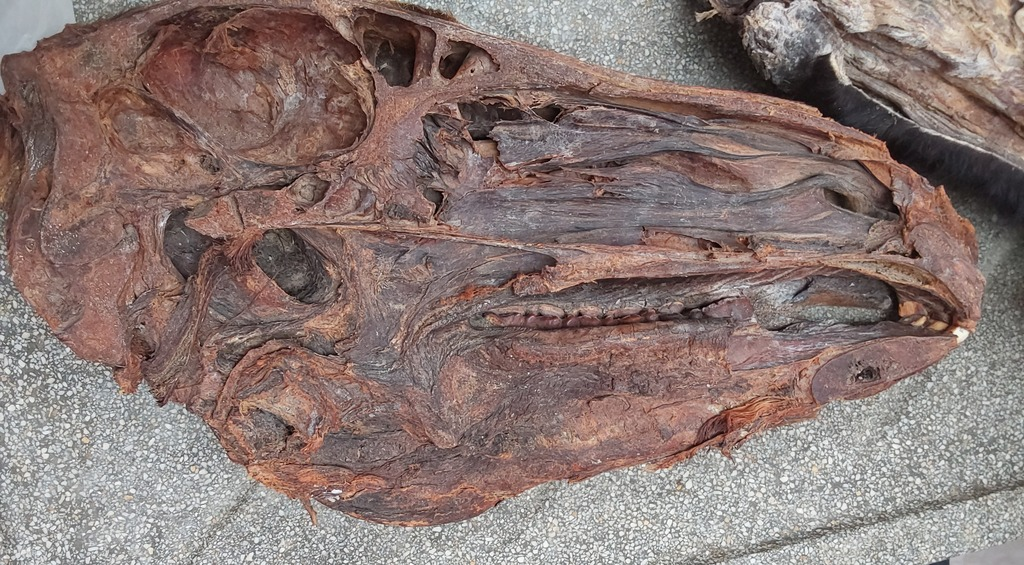
Pars laryngea pharyngis - caudally. With the structures auditus laryngis caudoventrally and vestibulum esophagi caudodorsally. (1 of 3 main parts of pharynx)
This is what main part of pharynx?
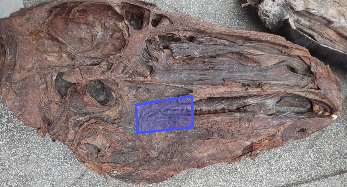
Pars oralis pharyngis - ventrally to palatum molle. (1 of 3 main of pharynx)
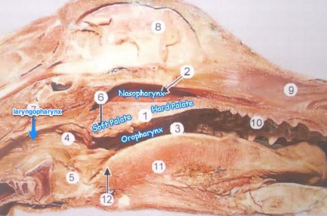
In what structure, does the pars nasalis pharyngis and pars oralis pharyngis meet?
Ostium intrapharyngeum - formed by caudal free border of palatum molle and paired arucs palatopharyngealis
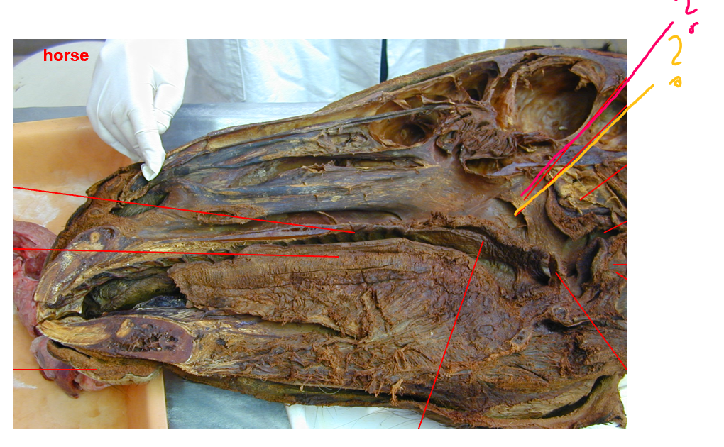
The roof of pars nasalis pharyngis is called?
Fornix pharyngis, divided dorsally in ru + su by septum pharyngis. It has the following openings:
choanae - open into nasal cavity
ostium pharyngeum tubae auditivae (yellow) - dorsolaterally - connecting pars nasalis pharyngis with tympanic cavity of middle air.
in eq: covered by mucosal fold known as the operculum (pink)
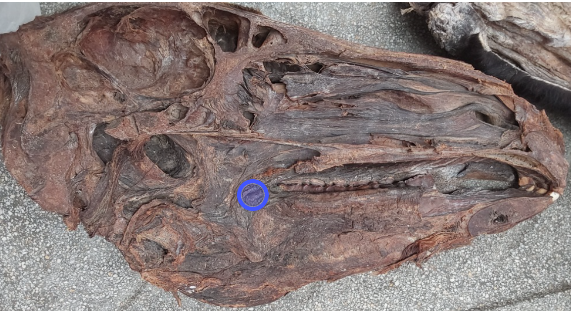
Pars oralis pharyngis, communicates with oral cavity by?
Isthmus faucium (bordered by soft palate, ventrally by roof of tongue, laterally by palatoglossal arch)
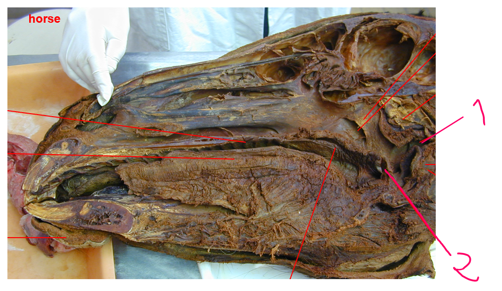
these structures of pars laryngea pharyngis is?
Vestibulum esophagi - caudoventrally, we also have auditus laryngis (openings at pars laryngea pharyngis)
in car: vestibulum esophagi is surrounded by a circular fold, the limen pharyngoesophageum with glands
epiglottis - on each side, is recessus piriformis which continues on both sides from floor or pars oralis pharyngis to the laryngeal entrance.
wall of pharynx consist of?
tunica mucosa → gll. pharyngeae + lymphoid tissue
fascia
a) fascia pharyngobasilaris - attached to raphe pharyngis serving for insertion of the muscles
muscles
a) striated muscles: mm. constrictores pharyngis rostrales, m. pterygopharyngeus, m. palatopharyngeus, m. constrictor pharyngis medius → m. hyopharyngeus, mm. constrictores pharyngis caudales → m. thyropharyngeus, m. cricopharyngeus.
b) one dilatator of pharynx: m. stylopharyngeus caudalis.
adventitia
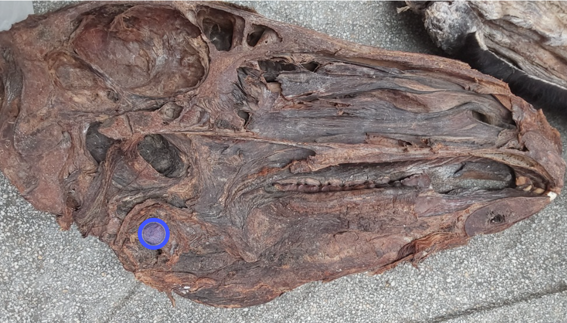
this is of pharynx?
Auditus laryngis - caudoventrally to pars laryngea pharyngis
The tonsillae of pharynx?
Independent lymphatic organs, important in defense against toxic substances and microorganisms. Makes lymphocytes, Ab and secrete protective substances into pharynx cavity.
tonsilla lingualis
tonsilla palatina
tonsilla veli palatini (on soft palate)
tonsilla paraepiglottica
tonsilla tubaria - on ostium pharyngeum tubae auditivae wall (absent in car)
tonsilla pharyngea
The 3 parts of esophageus?
Esophageus is divided into 3:
pars cervicalis - first dorsally to trachea, shifting position to left surface of trachea (vestibulum esophagi -pharynx)
pars thoracica - begins at thoracic inlet, then returning to dorsal position to trachea
pars abdominalis - very short at cardia of stomach (ostium cardiacum)
Wall of esophageus - 3 layers from outside to inside:
tunica adventitia (CT - Adventitia) - in cervical region and tunica serosa in thoracic and abdominal part.
tunica muscularis (stratum circulare, stratum longitudinale)
tunica mucosa (in ca, submucosa contains gll. esophageae over whole esophageus, in su only in cranial half, and in eq, ru + fe only in vestibulum esophagi)
cavum abdominis, internally covered by mucous serous membrane. (Theory)
cavum abdominis: largest cavity, containing most digestive organs, it is held by lumbar vertebrae dorsally, by abdominal muscles laterally and ventrally, and by diaphragm cranially. Continues caudally into pelvic cavity.
Internally - covered by mucous serous membrane: Peritoneum consisting of:
1. Peritoneum parietale (part that lines walls of cavity)
2. peritoneum viscerale (covering viscera inside)
The surface of peritoneum is smooth, moistened by liquor peritonei.
Mesentery: fold that attaches the intestine to the cavity roof, arising as:
a) radix mesentery communis, which divides into radix mesenterii cranialis et caudalis. The cranial one gives folds of duodenum into colon transversum, while caudal one is the folds of colon descendens going into the rectum. (like mesorectum)
organs are classified according to position in the abdominal cavity as?
intraperitoneal - stomach, intestine, liver
Retroperitoneal - kidneys or adrenal glands
Caudal part of peritoneum stops within cranial part of pelvic cavity forming three pouches, the?
Excavationes:
excavatio rectogenitalis - on both sides of rectum is the fossae pararectales
excavatio vesicogenitalis - bw. uterus + urinary bladder in female, bw. bladder + genital fold in male
excavatio pubovesicalis
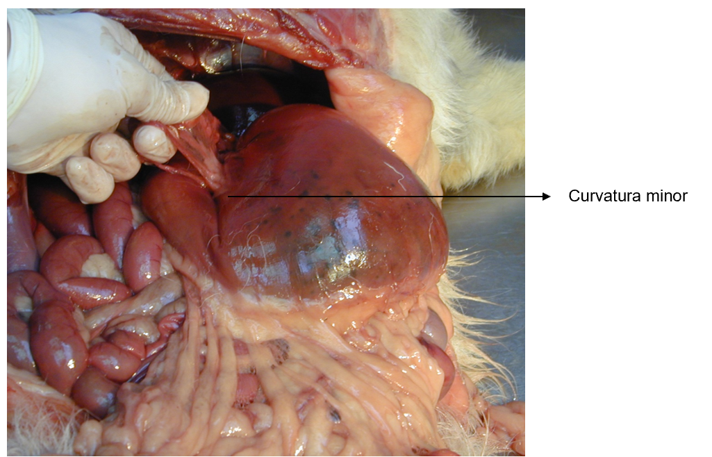
Omentum is?
Serosal fold passing from stomach to the other organs, we can recognize:
omentum minus → lig. hepatogastricum + lig. hepatoduodenale (picture from pres, from slide omentum minus)
omentum majus (epiploon) - serous fold, folding itself and making paries surperficialis et profundus
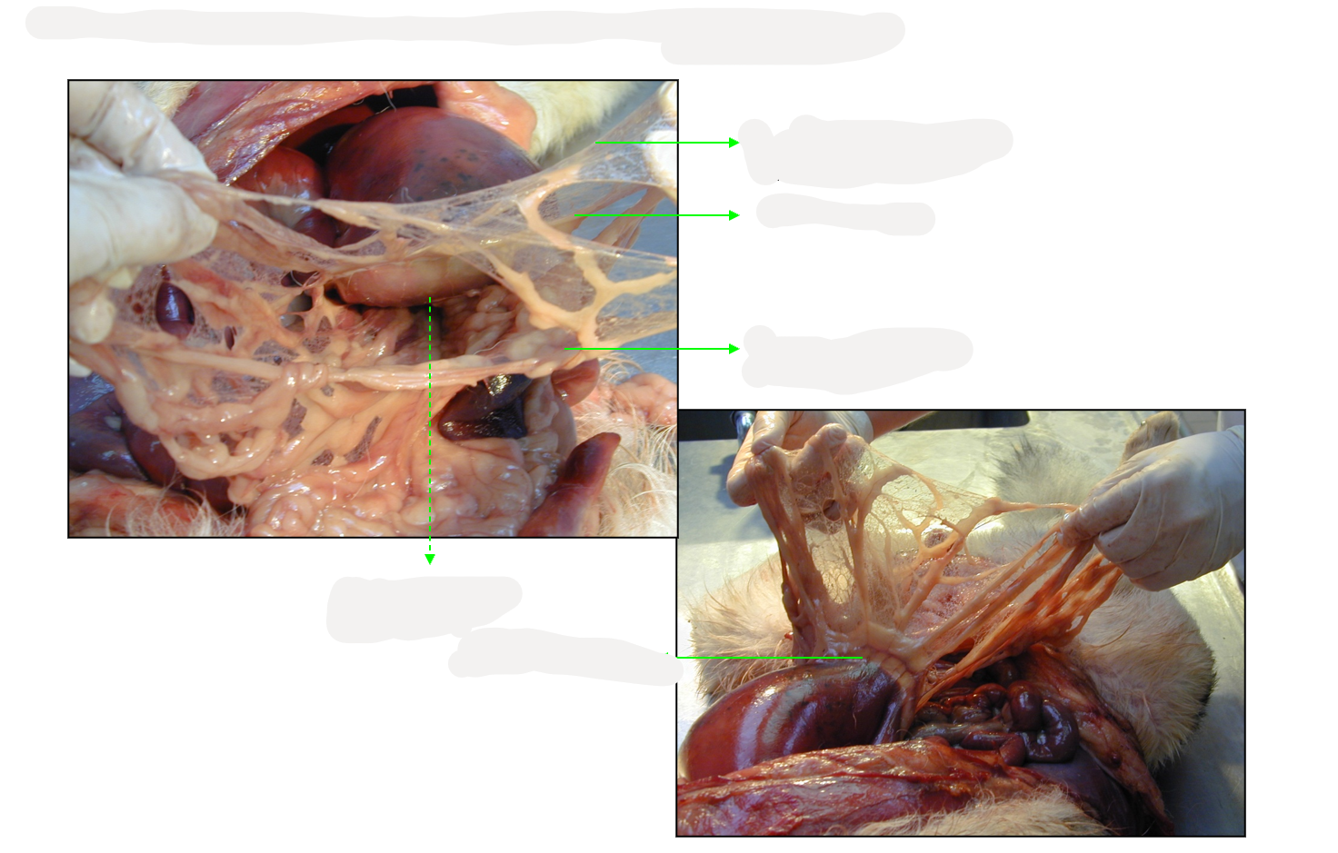
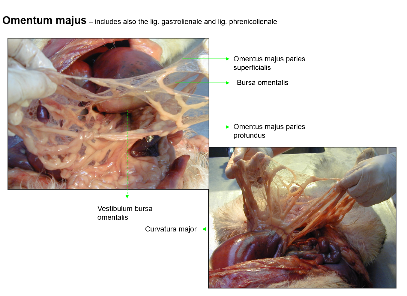
Omentum majus consist of?
Includes also the lig. gastrolienale and lig. phrenicolienale
omentum majus paries superficialis
bursa omentalis
omentus majus paries profundus
vestibulum bursa omentalis
curvatura major
Intestine goes from?
goes from pars pylorica ventriculi to the anus, consisting of tenue and crassum part. The canalis analis as its termination part with anus.
The difference of intenstinum tenue vs. crassum?
tenue - beginning at pylorus of stomach and ends at cecum:
duodenum
jejunum
ileum
crassum:
cecum
colon: colon ascendens, transversum et descendens
rectum
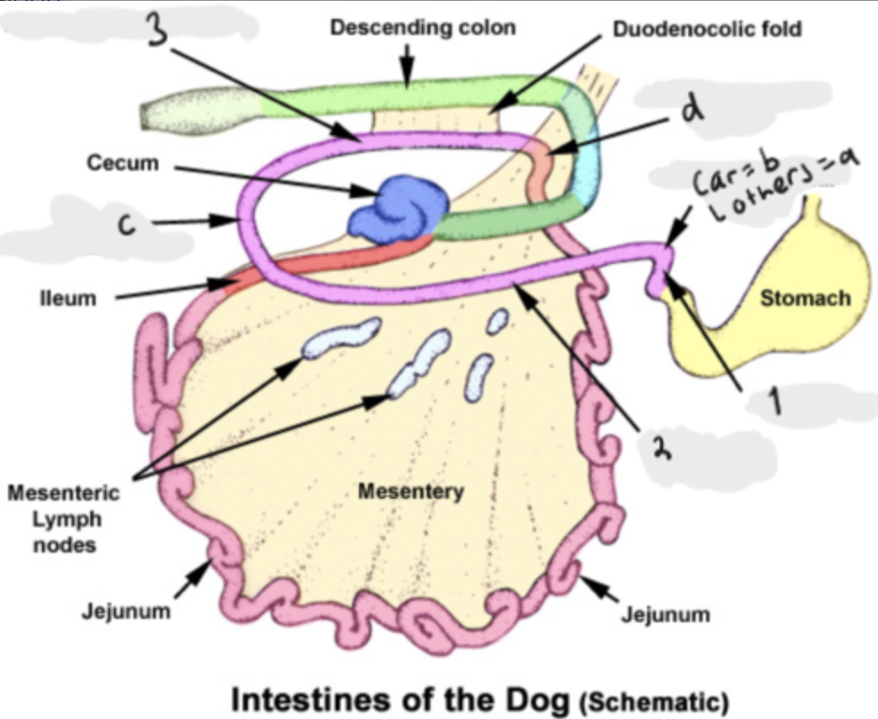
The parts of duodenum?
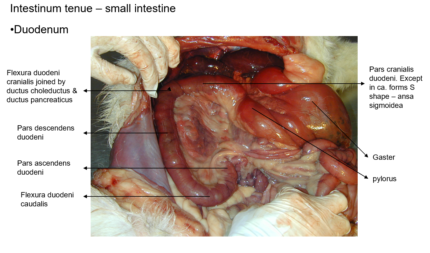
Position: dorsally, to the right in abdominal cavity, going from pars pylorica ventriculi to the jejunum.
A part of: Intestinum tenue
pars cranialis (between pars pylorica ventriculi and flexura duodeni cranialis)
forms ansa sigmoidea (a) which is absent in car, car =flexura duodeni cranialis (b)
pars descendens (goes caudally along cavity)
C) forms flexura duodeni caudalis
pars ascendens (runs cranially)
d) continuing by means of flexura duodenojejunalis, ventrally to jejunum, at this flexura, the colon descendens is attached to duodenum ascendens by means of plica duodenocolica.
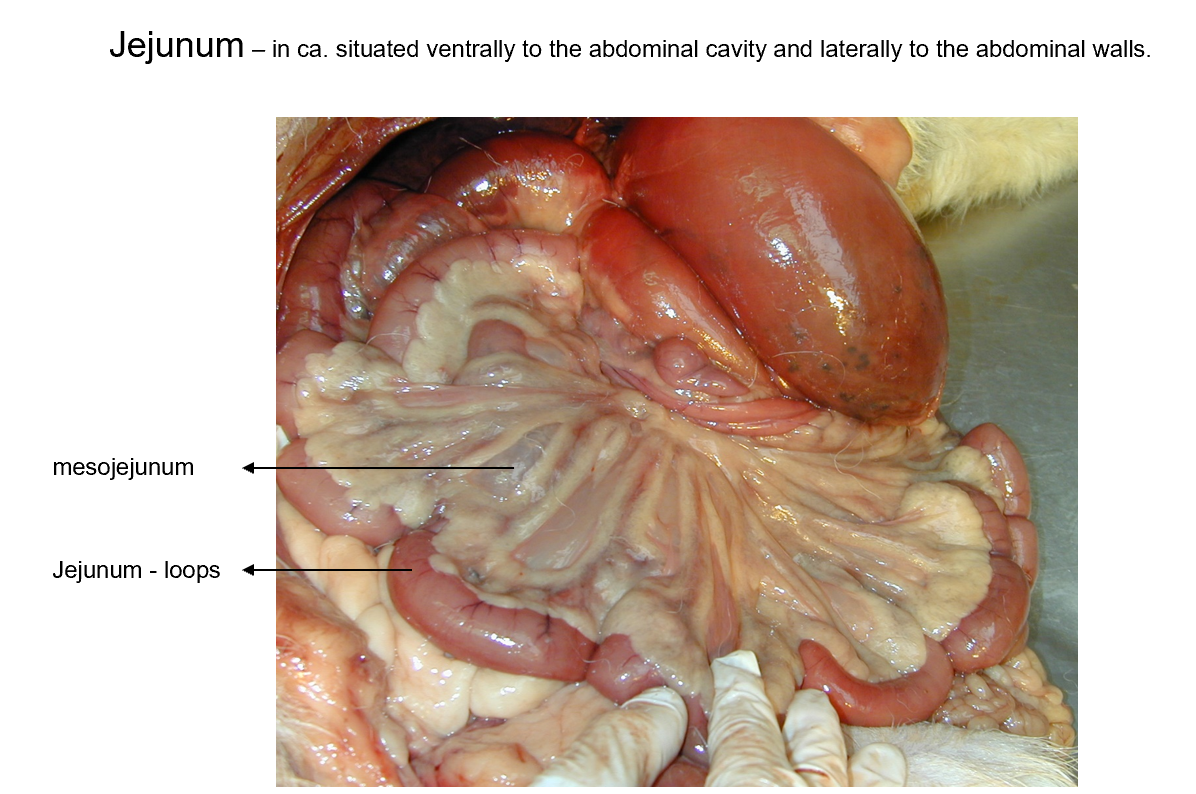
The position of jejunum?
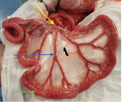
Position: longest part of intestinum tenue, coming after the flexura duodenojejunalis.
differences:
- car: ventrally situated
- eq: no regular position
- ru + su: in right half of cavum abdominalis
1. mesentery, blue arrow: mesojejunum, green arrow: jejunum loops
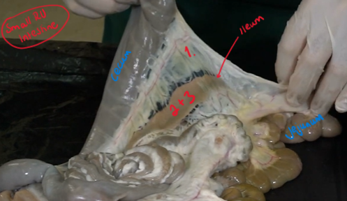
The structures of ileum?
shortest part of intestinum tenue
POP:
1. plica iliocecalis (serous fold between ileum and cecum)
2. ostium ileale (opening to cecum, around it is the m. sphincter ilei)
3. papilla ilealis (the opening is on this, internally)
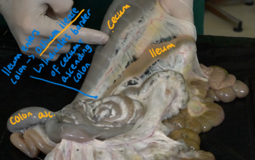
The position of cecum?
Blind part of intestine.
eq, ru +car: lies in right half of abd. cavity
su: left half
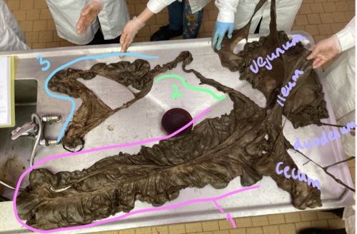
The parts of colon?
1. colon ascendens (cranially)
2. colon transversum (runs from right to left side)
3. colon descendens (caudally on left side)
Eq + RU: sacculations - Haustra structures, with teniae (longitudinal muscle fibers)
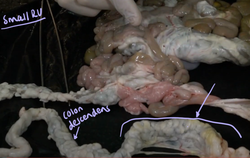
The position of rectum?
caudal continuation of colon descendens in the pelvic cavity
lies on roof of pelvic cavity, continuing with canalis analis and ending with anus.
except of small ru + fe: rectum dilates before joining the canalis analis and forming the ampulla recti.
Anus is closed by which 2 muscles?
m. spinchter ani internus
m. sphinchter ani externus
mucous membrane of canalis analis may be divided into 3 segments?
zona columnaris ani (only in su + car) → forming:
zona intermedia
zona cutanea - surrounding anus
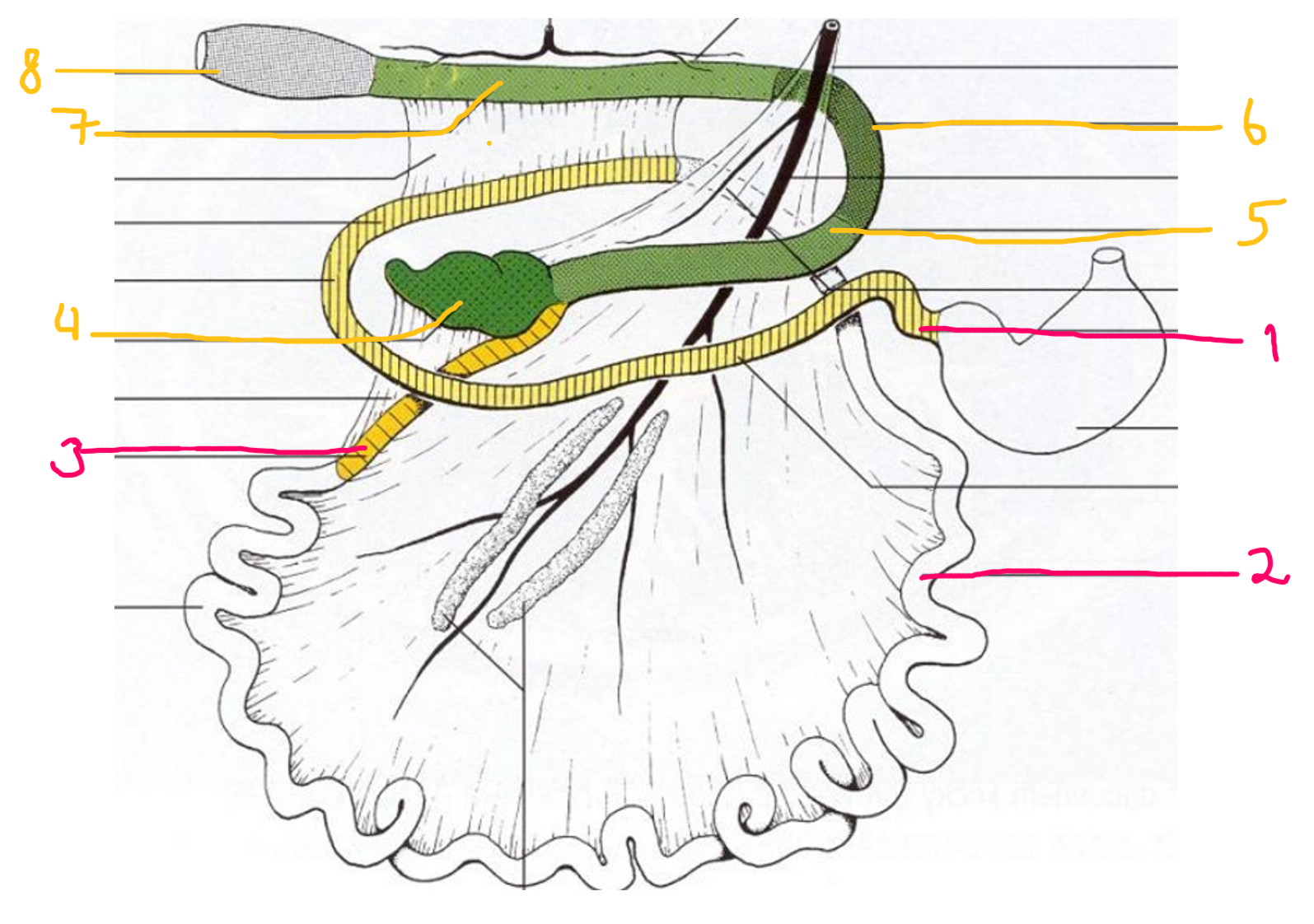
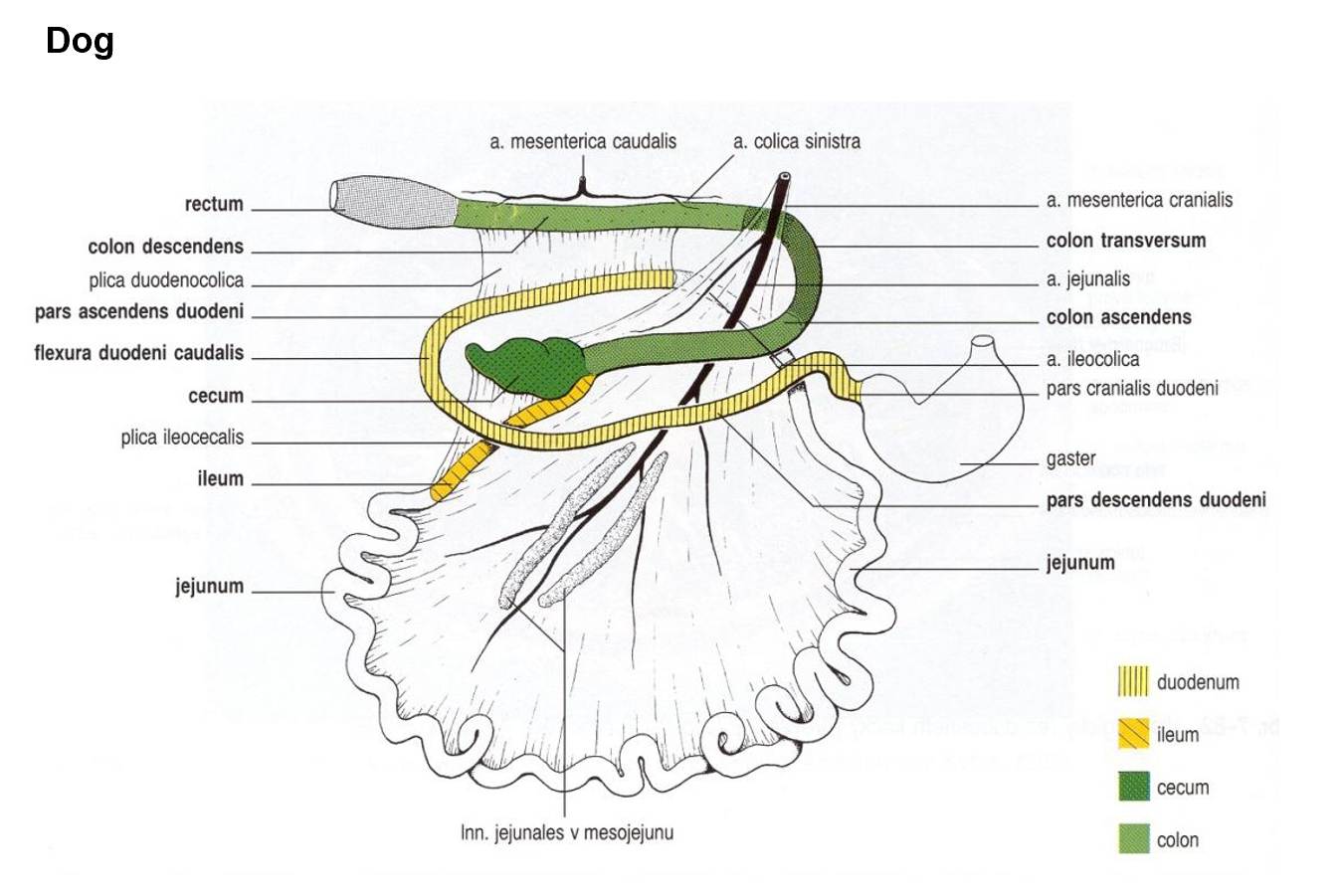
The large intestine of carnivores parts? (pink - small, yellow-big)
duodenum: pars cranialis, from pars pylorica ventriculi. Forming flexura duodeni cranialis, pars descendens, running causally, forming flexura duodeni caudalis., pars ascendens, running cranially and forms the flexura duodenojejunalis which is ventrally to jejunum.
jejunum: continuing from flexura duodenujejunalis,
ileu
cecum (right half of cavity)
colon ascendens (shortest part, right on roof of cavitas abdominalis)
colon transversum (turning left)
colon descendens (caudally on roof of left half of cavity)
rectum + canalis analis short → amupulla recti is formed.
Zona cutanea contains gll. circumanales in ca.
sinus paranales with gll. sinus paranalis - in bw. the m. spinchter ani internus et externus (for scent marking)
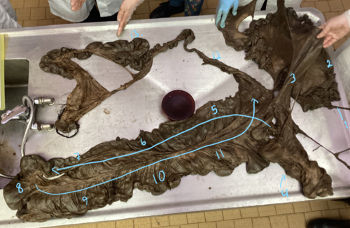
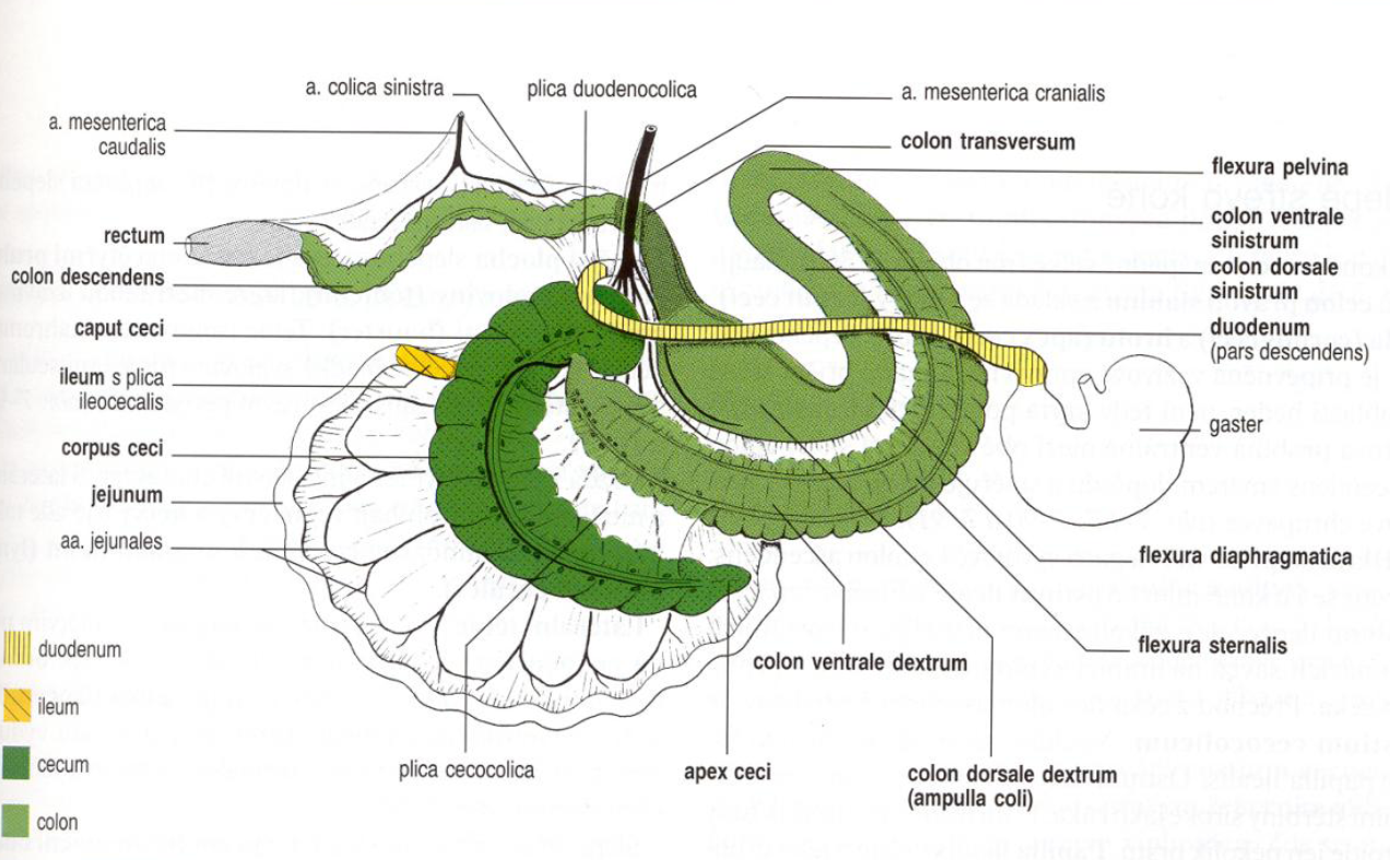
Describe large intestine of horse. (also parts of small intestine)
duodenum: pars cranialis with flexura duodeni cranialis forming ansa sigmoidea, pars descendens forming flexura duodeni caudalis and pars ascendens with the flexura duodenojejunalis (meeting with jejunum)
jejunum: jejunal folds, no regular position in eq.
ileum: plica ileocecalis, ostium ileale on papilla ilealis.
cecum: apex, corpus and basis ceci, consisting of 4 rows of Haustra and 4 teniae, internally of plica semilunares ceci. Has ostium cecocolicum and ostium ileale ventrally on papilla ileale. Has the curvatura ceci major et minor on the sides.
ostium cecocolicum is closed by valva cecocolica
plicae semilunares seci (folds of cecum)
colon: consisting of colon crassum et tenue. The colon crassum starts as colon ventrale dexter (4 rows of Haustra + 4 teniae)
plica intercolica - ventral loop is attached to dorsal loop by this
colon crassum starts at ostium cecocolicum, at collum colli, continuing as the colon ventrale dextrum.
flexura sternalis (4 rows of haustra and 4 teniae)
plica cecocolica - colon ventrale dextrum turns, and comes in contact with cecum by this.
colon ventrale sinister (4 rows of haustra and 4 teniae)
flexura pelvina (1 teniae)
colon dorsale sinister (1 teniae)
Flexura diaphragmatica (3 teniae)
colon dorsale dexter (3 teniae) - also known as ampulla colli.
colon transversum (from right to left)
colon tenue (colon descendens) consisting of 2 teniae.
teniae libera + mesocolica
lastly rectum, not seen here.
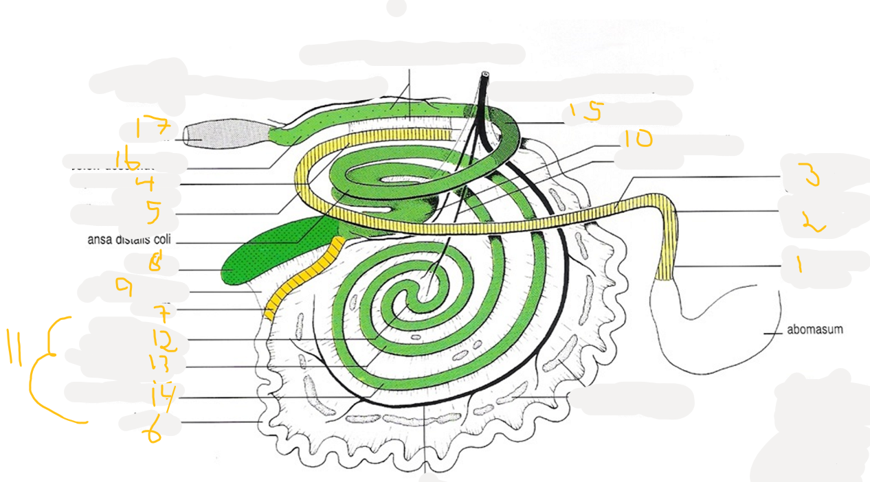
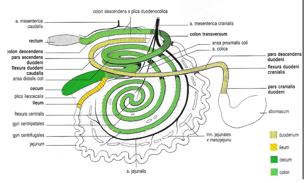
Describe the large intestine of ruminants.
Pars cranialis duodeni - with ansa sigmoidea
flexura duodeni cranialis
pars descendens
pars ascendens with flexura duodenojejunalis
flexura duodeni caudalis (before pars ascendens)
Jejunum
ileum - can see its ileum as the plica ileocecalis (a fold) is seen between this structure and the cecum.
cecum
plica ileocecalis (fold)
ansa proximalis colli (first part of colon ascendens in ru)
sub-part of colon ascendens. Consist of S-shaped curve with 3 segments, gyrus ventralis, gyrus medius and gyrus dorsalis.
ansa spiralis colli (second part of colon ascendens in ru)
sub-part of colon ascendens. It forms an elliptical disc, consisting of gyri centripetalis (13), flexura centralis and gyri centrifugales (14). And flexura centralis (12)
Part before nr. 15 → ansa distalis colli
colon transversum (from right to left)
Colon descendens - forms some loops “colon sigmoideum”
rectum (forms ampulla recti., absent in sheep and goat)
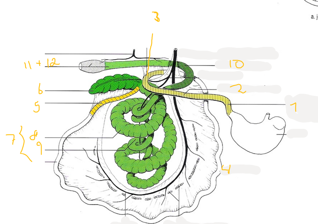
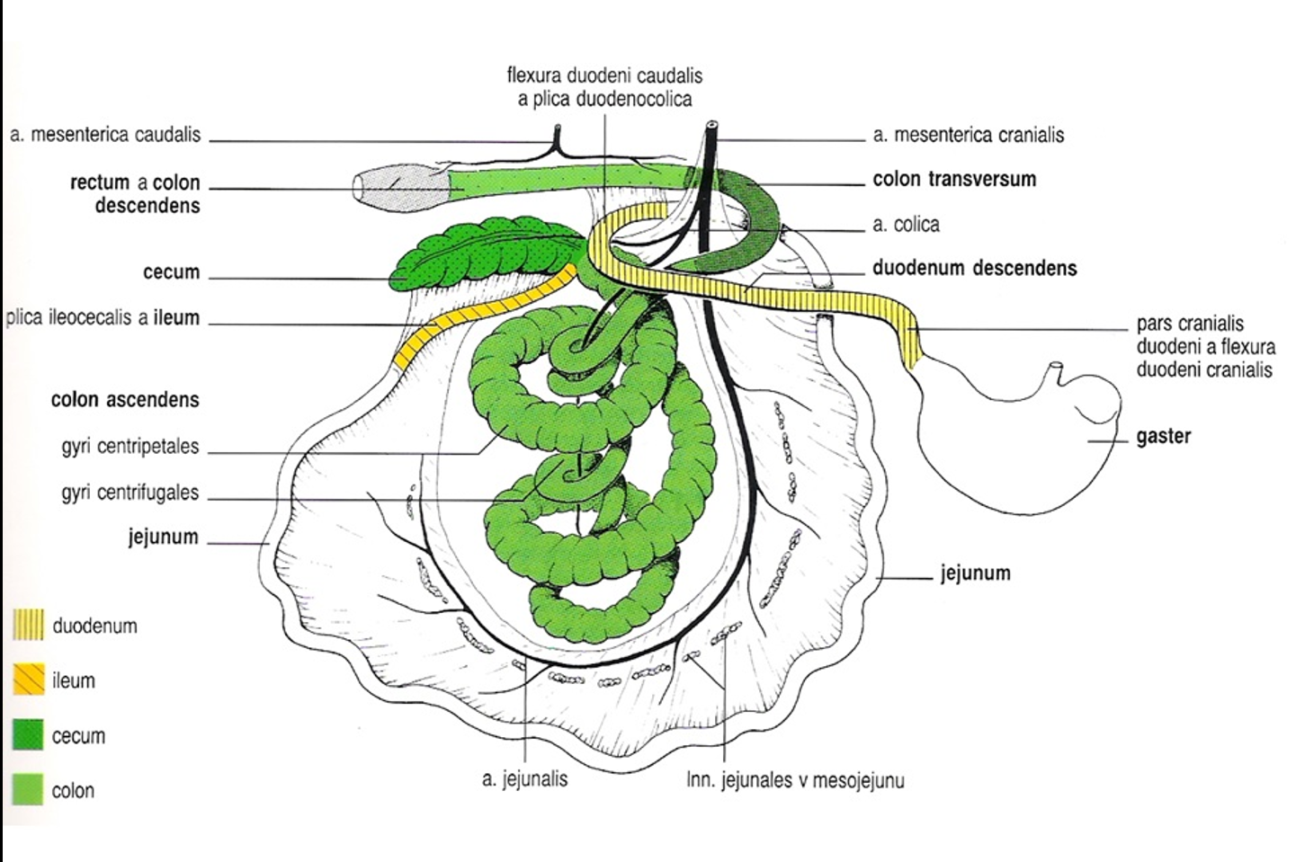
Describe the large intestine of the pig.
pars cranialis duodeni and the flexura duodeni cranialis
pars descendens duodeni
flexura duodeni caudalis
jejunum
ileum
cecum (left half of cavity, 3 hausta + 3 teniae)
Ansa spiralis colli (colon ascendens) - has the structures: (8) gyri centripetales (has 2 rows of haustra and 2 teniae), flexura centralis and (9) gyri centrifugales.
colon transversum
colon descendens
rectum
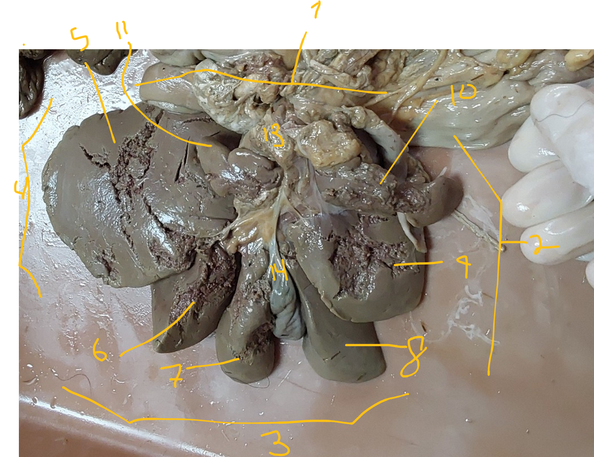
Describe the liver of dog (carnivores)
margo dorsalis
margo dexter
margo ventralis
margo sinister
lobus hepatis sinister lateralis
lobus hepatis sinister medialis
lobus quadratus
lobus hepatis dexter medialis
lobus hepatis dexter lateralis
processus cadatus (of lobus caudatus)
processus papillaris (of lobus caudatus)
porta hepatis - centre of facies visceralis, hepatic arteries, veins and ducts go through here.
we continue from here into ductus hepaticus sinister et dexter which fuse to form ductus communis further down, sends bile from parenchyma, and leaves through porta hepatis → collum vesicae felleae → ductus cysticus continues from here - which together with ductus communis forms ductus choleductus.
vesicae felleae, with fundus vesicae felleae
formed in the fossa vesicae felleae - on facies visceralis of liver.
absent in eq.
Facies visceralis is shown - touches other organs, while the opposite side, facies diaphragmatica touches the diaphragm.
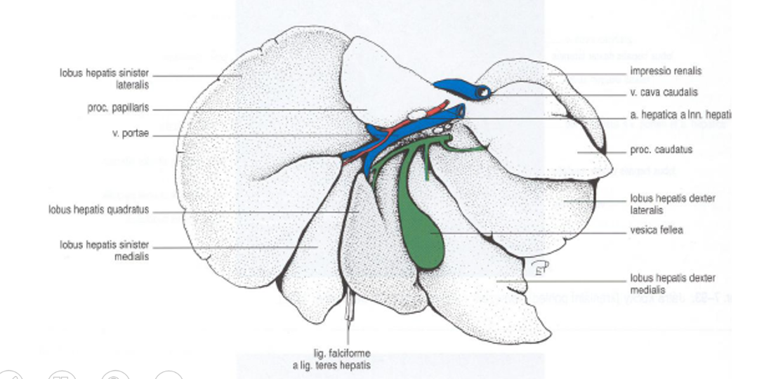
Around the entrance of bile duct into the duodenum, circular muscle fibres of the duct forms what?
m. sphincter ductus choledochi
what is the general ligaments of liver?
lig. teres hepatis + lig. falciforme → running in bw. lobus hepatis sinister and the lobus quadratus.
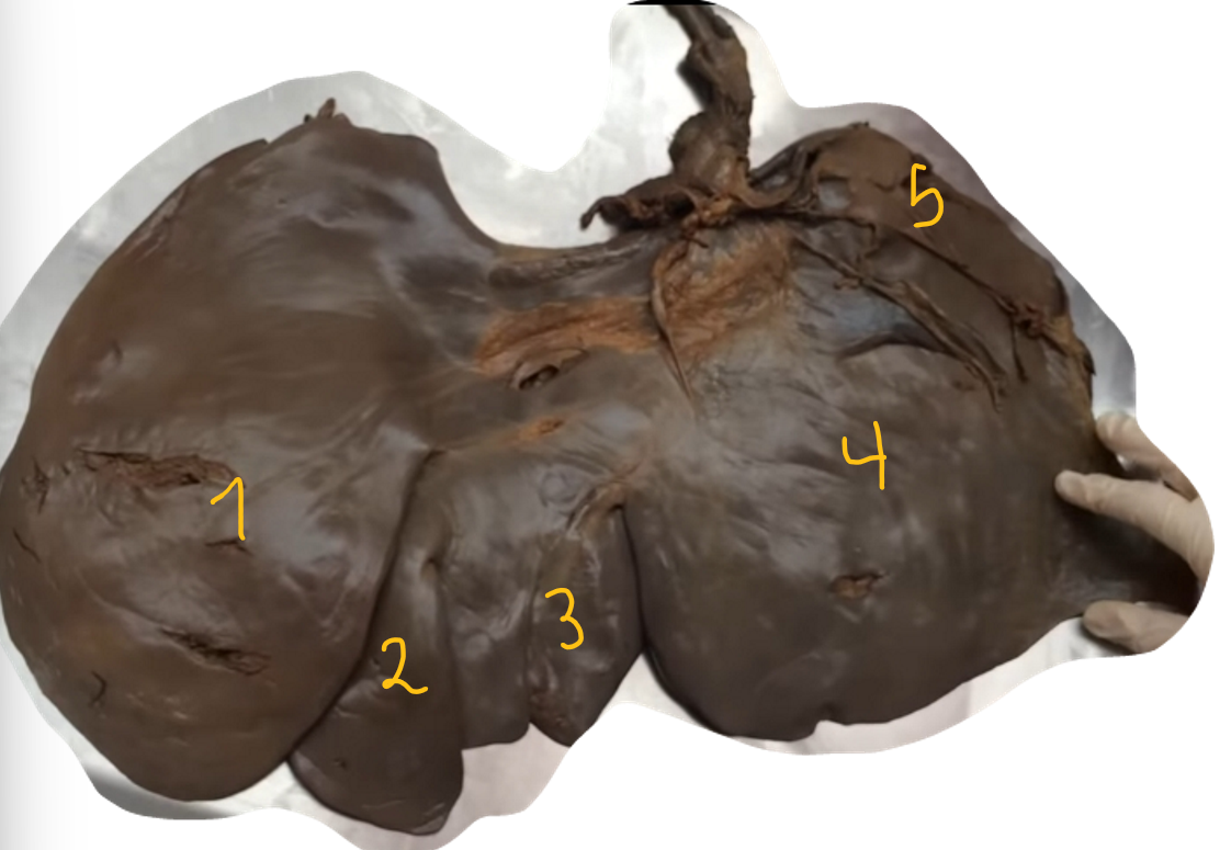
Lobulation of horse liver?
lobus hepatis sinister lateralis
lobus hepatis sinister medialis
lobus hepatis sinister quadratus
lobus hepatis dexter → not divided in eq.
processus caudatus (of lobus caudatus)
Facies visceralis seen, opposite is facies diaphragmatica. Gall bladder is absent in horse liver.
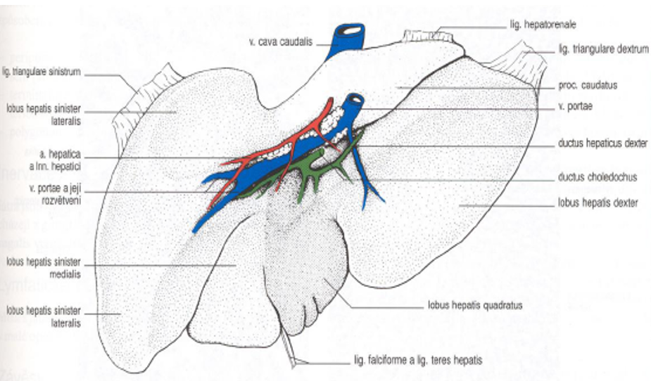
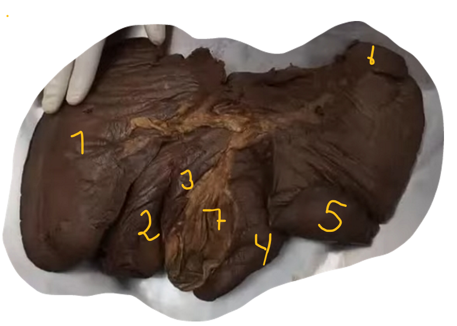
Lobulations in pig liver.
lobus hepatis sinister lateralis
lobus hepatis sinister medialis
lobus quadratus
lobus hepatis dexter medialis
lobus hepatis dexter lateralis
processus caudatus (of lobus caudatus)
vesica fellea
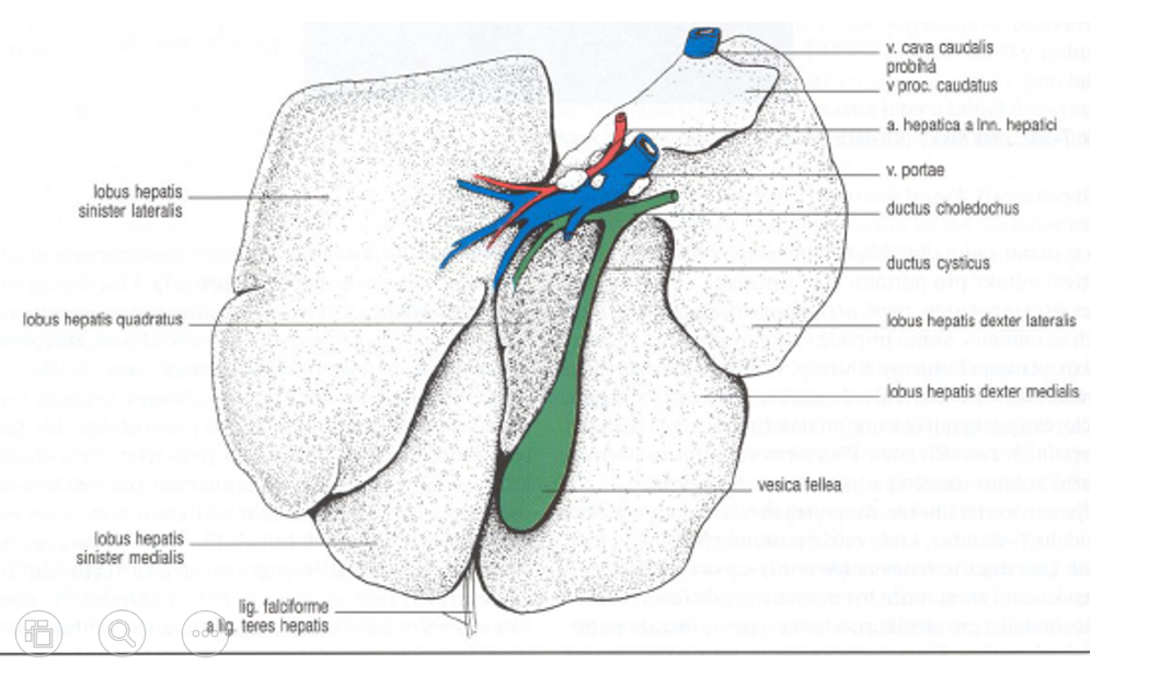
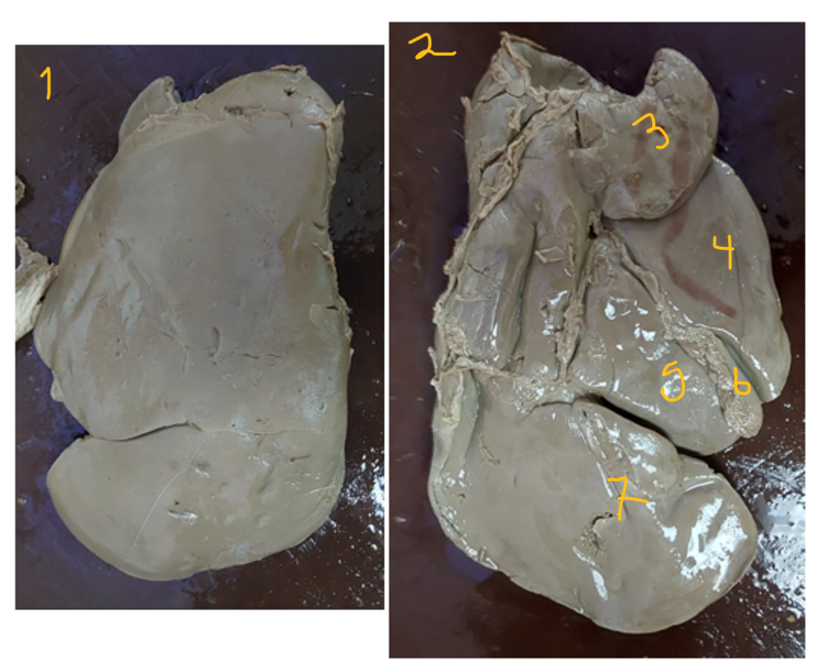
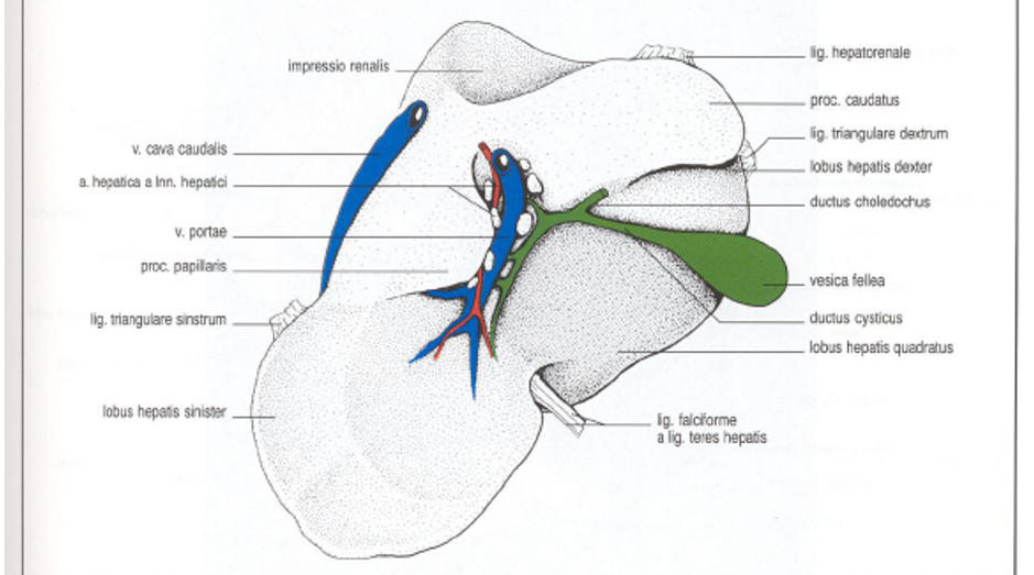
Lobulation of ruminant liver.
facies diaphragmatica
facies visceralis
processus caudatus + processus papillaris (on the side there, towards margo dorsalis)
lobus hepatis dexter
lobus quadratus
vesica fellea
lobus hepatis sinister
surfaces of liver are covered by?
Peritoneum, which forms the tunica mucosa.
Fused with tunica fibrosa, that enclosed the whole organ.
lobuli hepatis - the smaller units of liver parenchyma.
The ligaments of liver? (attachments)
lig. falciforme (at facies diaphragmatica)
lig. teres hepatis (at facies diaphragmatica)
lig. hepatoduodenale
lig. hepatogastricum
this together with lig. hepatoduodenale forms omentum minus
lig. coronarium (at margo dorsalis)
lig. tranngulare sinistrum et dextrum (margo dorsalis)
area nuda - area on facies diaphragmatica of the liver, for the diaphragm and liver to lie in direct contact and adhere.
extra: lig. hepatorenale (proc. caudatus)
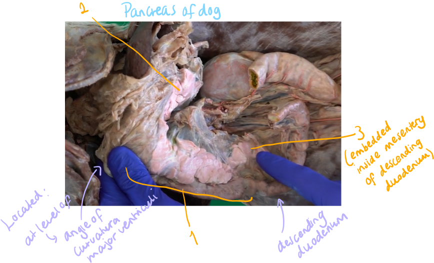
The pancreas of a dog.
corpus pancreatis → pancreas: locates beside duodenum, going from pars pylorica ventriculi to pars descendens duodeni.
incisura pancreatis (ru, car)
anulus pancreatis (eq, su
through either incisura or anulus, runs the portal vein.
lobus pancreatis sinister
lobus pancreatis dexter - embedded inside mesentery of pars descendens duodeni.
Ducts which transports the juice into duodenum:
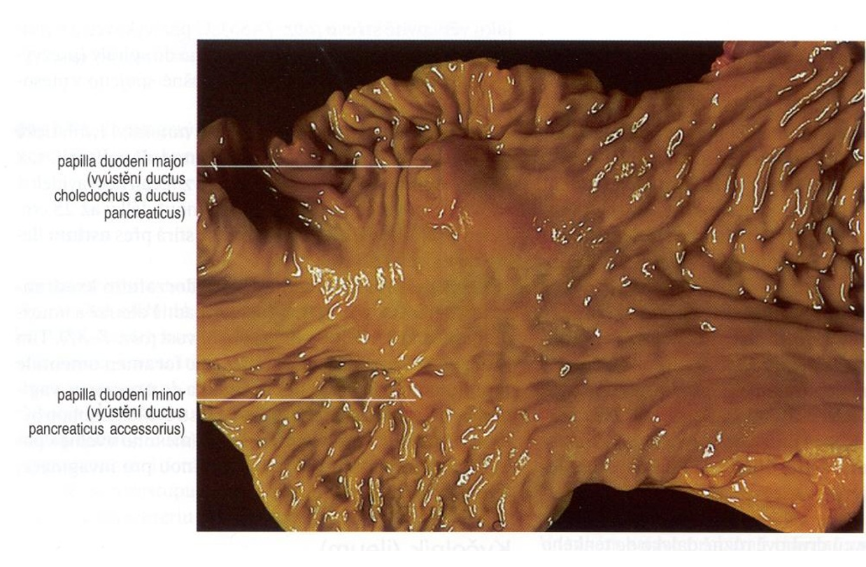
ductus pancreaticus, opening together with ductus choledochus on papilla duodeni major.
ducturs pnacreaticus accessorius , enters on papilla duodeni minor
car: usually two ducts carrying the juice, also in horse, bovine only 1, su only 1.
function: exocrine function, produces pancreatic juice, and endocrine function by langerhan`s islands, producing hormones.
