BRACE YOURSELF!!! Its about to get rocky!! (Extracellular accumulations)
1/79
There's no tags or description
Looks like no tags are added yet.
Name | Mastery | Learn | Test | Matching | Spaced |
|---|
No study sessions yet.
80 Terms
extracellular accumulations
what are all of these?: Hyaline Substances, Amyloid, Fibrinoid Necrosis/Change, Collagen/Fibrosis, Fatty Infiltration, Gout/Pseudogout, Cholesterol, Calcification, Heterotopic Ossification
hyaline substances
homogenous, eosinophilic, and translucent appearance to a cellular or extracellular substance
EOSINOPHILIC
Proteins are stained _________ in hyaline substances?
True
true/false: Hyaline is an ADJECTIVE to describe a substance, but often means theres protein somewhere
protein in renal tubules, serum/plasma in vessels, collagen fibers, thickened basement membranes, corpora amylacea
What are some examples of hyaline substances?
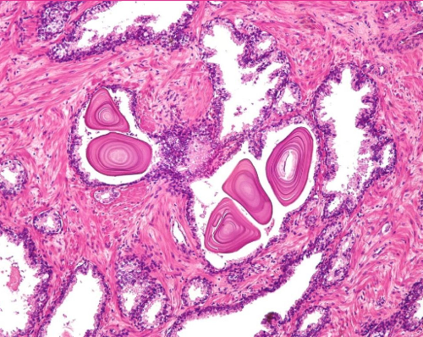
Concentric layers of glycoprotein in glands or CNS
Corpora amylacea? Wtf? Define:
hyaline casts
What extracellular inclusion?
corpora amylacea
What extracellular inclusion?
FALSE; named for it's ability to stain with iodine
true/false: amyloid is named for its "starch like" qualities
mis-folded proteins
What is amyloid?
highly ordered, fibrillar polypeptide chains, cross beta sheets, pathogenesis, morphological appearance
Amyloid is: Unfolded or partially unfolded proteins or peptide fragments
They are ___________, generic structure of ________________________
Rich in ______________________
Biochemically diverse with common __________ and ____________________
true
true/aflse: myloid proteins can self replicate
self-replicaiting template, failure to degrade, genetic mutations, overproduction from abnormality in synthesizing cell, loss of chaperone molecules in assembly process
There are 5 "causes" of amyloid on slide 10. Read if you want but they're all ways the body screws up proteins; listed VERY generally here:
precursor peptide/protein
Amyloid types are classified based on the biochemical identity of their _______________________
immunoglobulin light chains, plasma cells
AL type Amyloid is from __________________ derived from __________
Primary
is type AL amyloid primary or secondary?
dyscrasia, neoplasia, light chain fragments
AL is produced by plasma cell _________ or _______________. Where the abnormal plasma cells secrete the _________________ into circulation
serum amyloid A protein, hepatocytes, high-density lipoproteins
Type AA amylase, is a ________________________ produced by ________________ and bound to ____________________ in circulation
secondary; associated with other proteins
is Amyloid type AA primary or secondary?
Chronic inflamation!!! (AL is typically more localized)
Type AA amyloid is more associated with ________________
true
true/false: Amyloid type AA can have Hereditary or familial forms also recognized like in Shar-Pei dogs and Abyssinian cats
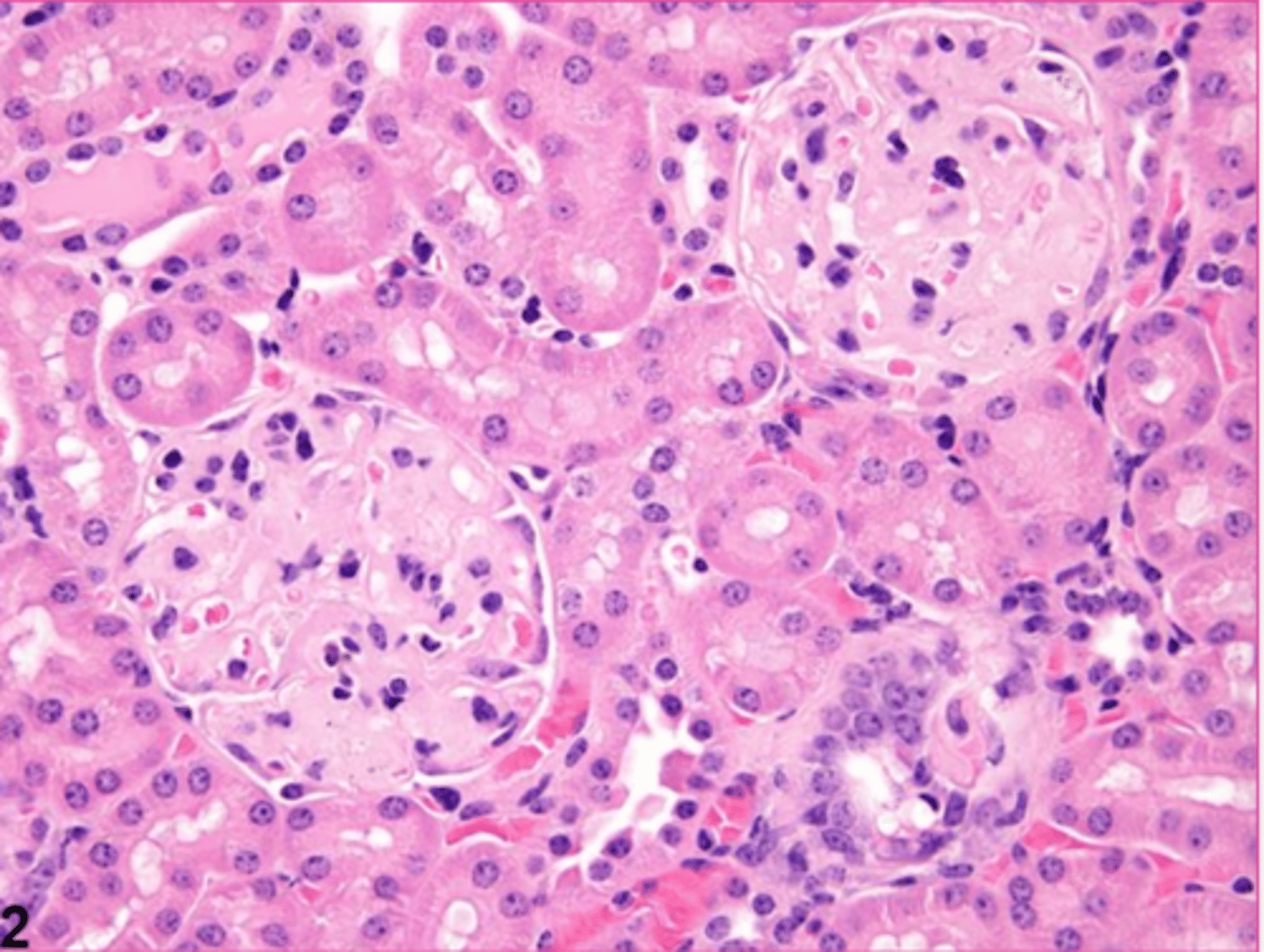
renal glomeruli, liver, spleen
AA amyloid is classically where?
read.
Read pls: (sorry) AA amyloid process:
taken up by cells and converted from alpha-helical confirmation to beta-sheets → forms oligomers that disrupt cell membranes and leave the cell → oligomers form fibrils that aggregate in deposits → disrupt tissue function
disrupts and damages
Deposition of amyloid ______- and ______________ tissues
systemic
what type of amyloid is Amyloid deposited around the body in multiple organs
localized
what type of amyloid is amyloid deposition restricted to tissues which produce the precursor protein or peptide
systemic
is systemic or localized amyloid more likely to be life threatening?
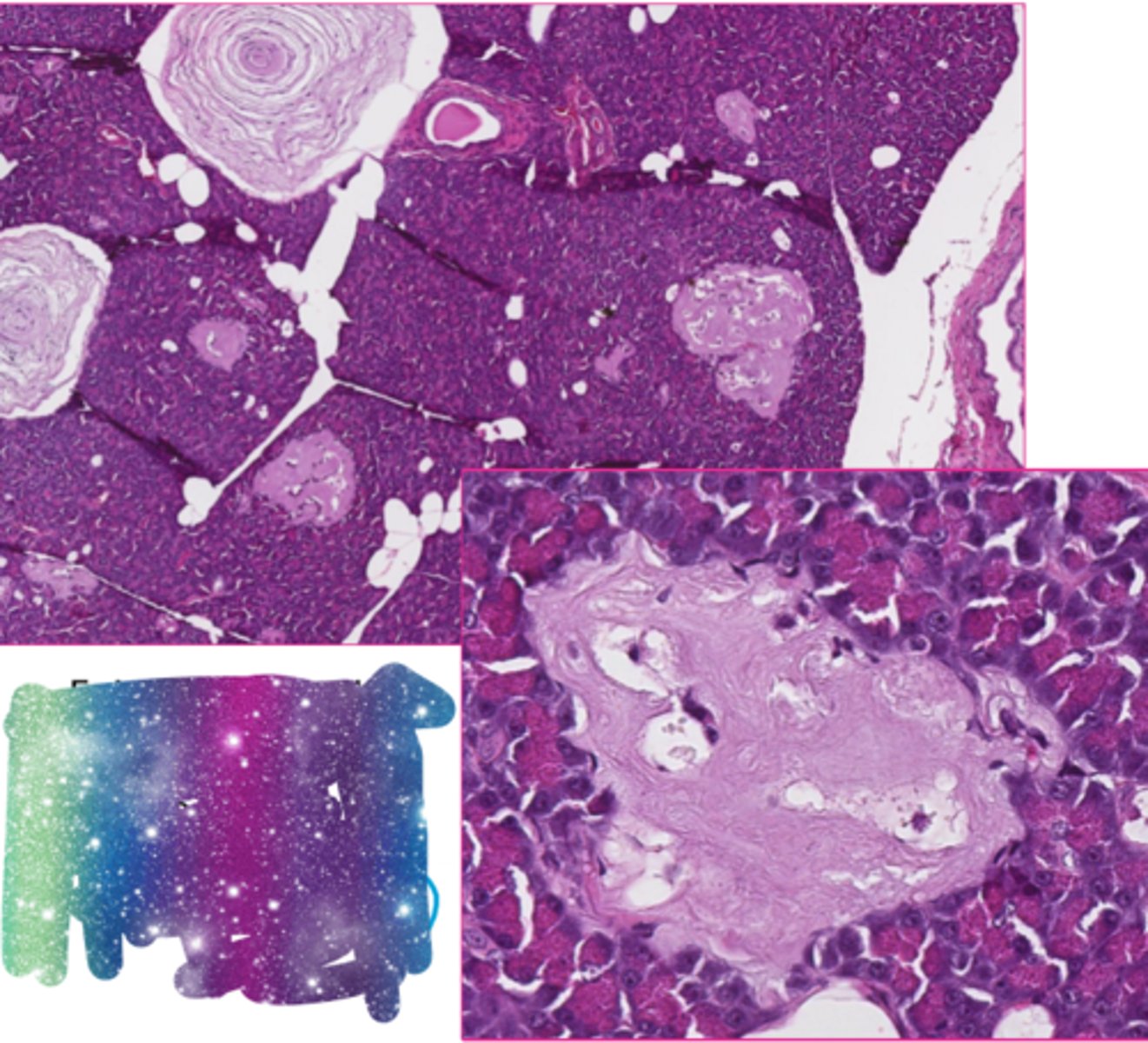
localized
These are both examples of what type of amyloid?
Islet amyloid peptide secreted by beta cells in pancreatic islets in cats
Beta-amyloid in cerebral cortex of aged dog with canine cognitive dysfunction
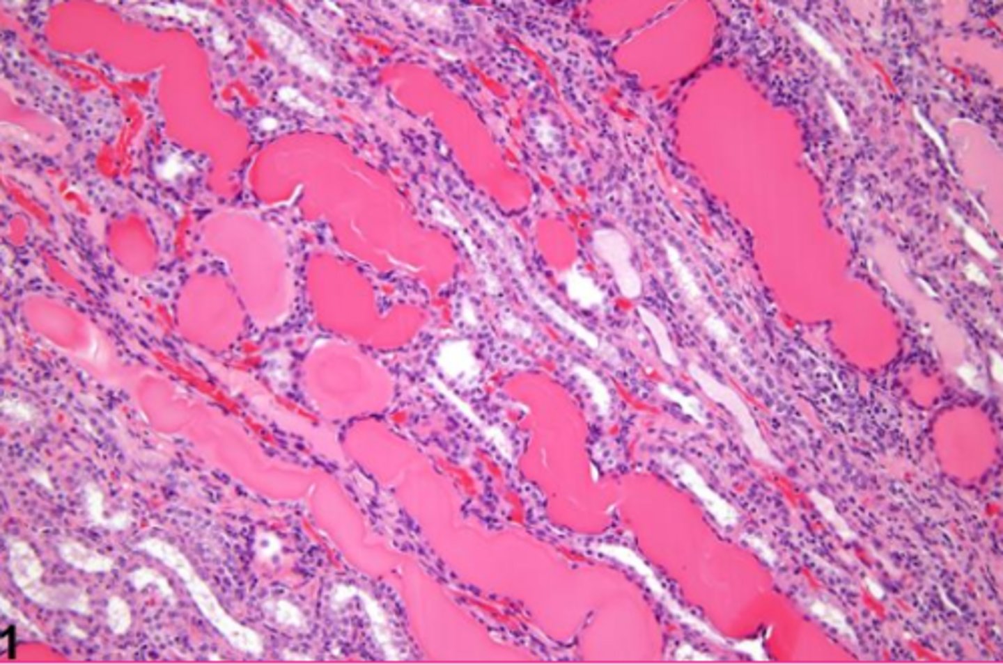
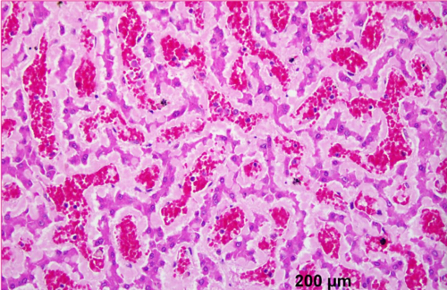
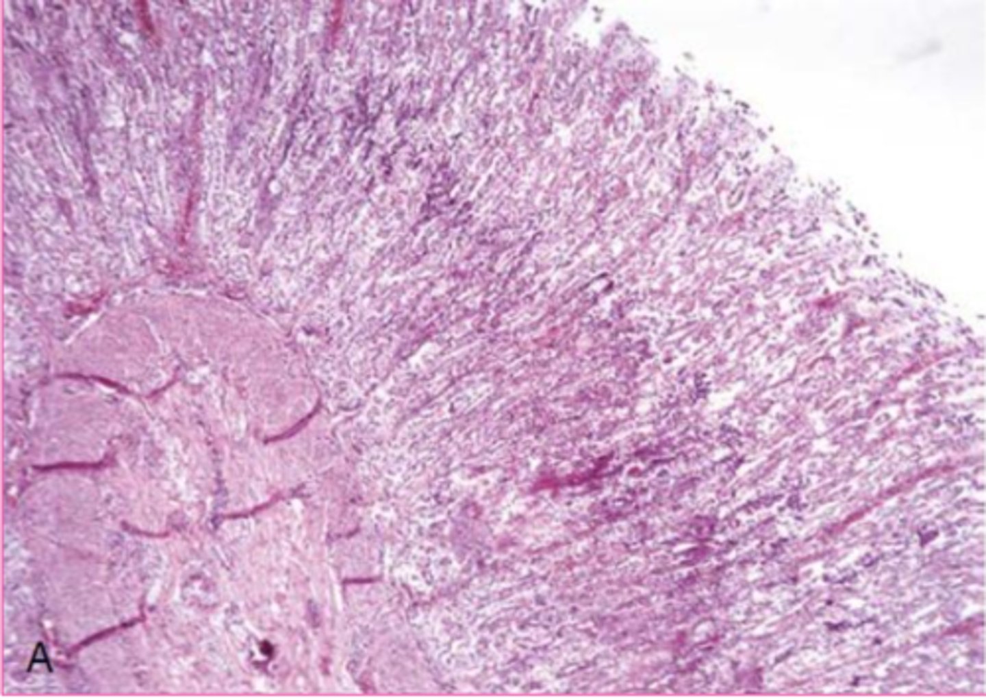
amyloid
describe the extracellular inclusion
amyloid
describe the extracellular inclusion
Congo red stain
what stain is uniquely associate with Amyloid inclusions?
apple green biferingence under polarized light
Congo red is orange-red on origional staining... but then you do something special to it. What does it look like and in what special conditions?
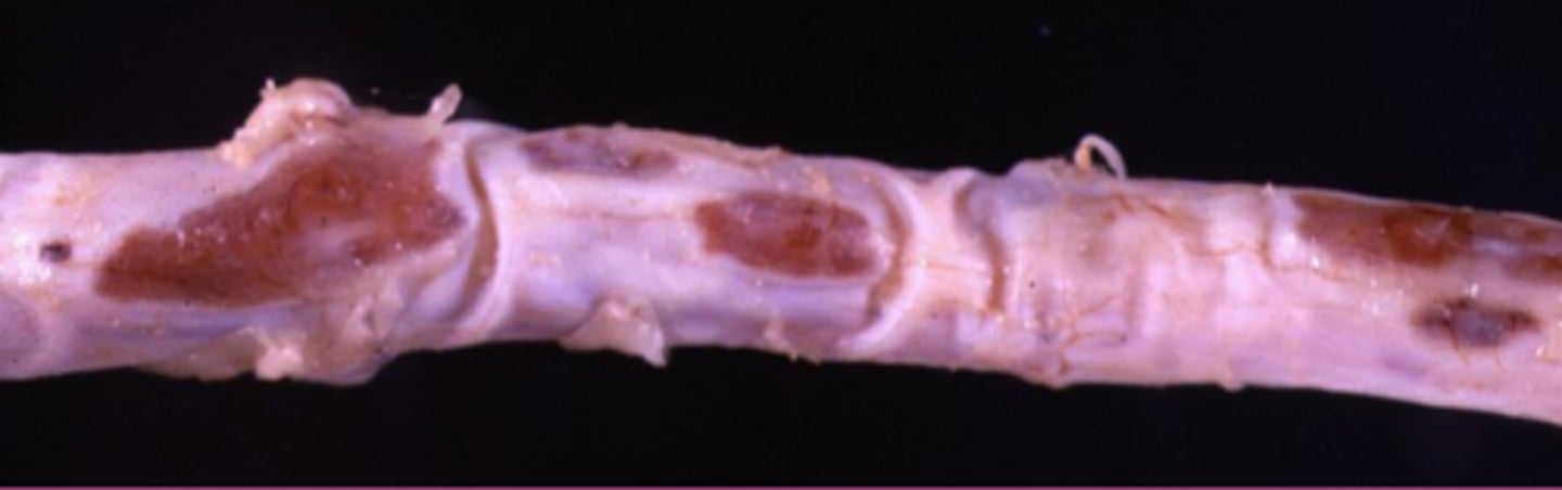
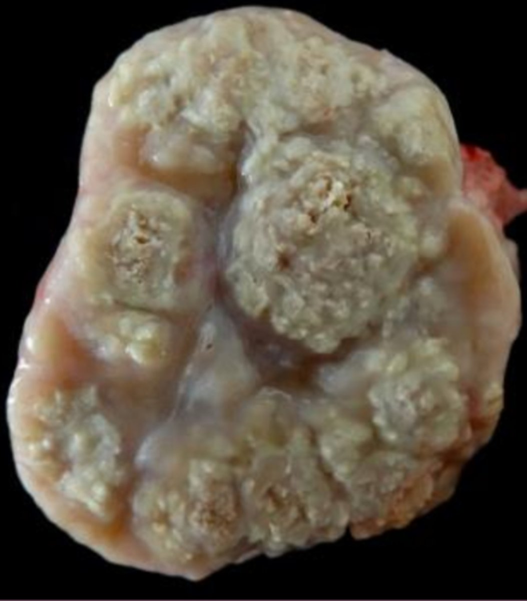
Firm, yellow, waxy, coalescing nodular or amorphous deposits
describe amyloid grossly
iodine, sulfuric acid
you pan amyloid lesions with ______ to get a yellow color, add _______ to get blue violet
conjunctival amyloidosis
Describe this lesion
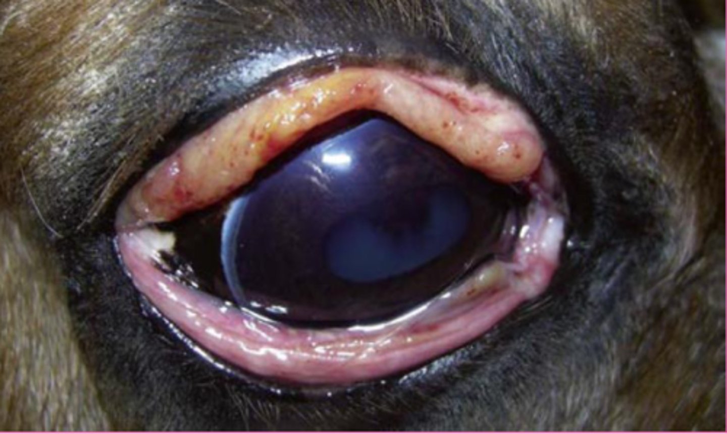
AA
Which type of amyloid?
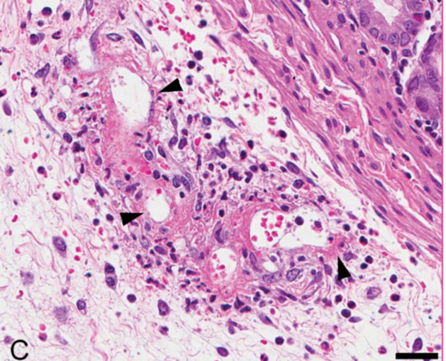
Fibrinoid necrosis
Type of extracellular change from the leakage of plasma proteins into the vessel wall
true
true/false: fibrinoid necrosis is related to inflammation, infection, trauma, or other injury
fibrinoid necrosis change
describe the extracellular accumulation
fibrosis
_________: excess in fibrous collagen (type I collagen) in the interstitium
fibroblasts, injury or inflammation
collagen/fibrosis is typically produced by __________________ after _________________ or ____________
scarring
collagen/fibrosis can also be present in ____________ that may compromise organ function
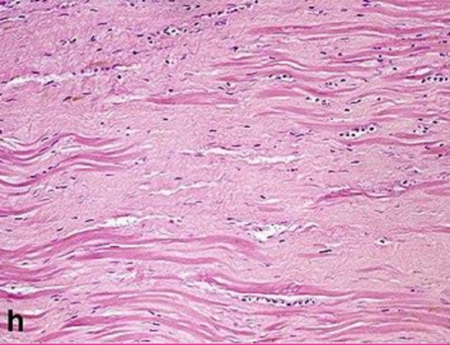
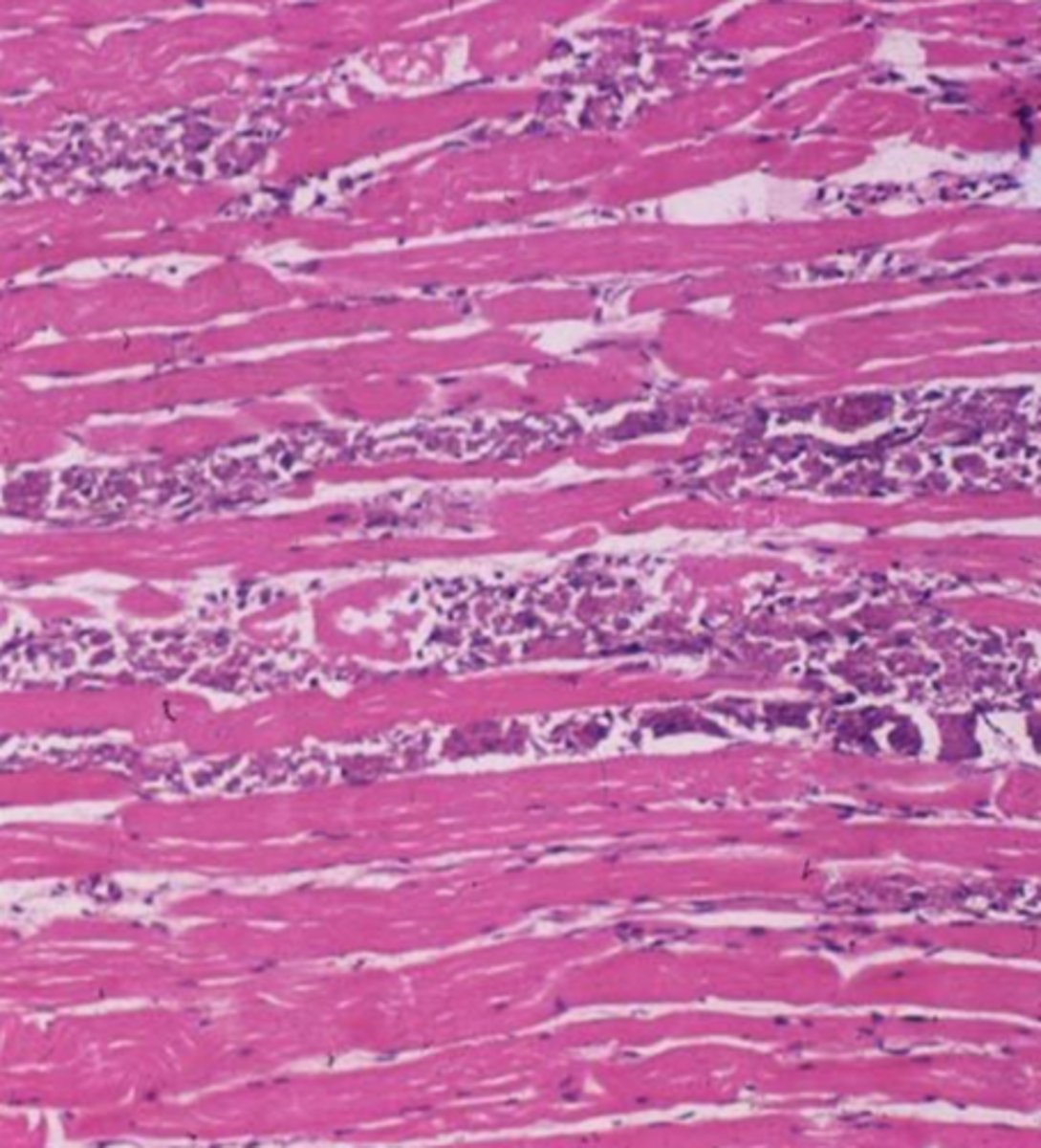
myocardial fibrosis
Describe this extracellular inclusion (heart muscle)
fatty infiltration
_______________: Increase in the number and/or volume of adipocytes in the interstitium of an organ or tissue due to obesity, cardio- or skeletal myopathies, or atrophied tissue
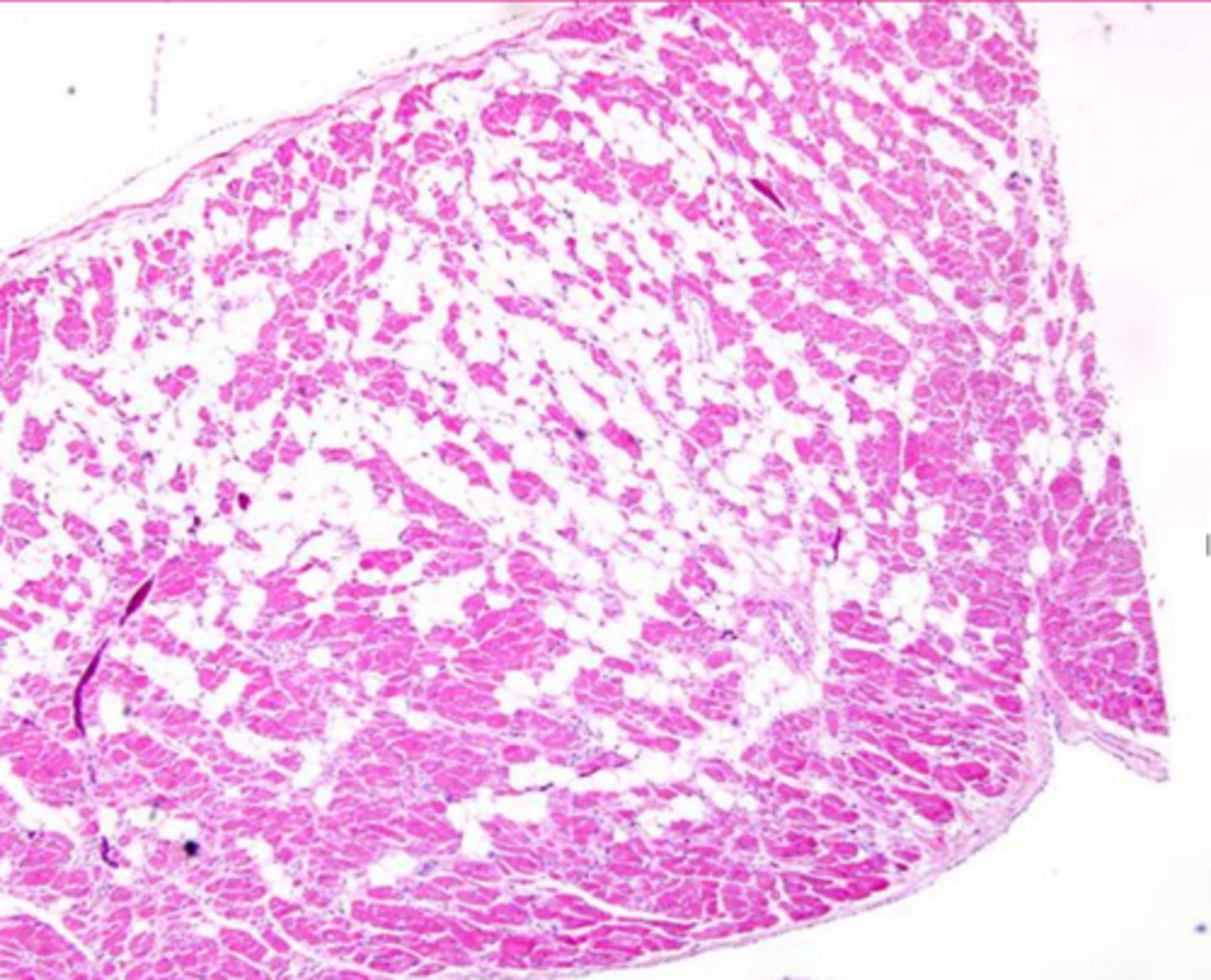
fatty infiltration
Describe the extracellular accumulation (this is a heart muscle)
gout
________: deposition of sodium urate crystals (urates) in tissues
birds, reptiles
gout is most commonly seen in animals in _______ and _______
pseudogout
___________________: deposition of calcium pyrophosphate crystals in tissues
tophi
What is the specific term for the inflammatory response ellicited from neutrophils/heterophils and macrophages by gout
articular, visceral
What are the two types of gout?
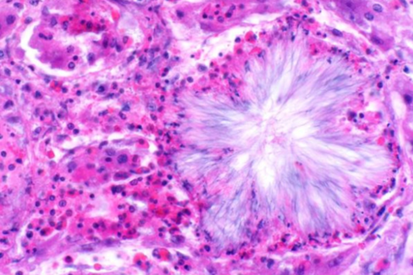
renal gout tophi
Describe this extracellular inclusion in the kidney
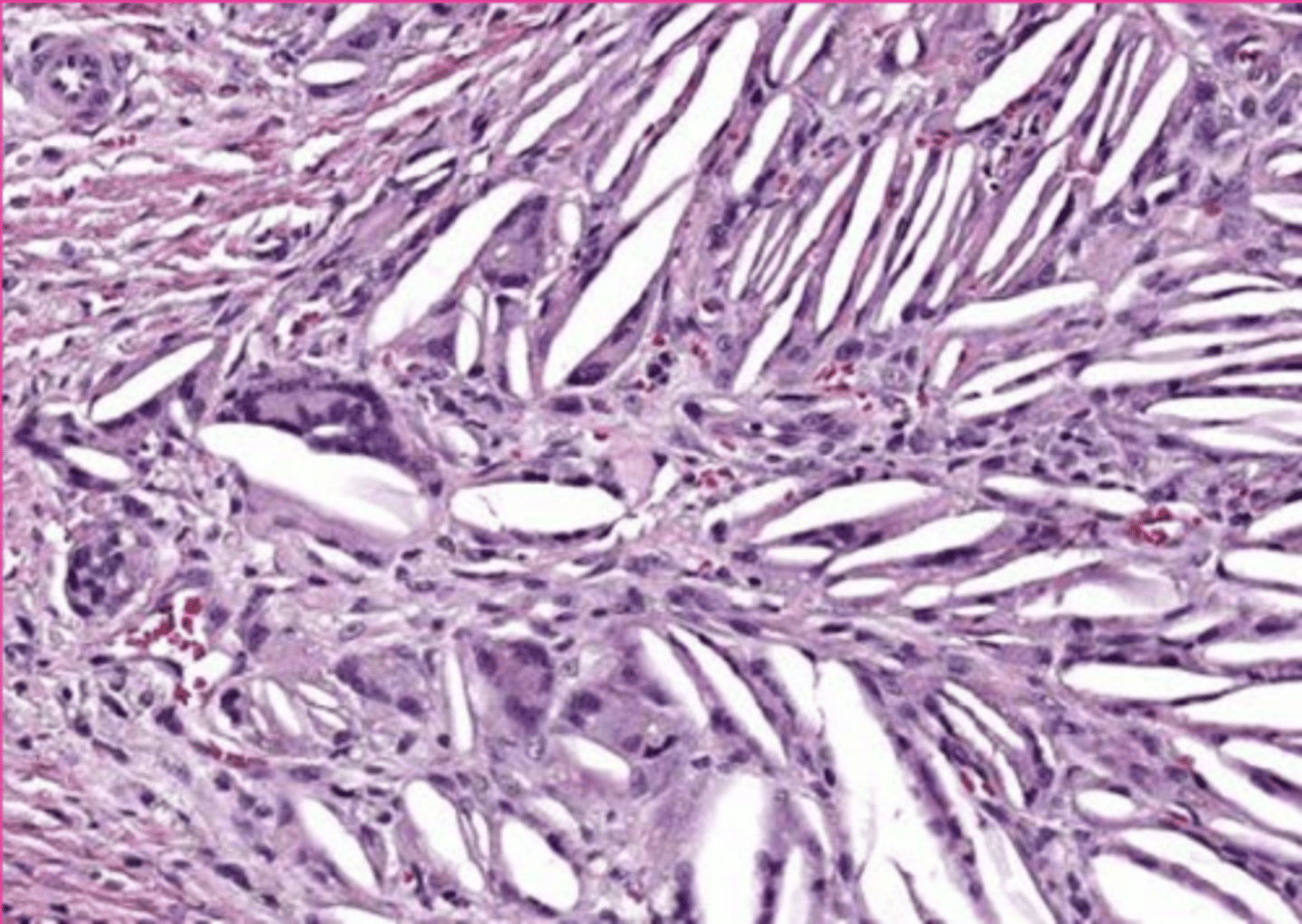
cholesterol
_______________ crystals can form at sites of hemorrhage or necrosis but dissolve during processing, forming acicular clefts seen in histologic sections
macrophages
what do cholesterol crystals attract?
cholesterol
what is this extracellular inclusion?
pathological calcification
____________________: deposition of calcium salts in soft tissues
metastatic calcification
type of calcification as the result of hypercalcemia
calcium and phosphorus
Metastatic calcification occurs as the result of an imbalance of _______ and _________
Primary hyperparathyroidism, Renal secondary hyperparathyroidism, Hypervitaminosis D (plant toxicosis, rodenticides), Paraneoplastic syndromes
What are some causes of metastatic calcification
intima, tunica media, basophilic stippling
Microscopically, metastatic calcification looks like deposits in the _______ and _________ of vessels, with a subtle ________________
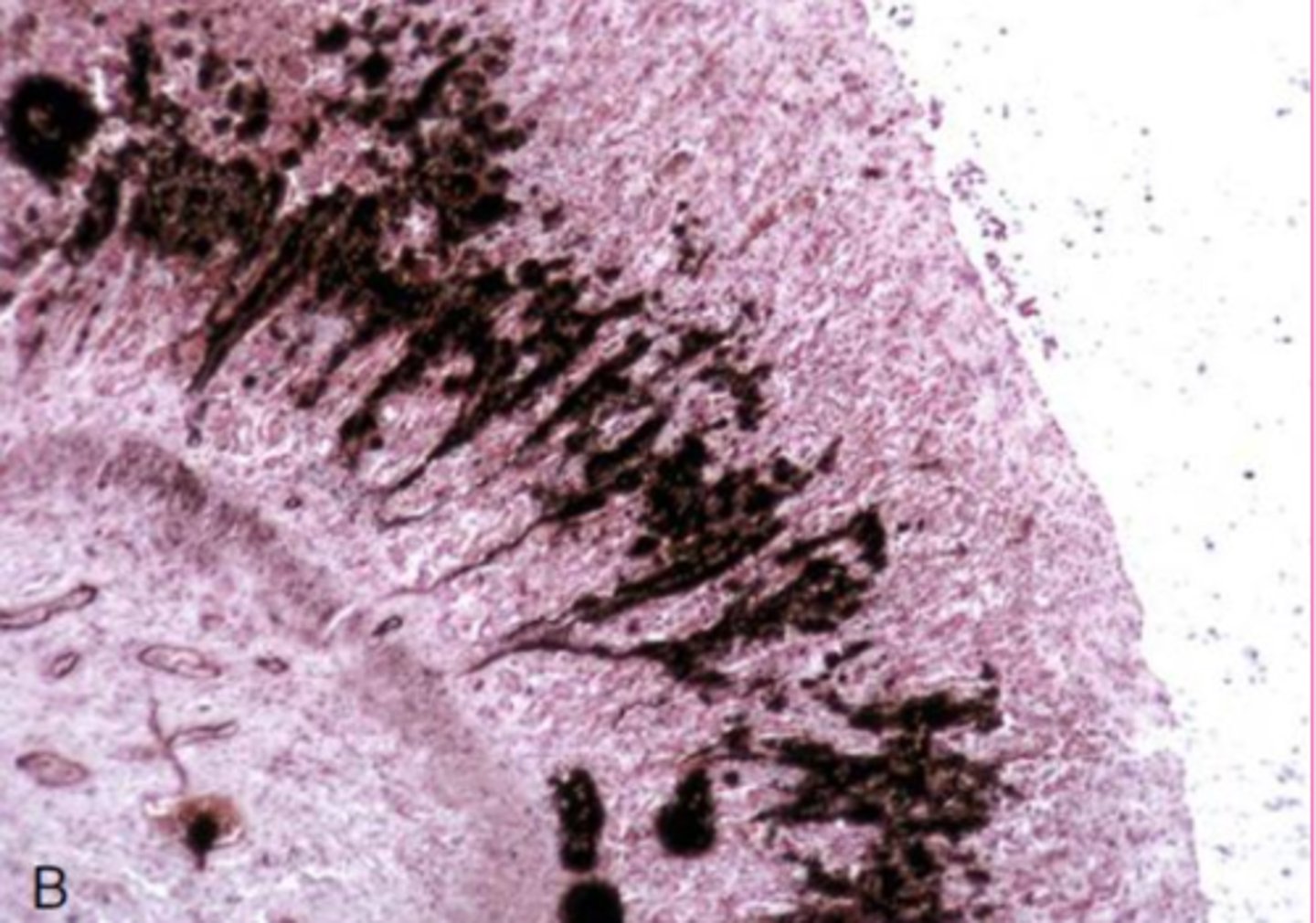
von kossa, the silver in it stains the calcium salts black
What is the specific stain associated with calcium? How does it work?
gastric calcification
Describe this extracellular inclusions?
Von kossa staining
this is calcification... why does it look like that?
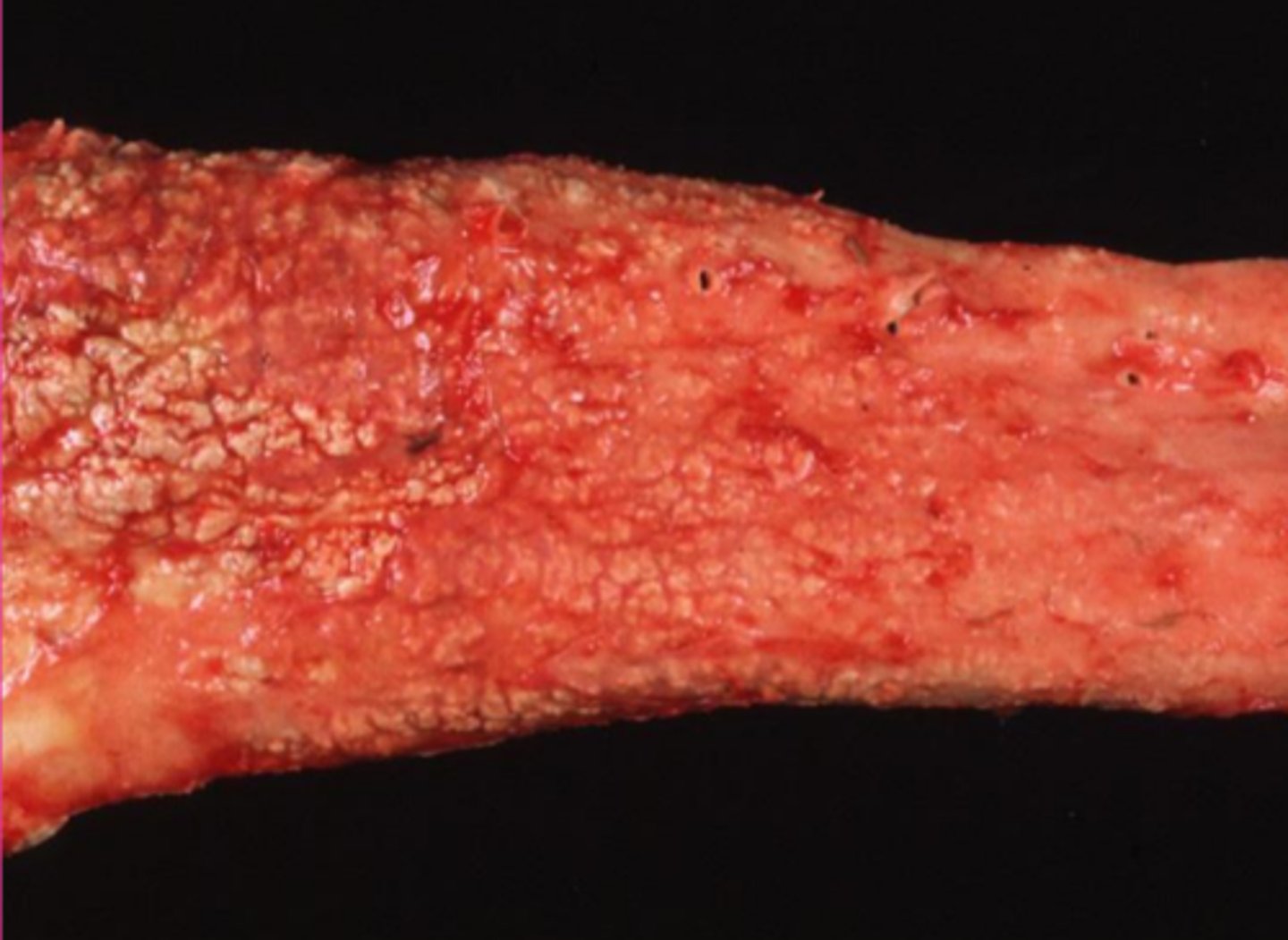
gritty white ish granules or streaks
Describe mestastatic calcification grossly
Metastatic calcification
what would make this aorta look like that?
dystrophic calcification
type of calcification of dead tissue as part of necrosis
loss of Ca balance during cell injury
What is the machanism for dystrophic calcification?
mitochondria, ER, cytosol
where is caclium sequestered in a healthy cell
white muscle disease
what is dystrophic calcification in striated muscle called? (from vitamin E/Se deficiency
necrosis, repetitive trauma
what are the main causes of dystrophic calcification?
basophilic stippling, mitochondria, basophilia
Dystrophic calcification is initially visible as _________ most profoundly in the [what organelle]. But then progresses to the whole cell and extracellular tissue as widespread intense ______________
dystrophic calcification
this is actually a caseous necrosis, but how would you describe the extracellular inclusions?
gritty white ish granules or streaks
describe dystrophic calcification grossly
dystrophic calcification
describe what extracellular inclusion you would expect to see with this lesion
dystrophic
is this dystrophic or metastatic?
heterotropic ossification
______________: formation of bony tissue at an extra-skeletal site
in chronic lesions of soft tissue calcification
Where do you find Heterotropic ossification?
False; does not always ossify
true/false: Pathological calcification, given enough time, will always calcify
hard spicules or nodules
What does heterotropic ossification look like grossly?
in lungs and dura of old dogs
Heterotrophic ossification can be an incidental finding on necropsy... for example, in:...
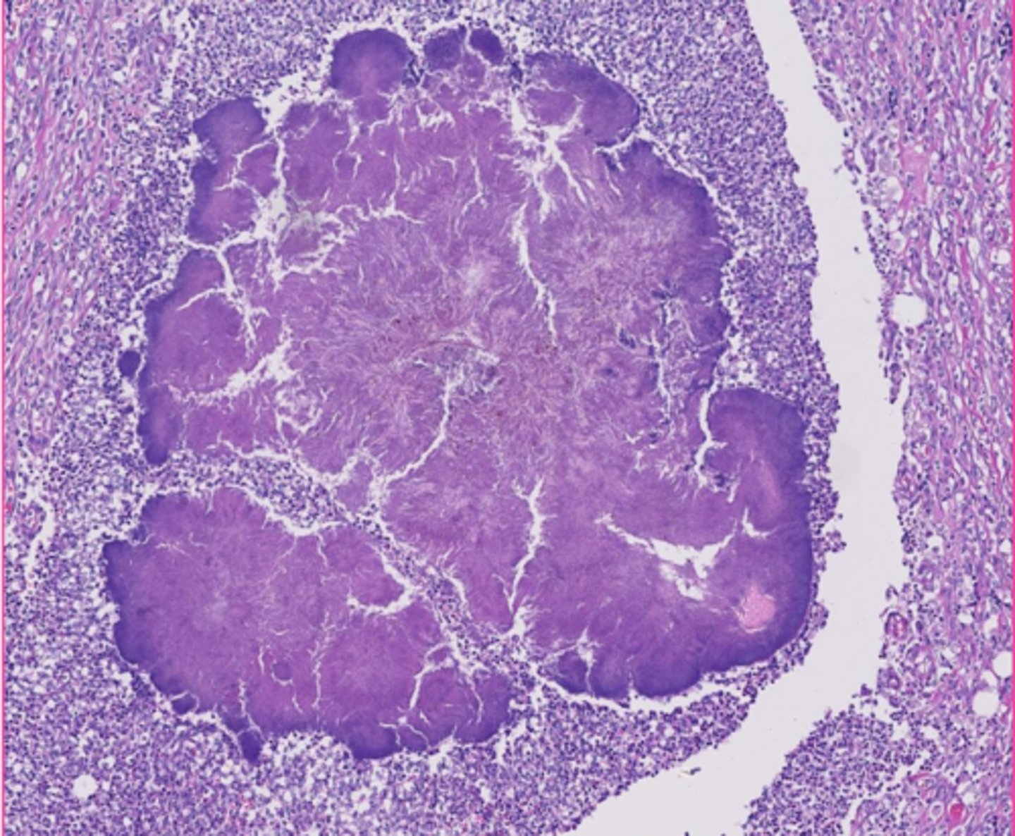
Dural ossification
What is this extracellular inclusion of the dura mater?