periodontal screening and recording ppt
1/42
There's no tags or description
Looks like no tags are added yet.
Name | Mastery | Learn | Test | Matching | Spaced |
|---|
No study sessions yet.
43 Terms
periodontal probing aids in
the diagnosis of gingival and periodontal diseases
what type of probe do we have
UNC
ideal probe
slender, blunt end- we use to examine depth and topography of gingival sulcus
materials probes can be made of
stainless steel or plastic
types of working end shapes for a probe
tapered, straight, curved, round, flat or rectangular
what probe is good for furcations
nabers probe
thinner probe means
more accurate reading
novatech probe
right angled probe for posterior teeth
what do you measure with probe
sulcus, pocket, recession, width of attached gingiva, size of lesions
diseases sulcus depth
4mm or greater
what are you looking for when using probe (not measuring)
consistency of gingival tissues, evaluate bleeding, post-treatment evaluation
WHO probe
screening probe
size of ball on end of WHO probe
.5 mm
for WHO probe from end of instrument to the beginning of black band is how many mm
.5-3.5 mm
for WHO probe end of black band to next marking is how many mm
5.5 to 8.5 mm
last markings on WHO probe is how many mm
8.5-11.5 mm
UNC 12 probe
used for a complete/comprehensive periodontal examination
for UNC 12 probe, how many mm are the larger black bands signifying
4-5, and 9-10 mm
length of probe is positioned…
as parallel as possible to the long axis of the tooth, and slightly angled under contact to access col
how to move probe
walking/bobbing stroke in 1mm steps
proper probing pressure
10-20 grams
how many sites on tooth do you measure and record
6
what does JE feel like when healthy
firm and resilient
parallelism (probing technique)
position probe as parallel as possible to long axis of tooth, must be parallel in proximal and smooth surface dimension
probing interproximally (probing technique)
postition probe parallel to proximal surface until touching contact area, then slant probe slightly to reach under contact area
adaptation (probing technique)
tip of probe should be kept in contact with tooth surface
walking stroke (probing technique)
cover entire circumference of base of sulcus pocket, keep probe against tooth surface whole time
how many areas is a tooth divided into
6
what is area one and 6 of tooth division
distofacial to midline of distal surface= 1, mesiolingual line angle to midline of mesial surface = 6
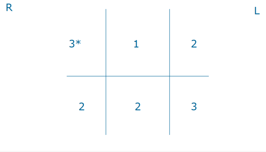
PSR
system for detecting PDZ and streamlining recordkeeping, screening technique
how to record probing depths
only deepest depth per sextant of tooth is recorded
example of PSR score
when to do PSR
initial visit for every new patient, and once a year for recall patients
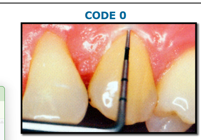
Code 0
colored area of probe completely visible
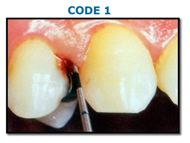
Code 1
colored area of probe completely visible, BOP
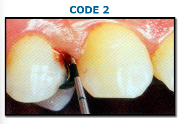
Code 2
colored area of probe completely visible, supra/ subgingival calculus or defective margins detected
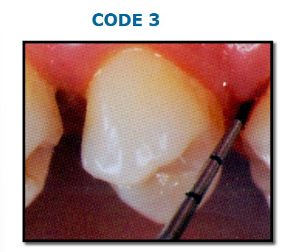
Code 3
colored area of probe partially visible, pockets depths between 3.5 to 5.5 mm
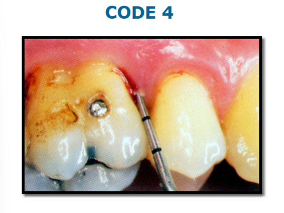
code 4
colored area of probe completely disappears, probing depths >5.5
when to add asterisk (*) to a PSR score
furcation, mobility, recession, mucogingival involvement
what to do when asterisk is added to sextant of 0,1, or 2
should make specific notation and/or treatment for condition as needed
what should you do when asterisk for code 3 or 4
comprehensive periodontal examination and charting are necessary to determine appropriate care plan
in a PSR score of 312/223 what would the first 2 on the second half indicate
mandibular right buccal
when to perform PSR
after dental charting