Foot & Ankle
 Call Kai
 Learn
Learn Practice Test
 Spaced Repetition
 Match
Match Flashcards
 Knowt Play
1/68

There's no tags or description
Looks like no tags are added yet.
Name | Mastery | Learn | Test | Matching | Spaced | Call with Kai | Chat |
|---|
No analytics yet
Send a link to your students to track their progress
69 Terms
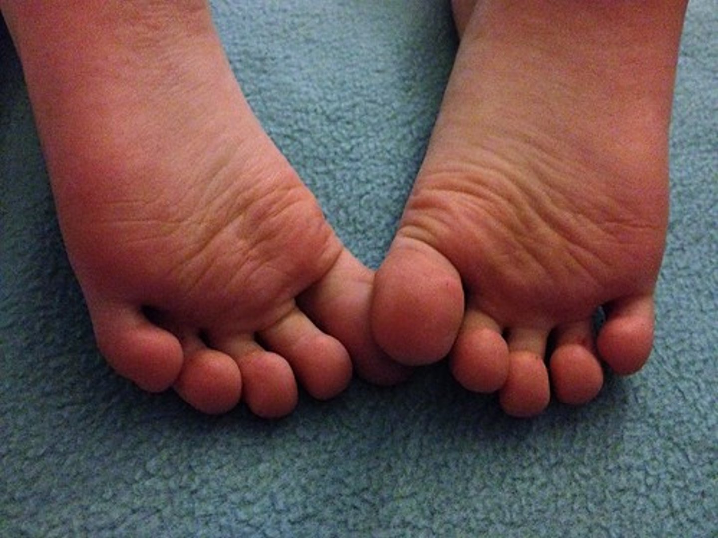
Plantar
the sole, or the bottom of the foot
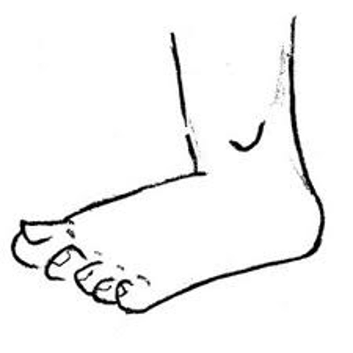
Dorsal
The top or superior portion of the foot
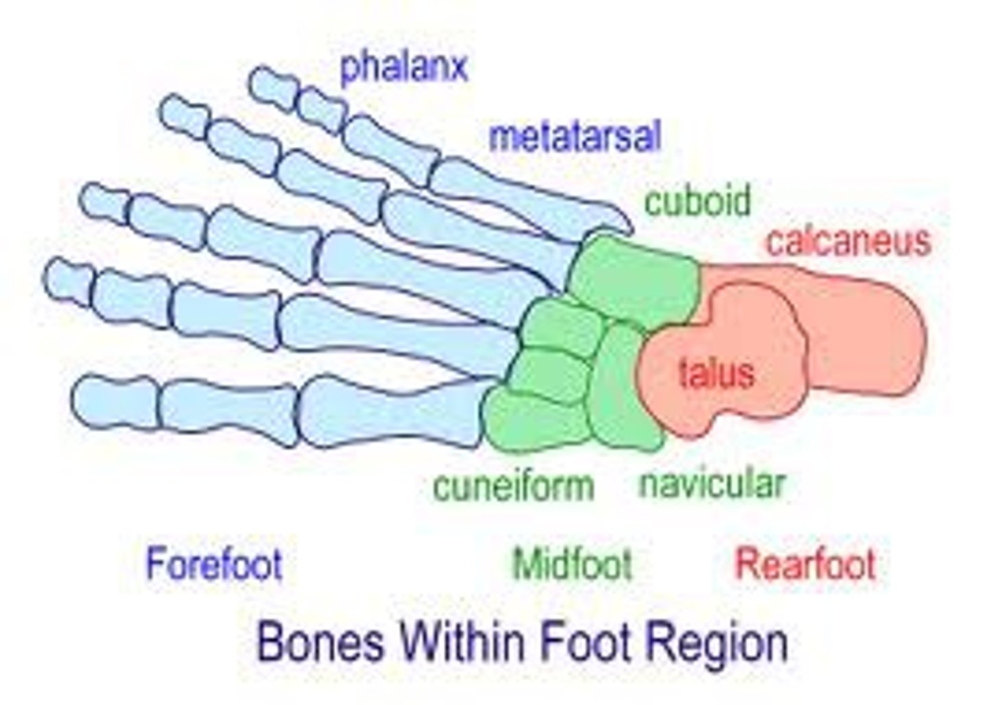
Hindfoot
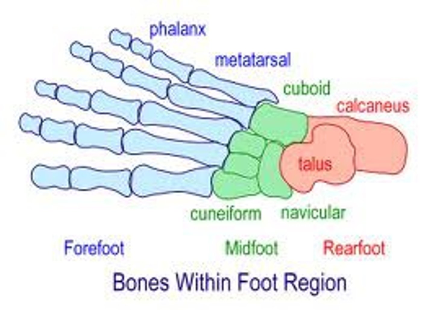
made up of the talus and calcaneus; the pink section of this picture
Midfoot
made up of the navicular, cuboid and the 3 cuneiform bones; the green section of this picture
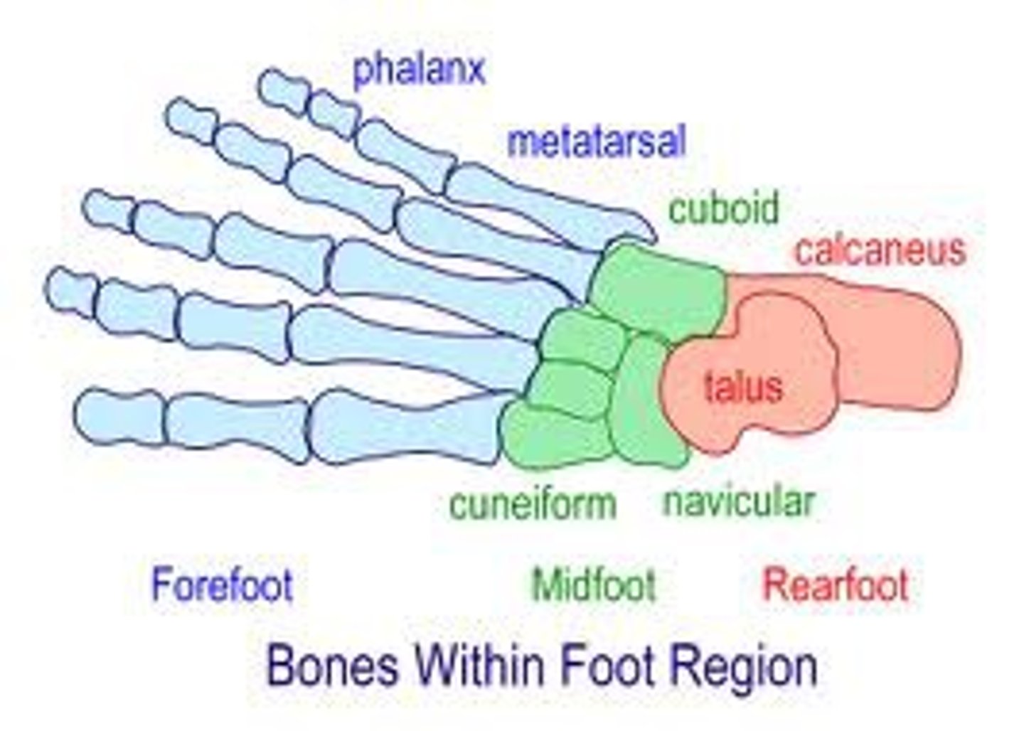
Forefoot
made up of the 5 metatarsals and all phalanges; the blue section of this picture
Metatarsals
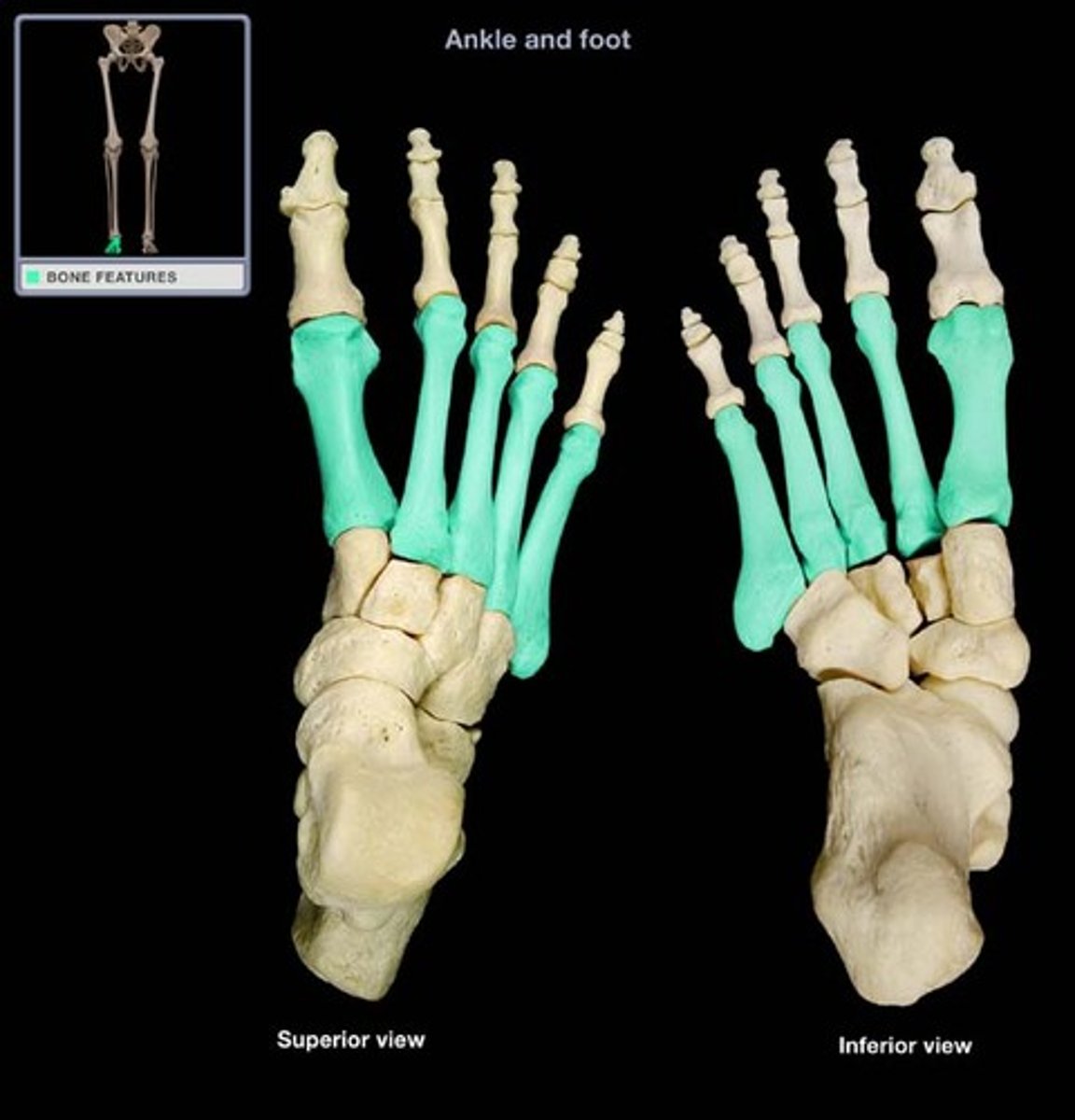
numbered 1-5 starting medially; the proximal end of each metatarsal is the base; the distal end of each metatarsal is the head; the 1st and 5th metatarsals are weight bearing bones
Metatarsal Articulations
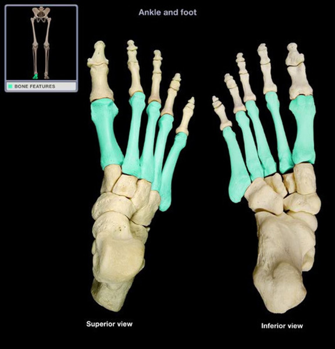
Distally, all metatarsals articulate with a proximal phalanx of the toe; Proximally: the 1st metatarsal articulates with the 1st cuneiform; the 2nd metatarsal articulates with the 2nd cuneiform; the 3rd metatarsal articulates with the 3rd cuneiform; and the 4th & 5th metatarsals articulate with the cuboid bone
Phalanges
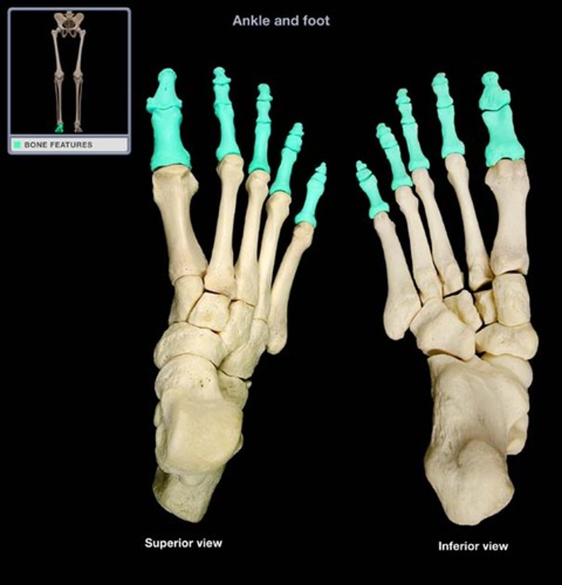
The bones of the toes. The first digit (the hallux or the great toe) has a proximal and distal , but no middle; Toes 2-5 (the lesser toes) each have a proximal, middle and distal
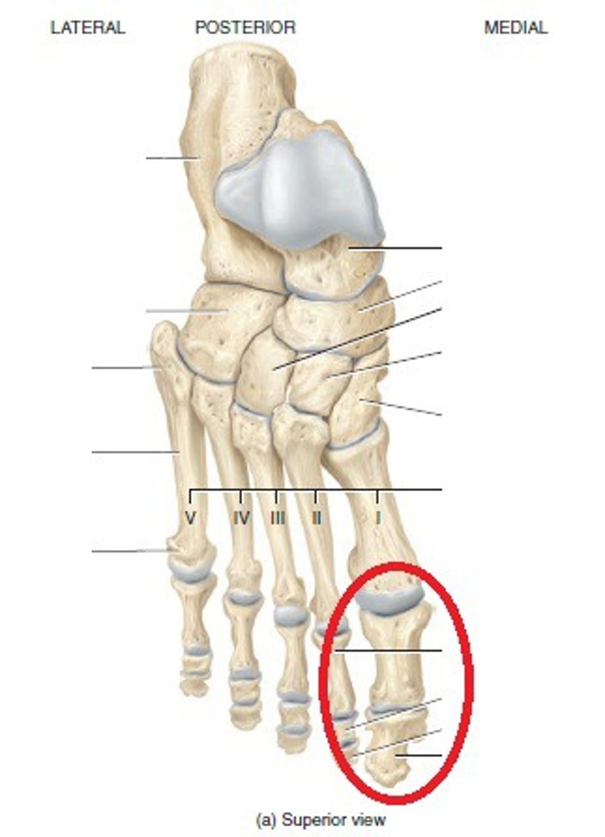
Hallux
The great toe; the most medial toe; the first toe
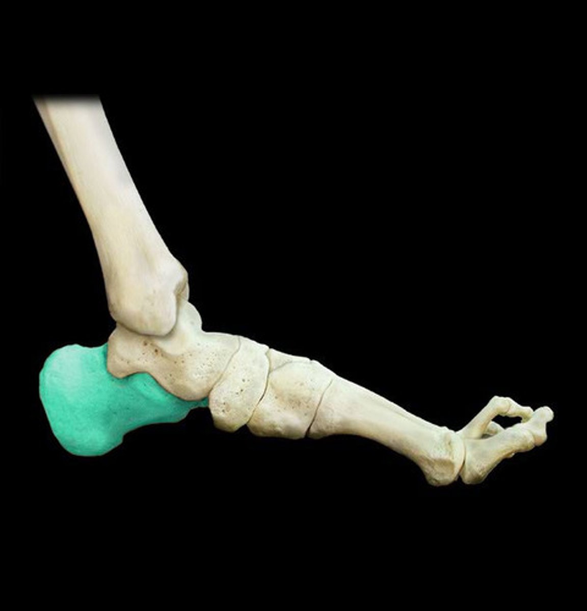
Calcaneus
the largest of the tarsal bones (heel bone)
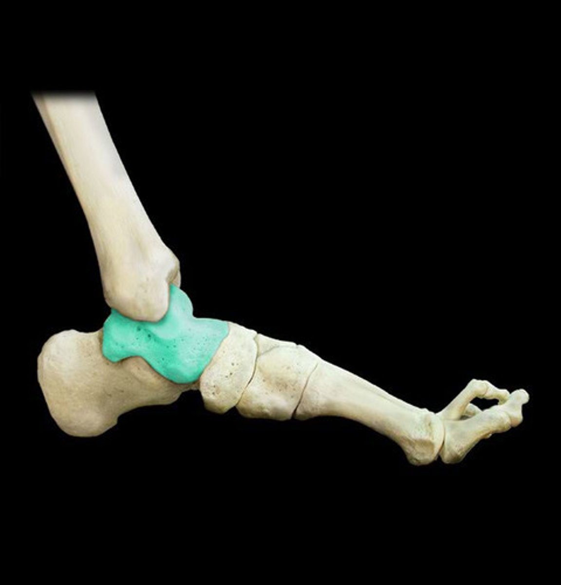
Talus
the anklebone that articulates with the tibia and fibula (proximally); and "sits on top of" the calcaneus
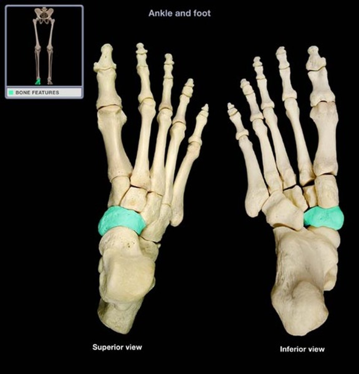
Navicular
One of the tarsal bones; it is located medially on the foot, just distal (or anterior) to the calcaneus
Cuboid
One of the tarsal bones; it is located laterally on the foot, just distal (or anterior) to the calcaneus
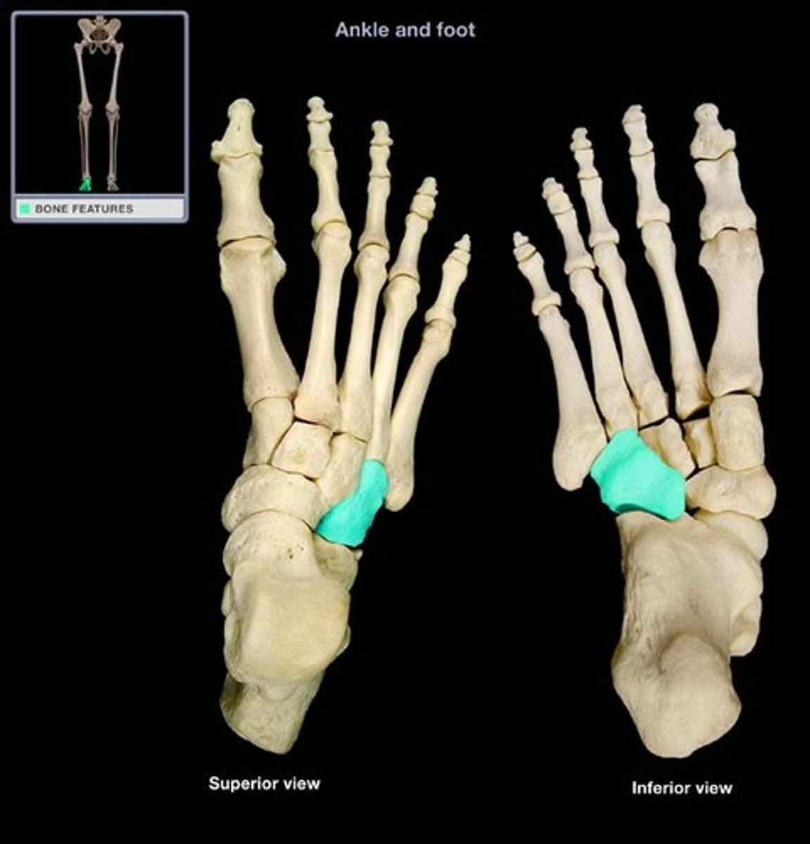
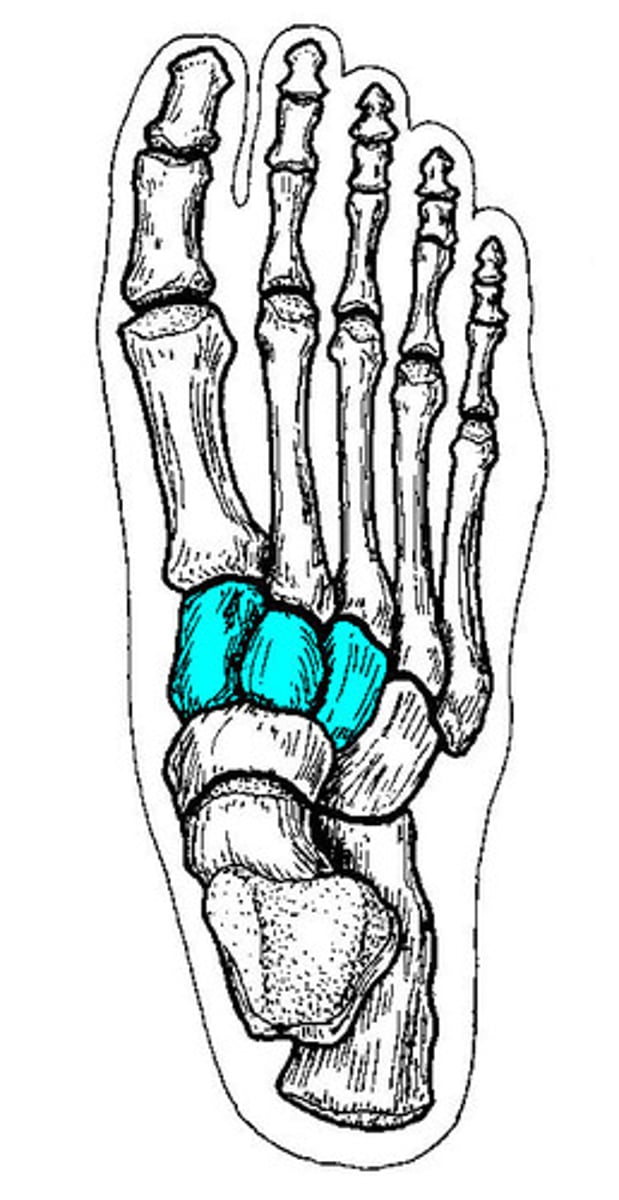
Cuneiforms
The 3 smallest tarsal bones; located medial to cuboid; distal articular surfaces articulate with base of metatarsals
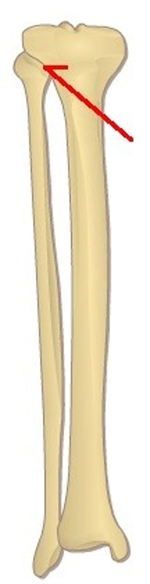
Superior Tibiofibular Joint
articulation between head of fibula and posterior lateral tibia; small amount of glide and rotation of fibula and tibia; has joint capsule
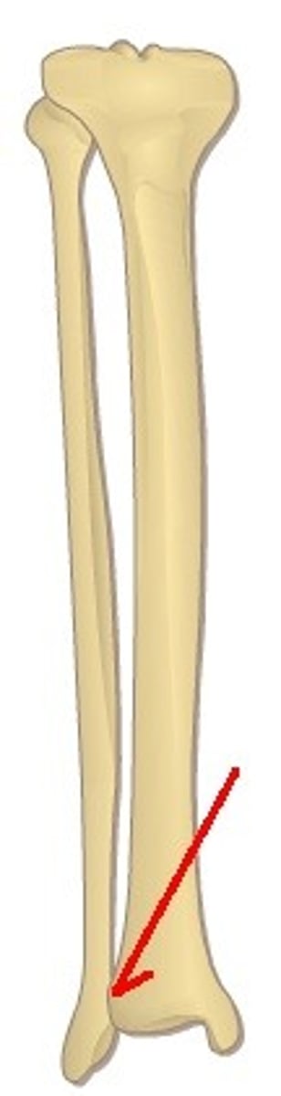
Inferior Tibiofibular Joint
syndesmosis (fibrous union); provides a very firm union between the tibia and fibula which adds significantly to the integrity of the talocrural joint; no joint capsule; articulation between the distal concave tibia and the convex distal fibula
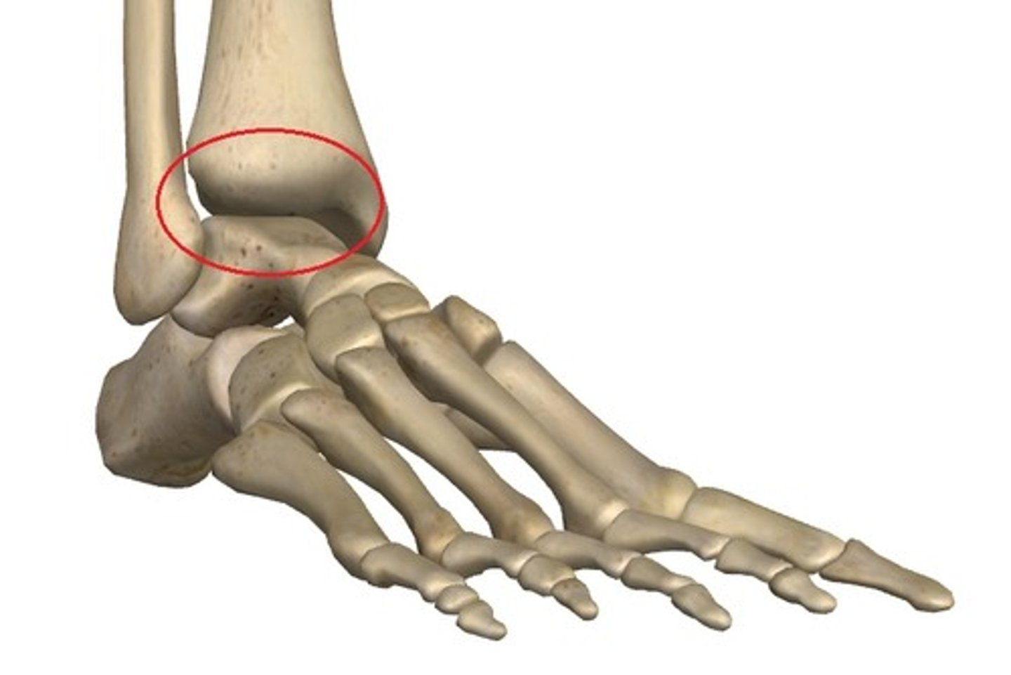
Talocrural Joint
An articulation between the distal ends of the tibia and fibula in the lower limb with the proximal end of the talus; dorsiflexion & plantarflexion occur at this joint
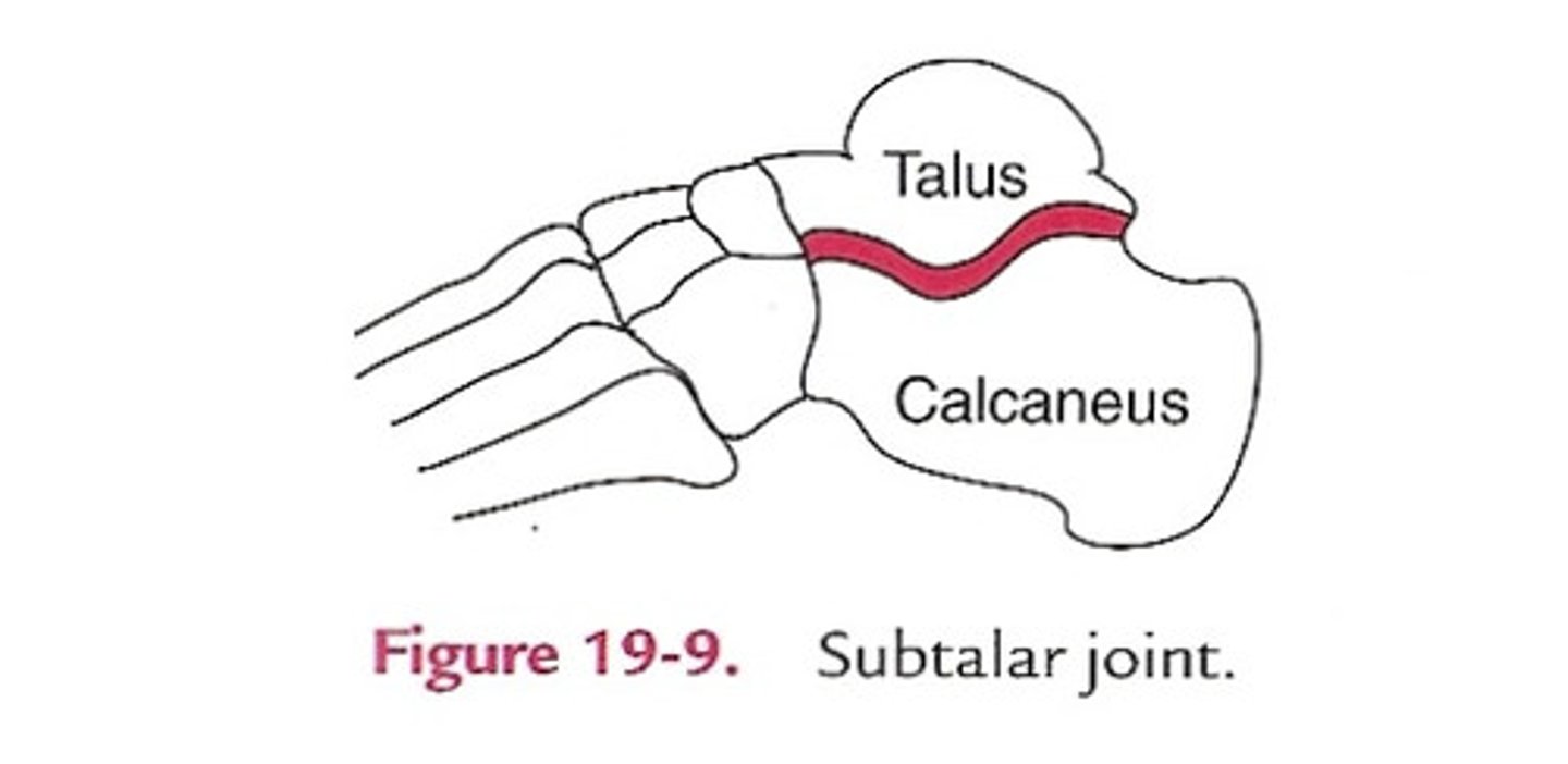
Subtalar Joint
Articulation between the inferior surface of the talus with the superior surface of the calcaneus; where inversion and eversion occur
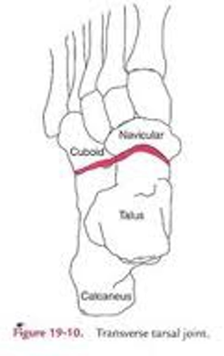
Transverse Tarsal Joint
AKA midtarsal joint; articulation between the anterior surfaces of the talus and calcaneus with the posterior surfaces of the navicular and cuboid; we cannot functionally separate this joint from the subtalar joint; so for the sake of simplicity > inversion and eversion are motions that occur at both joints
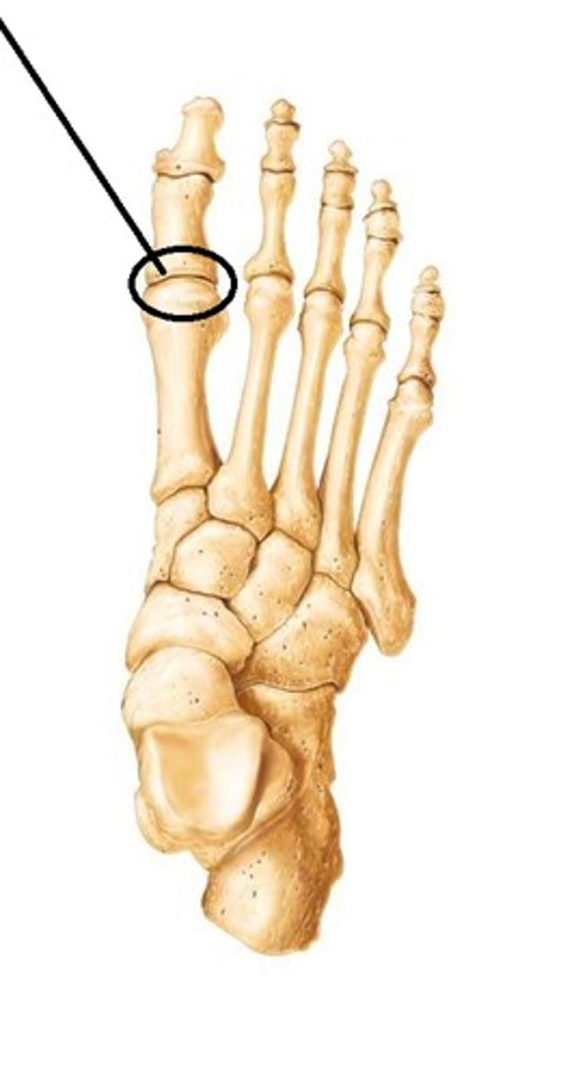
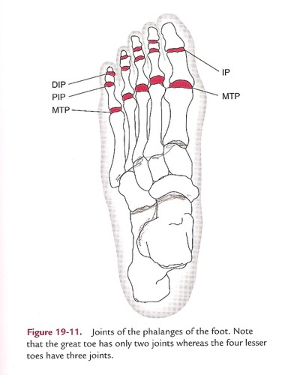
Metatarsophalangeal (MTP) Joints
Where the metatarsal heads articulate with the proximal phalanges of the toes; osteokinematics = flexion, extension, hyperextension, abduction, adduction
Foot point of reference for MTP abduction/adduction
The 2nd toe
IP, DIP, PIP joints
Articulations between the phalanges; osteokinematics = flexion and extension
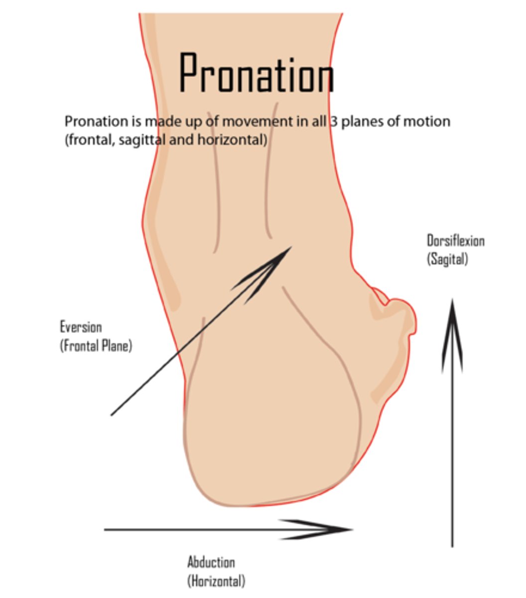
Ankle pronation
combination of dorsiflexion, eversion, abduction
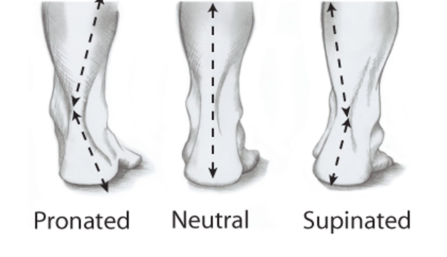
Ankle supination
combination of Plantar flexion, inversion, adduction
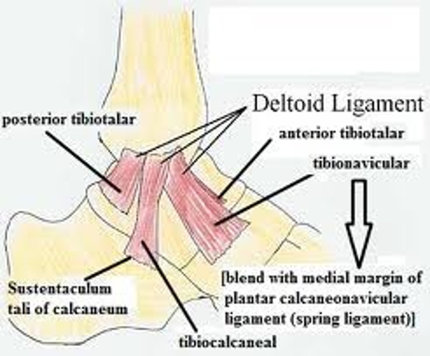
Deltoid Ligament
Triangle-shaped collateral ligament on the medial side of the ankle; limits eversion; has 3 sets of fibers (tibionavicular, tibiocalcaneal, tibiotalar)
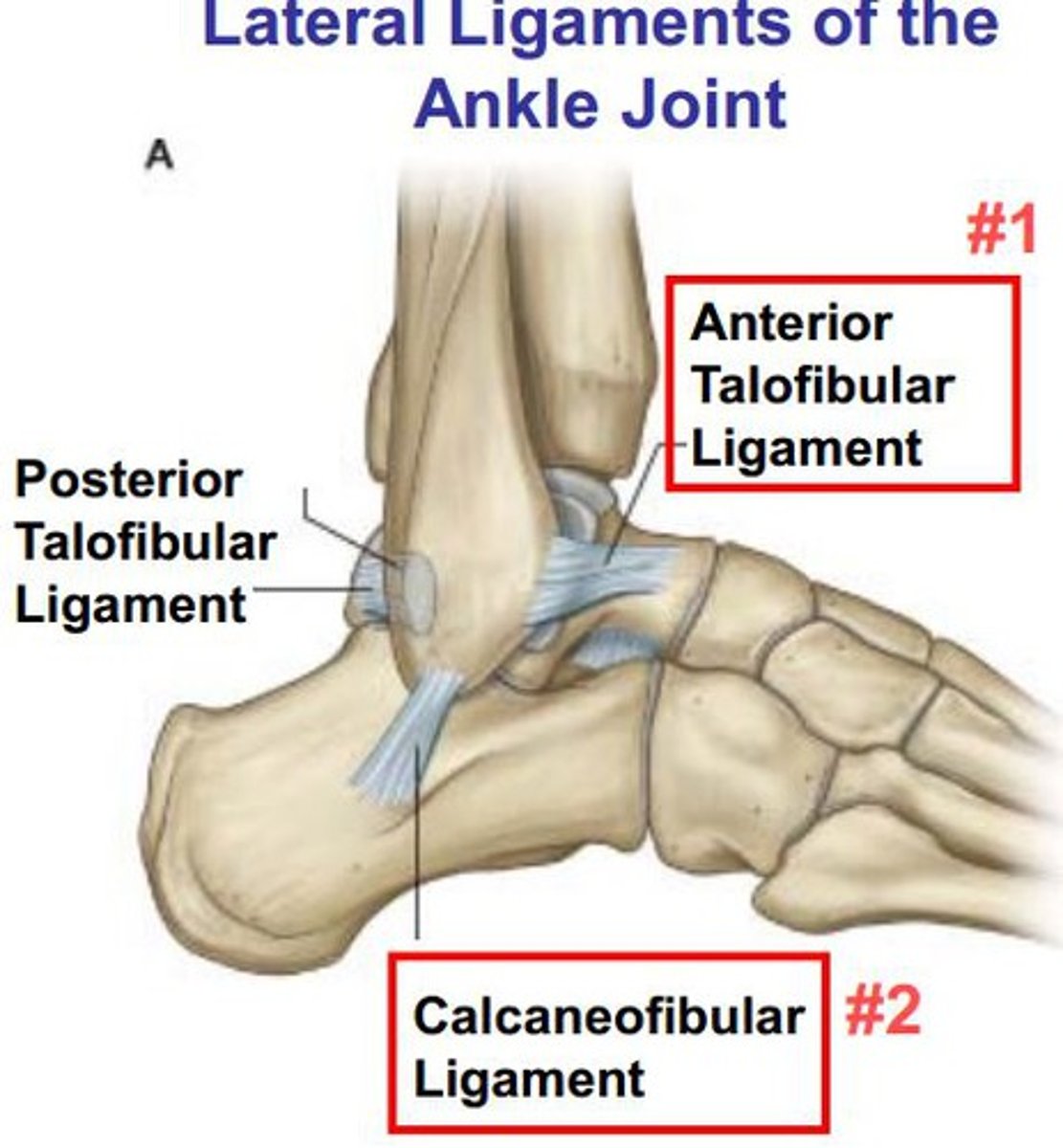
Lateral Collateral Ligaments
Collateral ligament on the lateral side of the ankle; limits inversion; consists of 3 ligaments (anterior & posterior talofibular and calcaneofibular)
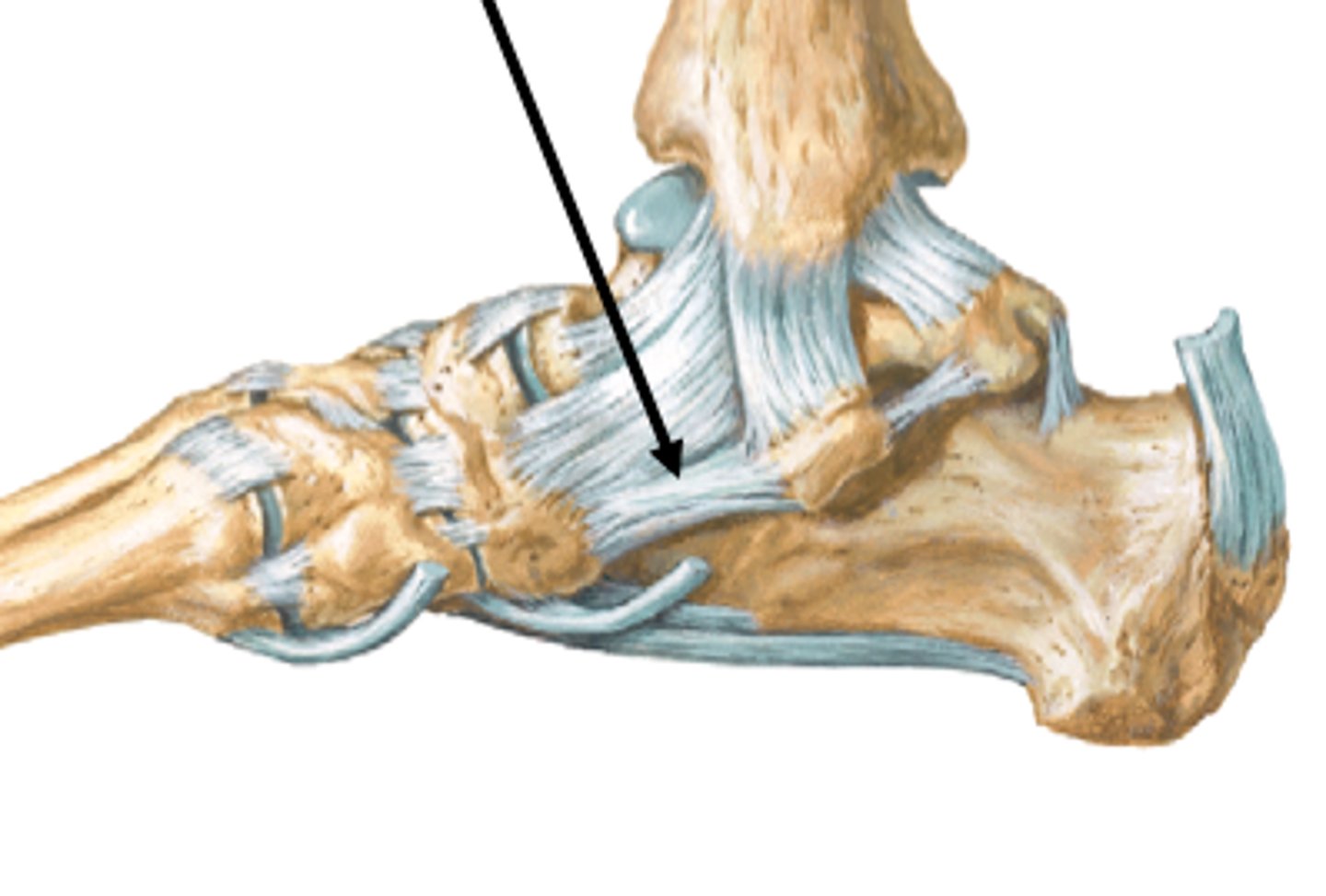
Spring Ligament
Ligament of the foot spanning the distance between the calcaneus and the navicular
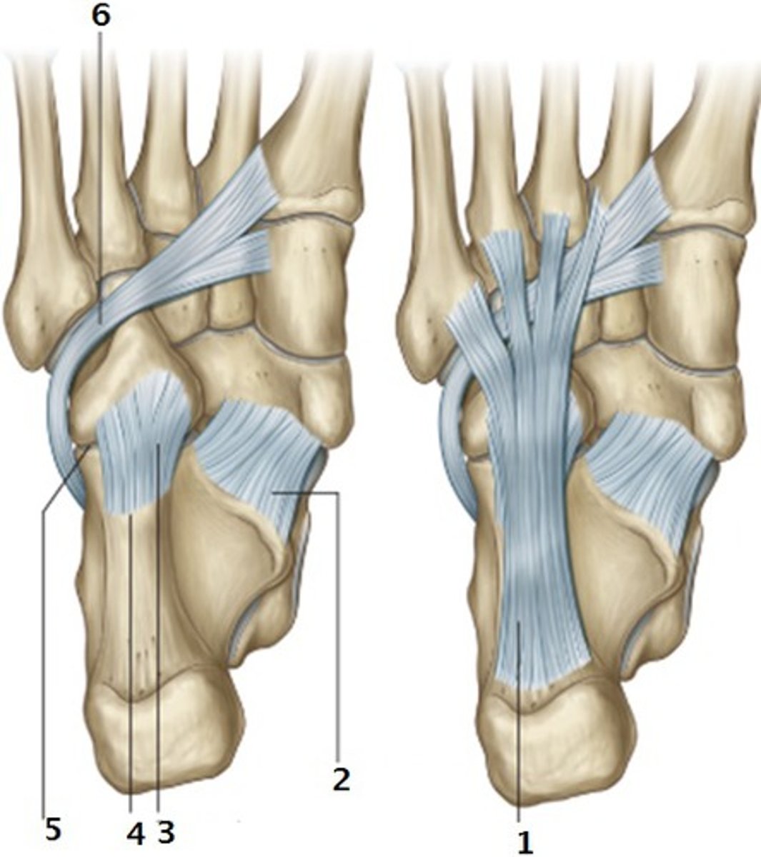
Long & Short Plantar Ligaments
Ligaments of the foot spanning the distance between the calcaneus and the cuboid and the bases of metatarsals 3, 4, 5
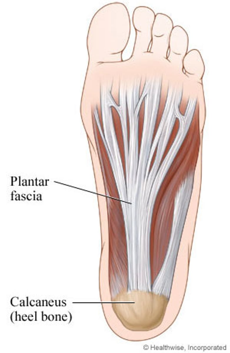
Plantar fascia
superficial fascia from the calcaneus to the proximal phalanes
Anterior Extrinsic Ankle Muscles
Tibialis anterior, extensor halluces longus, extensor digitorum longus, peroneus tertius; all 4 muscles innervated by Deep Peroneal Nerve; all 4 muscles perform dorsiflexion as one of their primary actions
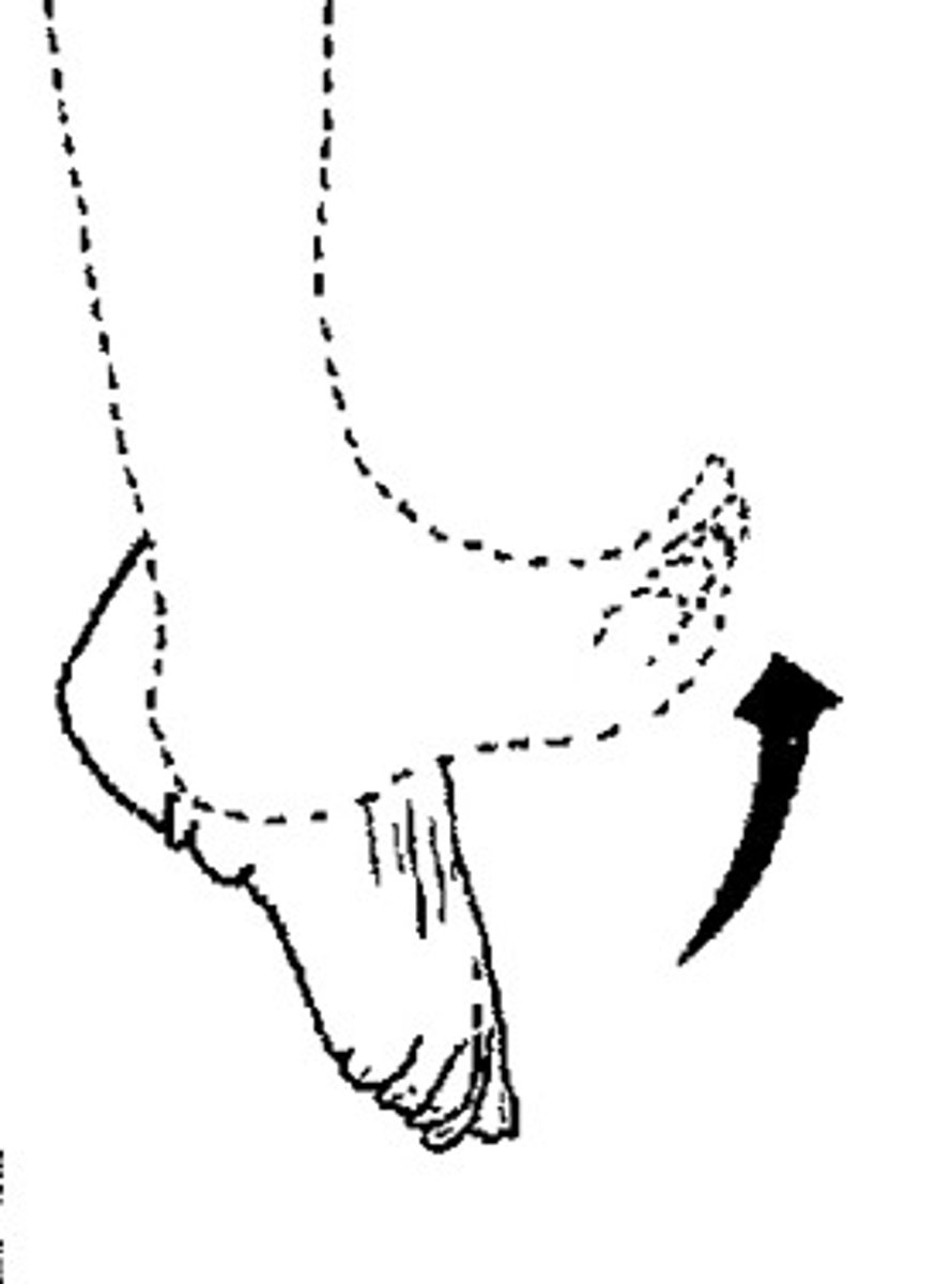
Lateral Extrinsic Ankle Muscles
peroneus longus, peroneus brevis; both muscles are innervated by the Superficial Peroneal Nerve; both muscles originate on the fibula; both muscles perform ankle eversion as one of their primary actions
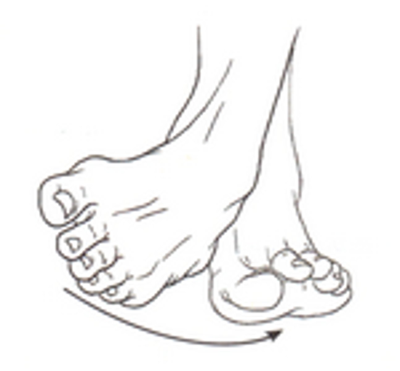
Deep Posterior Extrinsic Ankle Muscles
tibialis posterior, flexor digitorum longus, flexor hallucis longus; all muscles innervated by the Tibial Nerve; all muscles perform plantarflexion as one of their primary actions
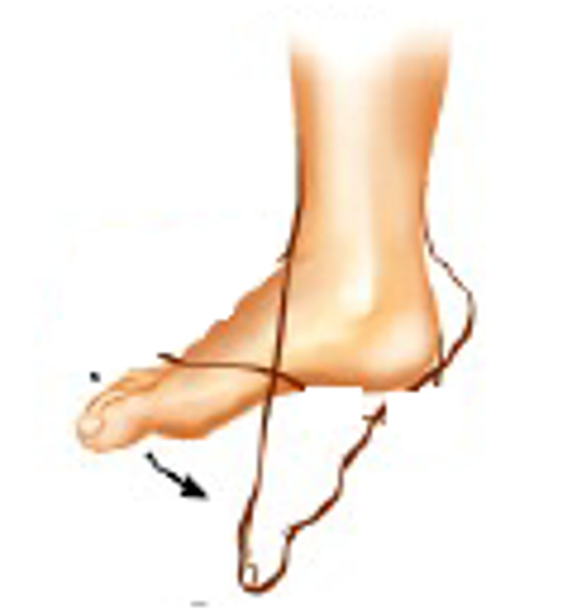
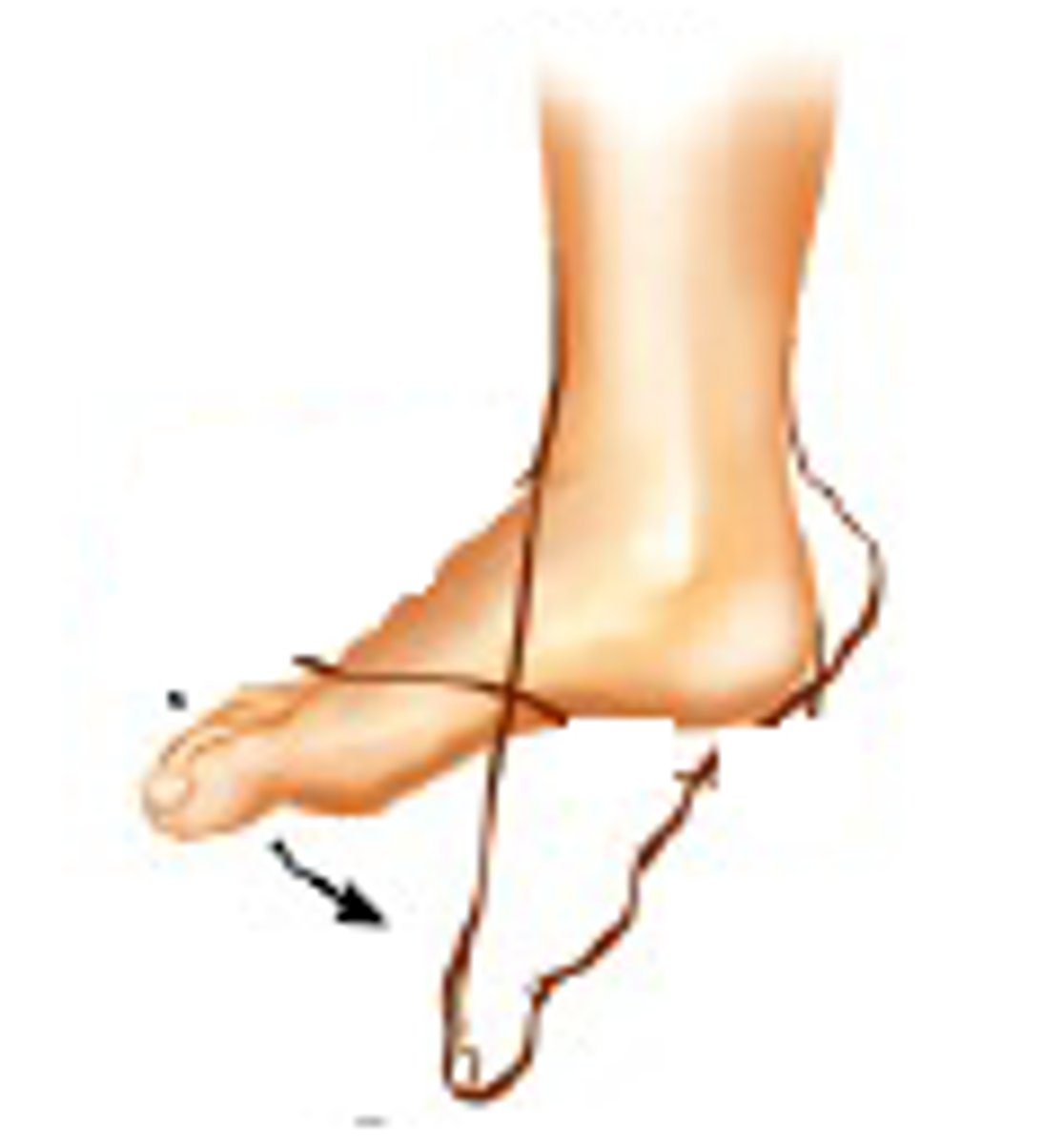
Superficial Posterior Extrinsic Ankle Muscles
gastrocnemius, soleus, plantaris; all muscles innervated by the Tibial Nerve; all muscles perform plantarflexion as one of their primary actions
Tibialis Anterior action
Ankle dorsiflexion, inversion
Tibialis Anterior location
Anterior ankle; originates on the proximal 2/3 of the lateral surface of the tibia and interosseus membrane; inserts on the medial and plantar aspects of the medial cuneiform and the base of the first metatarsal
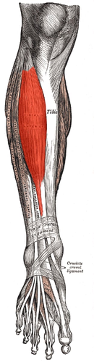
Tibialis Anterior innervation
Deep branch of the peroneal nerve
Extensor Hallucis Longus action
Ankle dorsiflexion; extension of the hallux (great toe)
Extensor Hallucis Longus location
originates on the middle section of the fibula and interosseus membrane; inserts on the dorsal base of the distal phalanx of the hallux (great toe)
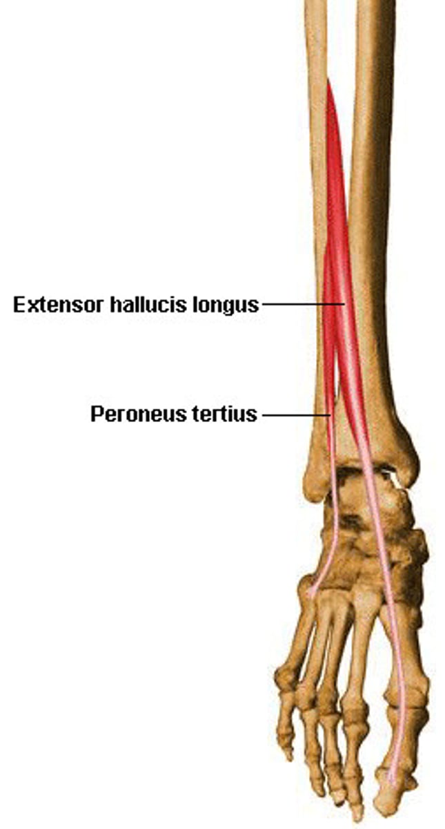
Extensor Hallucis Longus innervation
Deep branch of the peroneal nerve
Extensor Digitorum Longus action
Ankle dorsiflexion; extension of all joints (MTP, PIP and DIP joints) of lesser toes #2-5
Extensor Digitorum Longus location
originates on the lateral condyle of the tibia, proximal 2/3 of the medial surface of the fibula and interosseus membrane; inserts by splitting into 4 tendons that attach to the proximal base of the dorsal surface of the middle and distal phalanges of toes #2-5
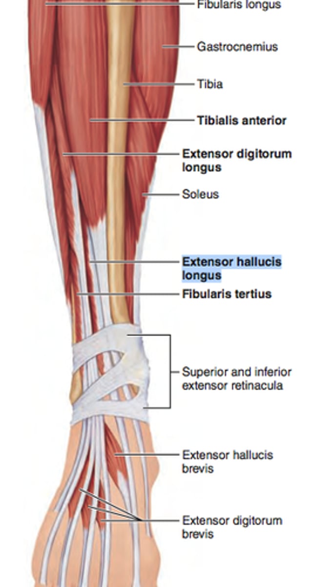
Extensor Digitorum Longus innervation
Deep branch of the peroneal nerve
Peroneus Tertius action
ankle dorsiflexion; eversion
Peroneus/Fibularis Tertius location
Originates on the distal third of the medial surface of the fibula and interosseus membrane; inserts on the dorsal surface of the base of the 5th metatarsal
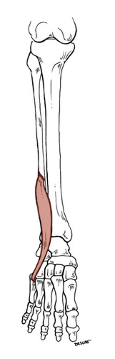
Peroneus Tertius innervation
Deep branch of the peroneal nerve
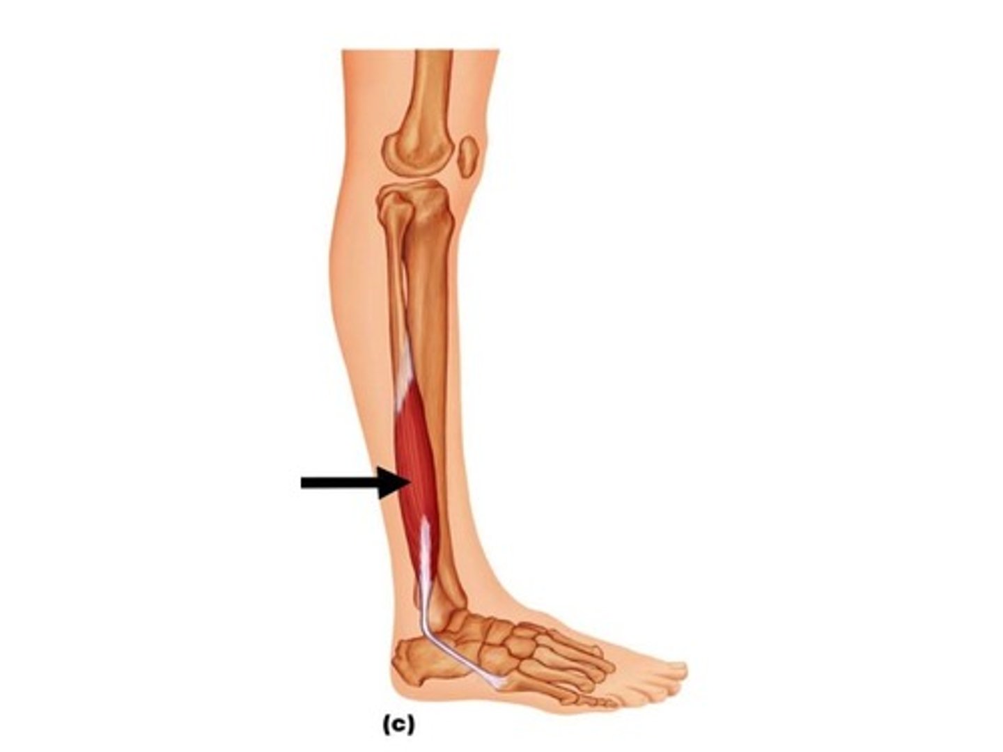
Peroneus Longus action
Ankle eversion, plantarflexion
Peroneus/Fibularis Longus location
Originates on the lateral fibular head and interosseus membrane; inserts on the lateral surface of the medial cuneiform and plantar base of the 1st metatarsal
Peroneus Longus innervation
Superficial Peroneal Nerve
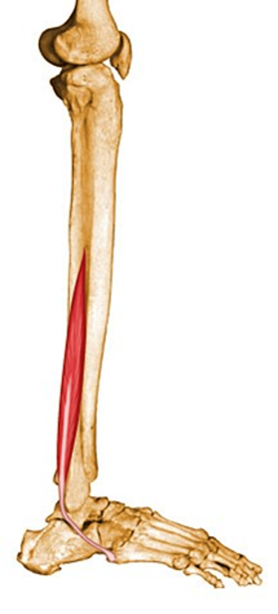
Peroneus Brevis action
Ankle eversion, plantarflexion
Peroneus/Fibularis Brevis location
Originates on the distal 2/3 of the lateral surface of the fibula; inserts on the styloid process of the 5th metatarsal
Peroneus Brevis innervation
Superficial Peroneal Nerve
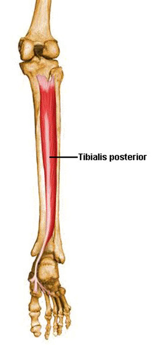
Tibialis Posterior action
ankle plantarflexion, inversion
Tibialis Posterior location
originates on the proximal 2/3 of the posterior aspect of the tibia, fibula and interosseus membrane; inserts via multiple attachments to every tarsal bone except the talus, also attaches to the bases of metatarsals 2-4; its most prominent insertion is on the navicular tuberosity
Tibialis Posterior innervation
Tibial Nerve
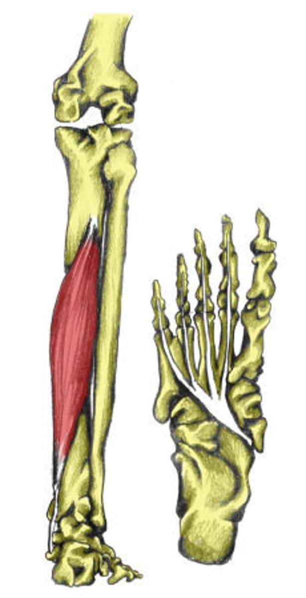
Flexor Digitorum Longus action
Ankle plantarflexion, inversion, flexion of toes 2-5
Flexor Digitorum Longus location
originates on the posterior surface of the middle third of the tibia; inserts by 4 separate tendons to the base of the distal phalanx of the 4 lesser toes
Flexor Digitorum Longus innervation
Tibial Nerve
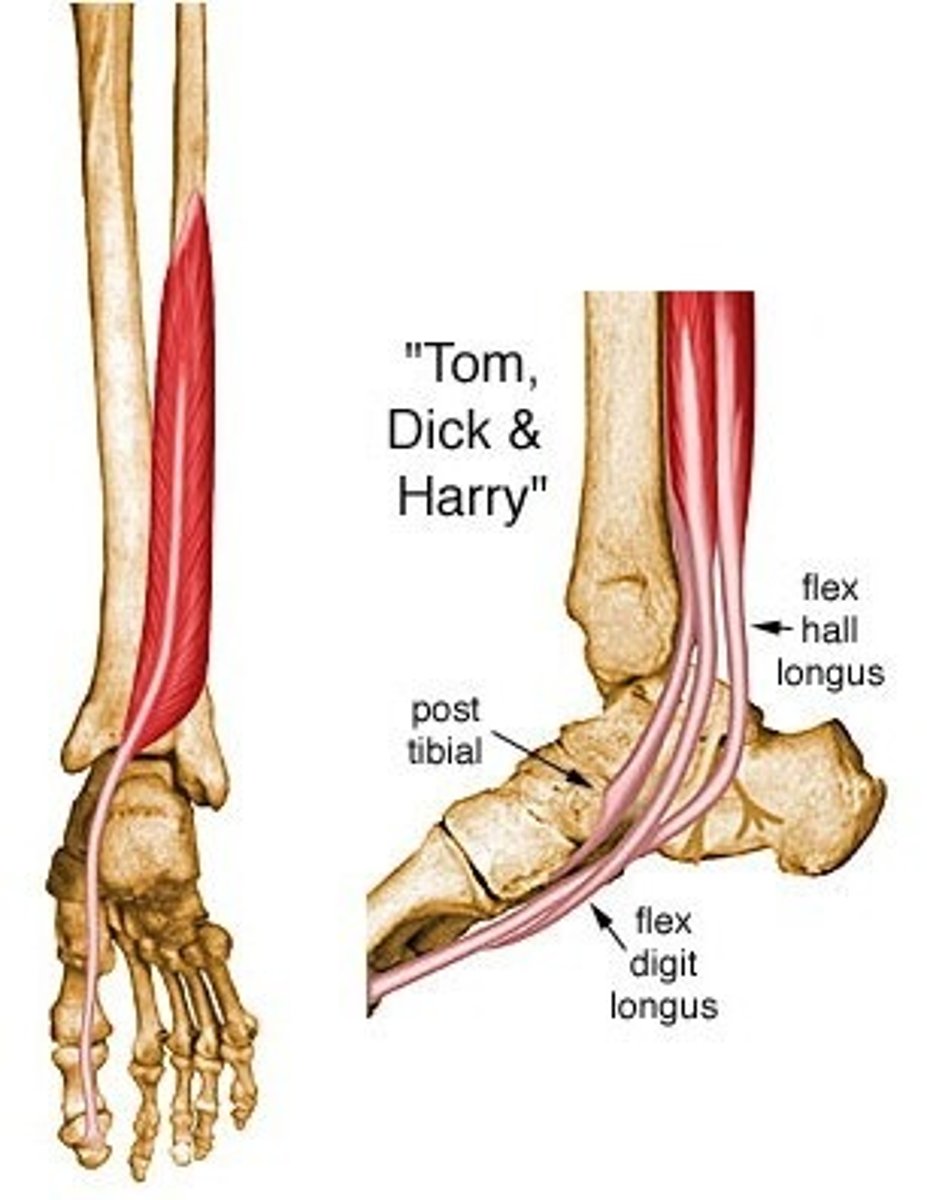
Flexor Hallucis Longus action
ankle plantarflexion, inversion, flexion of the hallux (great toe)
Flexor Hallucis Longus location
originates on the distal 2/3 of the posterior fibula; inserts on the plantar surface of the base of the distal phalanx of the hallux (great toe)
Flexor Hallucis Longus innervation
Tibial Nerve
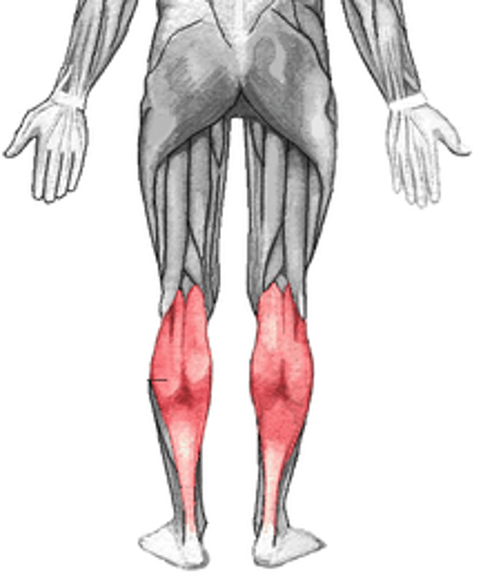
Gastrocnemius action
ankle plantarflexion; knee flexion
Gastrocnemius location
medial head originates on the posterior aspect of the medial femoral condyle; lateral head originates on the posterior aspect of the lateral femoral condyle; inserts on the calcaneal tuberosity of the calcaneus via the Achilles tendon
Gastrocnemius innervation
Tibial Nerve
Soleus action
Ankle Plantarflexion
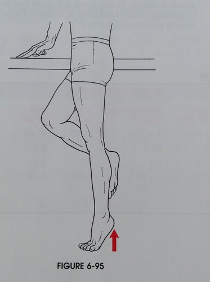
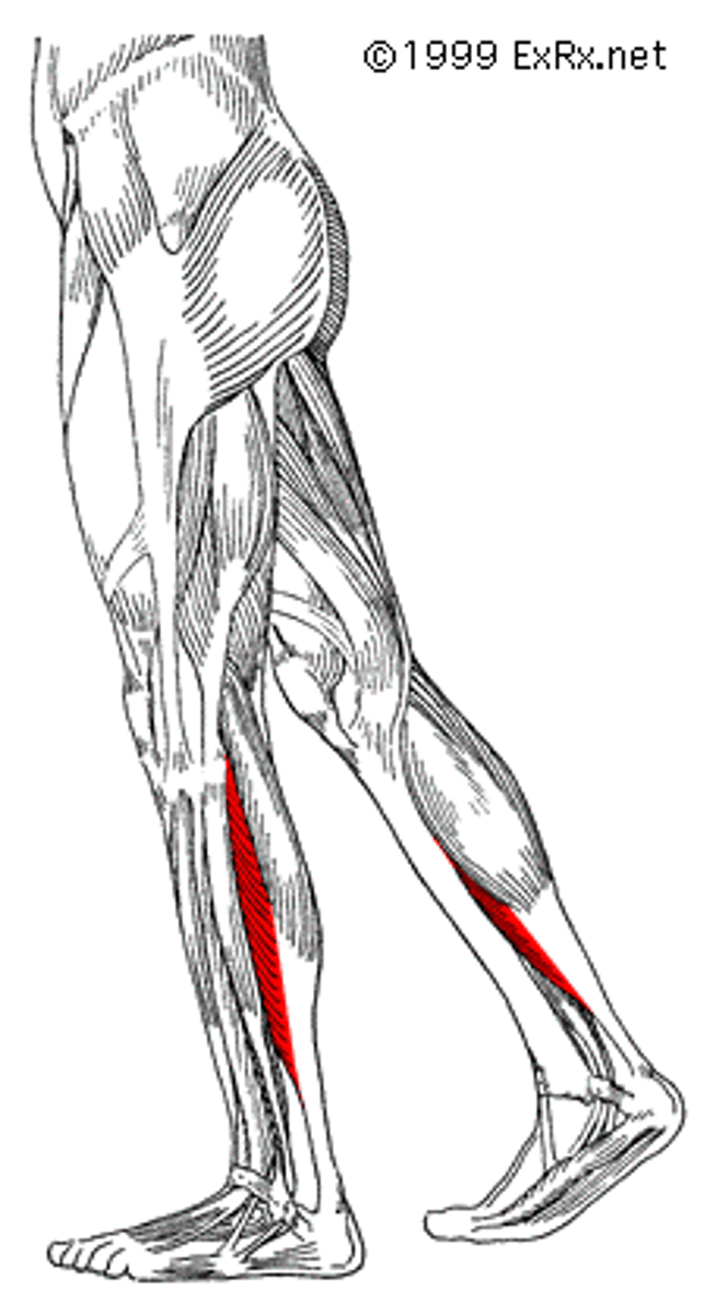
Soleus location
Originates on the proximal third of the posterior fibula, fibular head and posterior tibia; inserts on the calcaneal tuberosity of the calcaneus via the Achilles tendon
Soleus innervation
Tibial Nerve
Plantaris action
ankle plantarflexion; initiates knee flexion
Plantaris location
originates on the posterior surface of the pateral epicondyle of the femur; inserts on the medial aspect of the Achilles tendon to insert on the calcaneal tuberosity
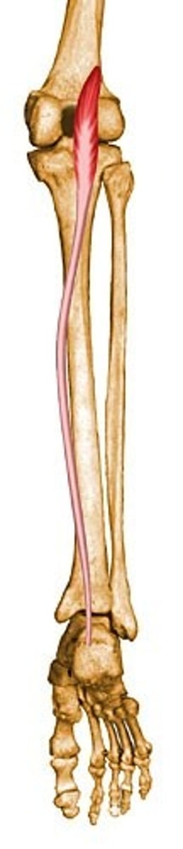
Plantaris innervation
Tibial Nerve