Post-Lab Quiz 5 - 232 Lab
 Call Kai
 Learn
Learn Practice Test
 Spaced Repetition
 Match
Match Flashcards
 Knowt Play
1/52

There's no tags or description
Looks like no tags are added yet.
Name | Mastery | Learn | Test | Matching | Spaced | Call with Kai | Chat |
|---|
No analytics yet
Send a link to your students to track their progress
53 Terms
Kidneys
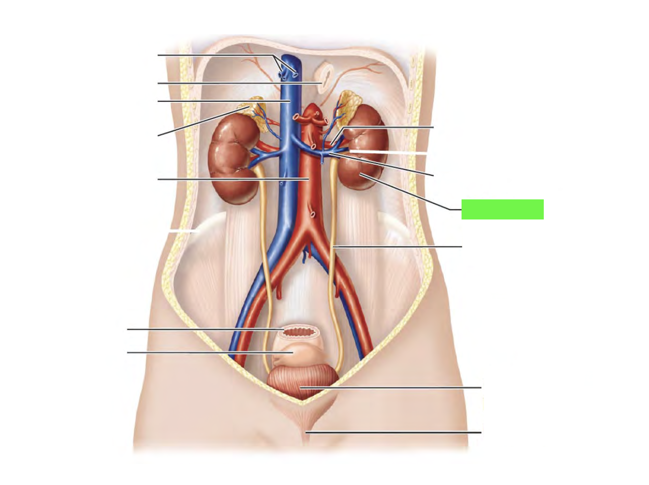
Ureters
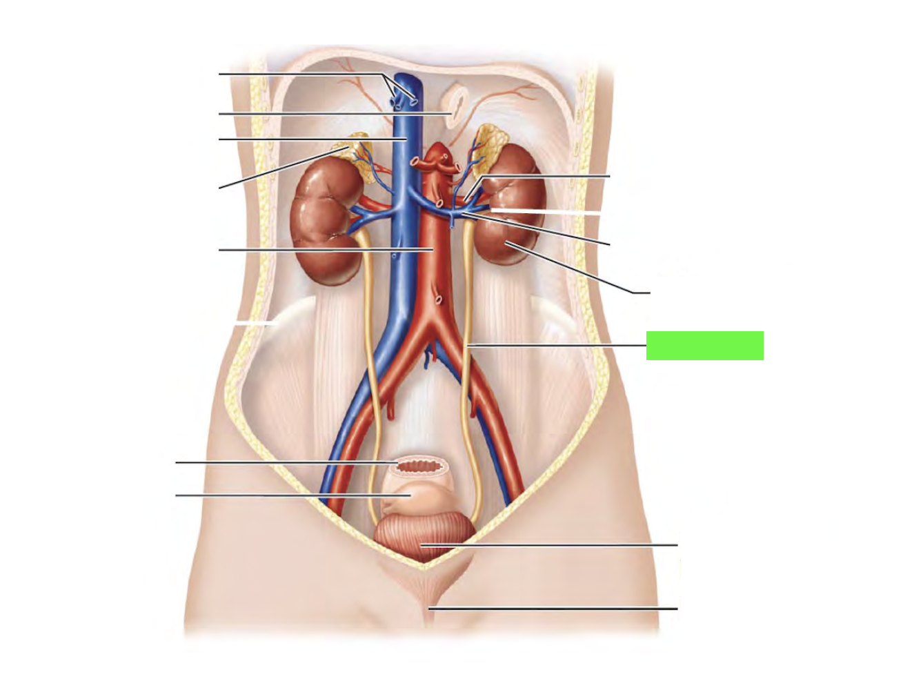
Urinary bladder
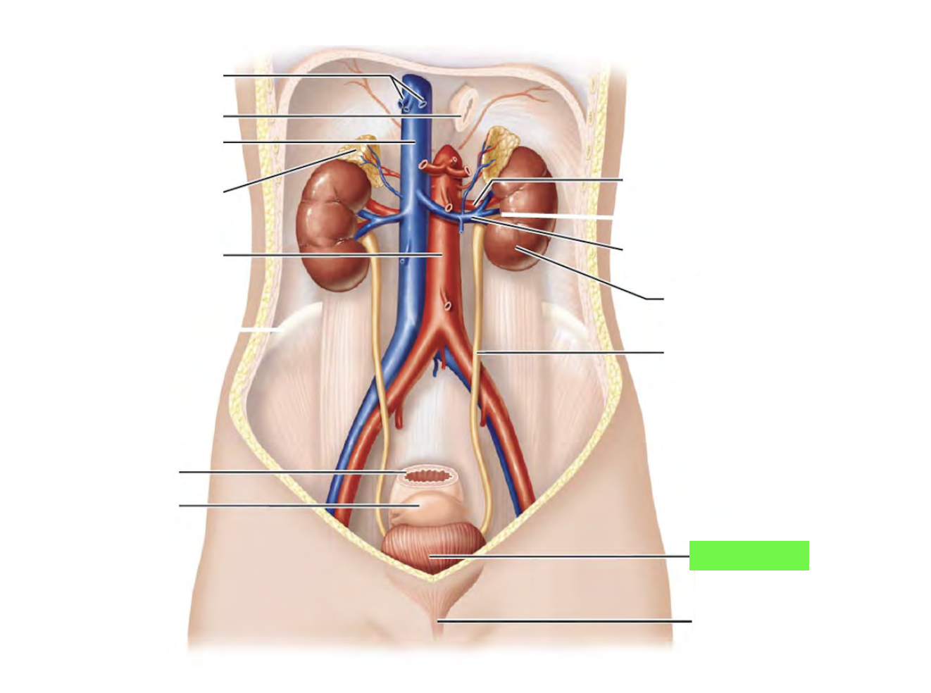
Urethra
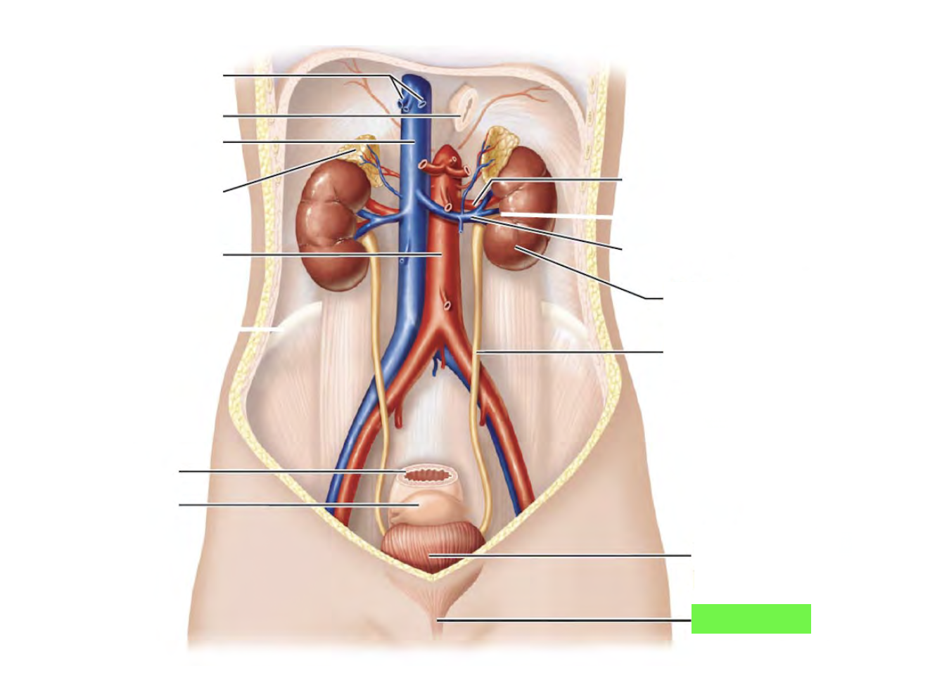
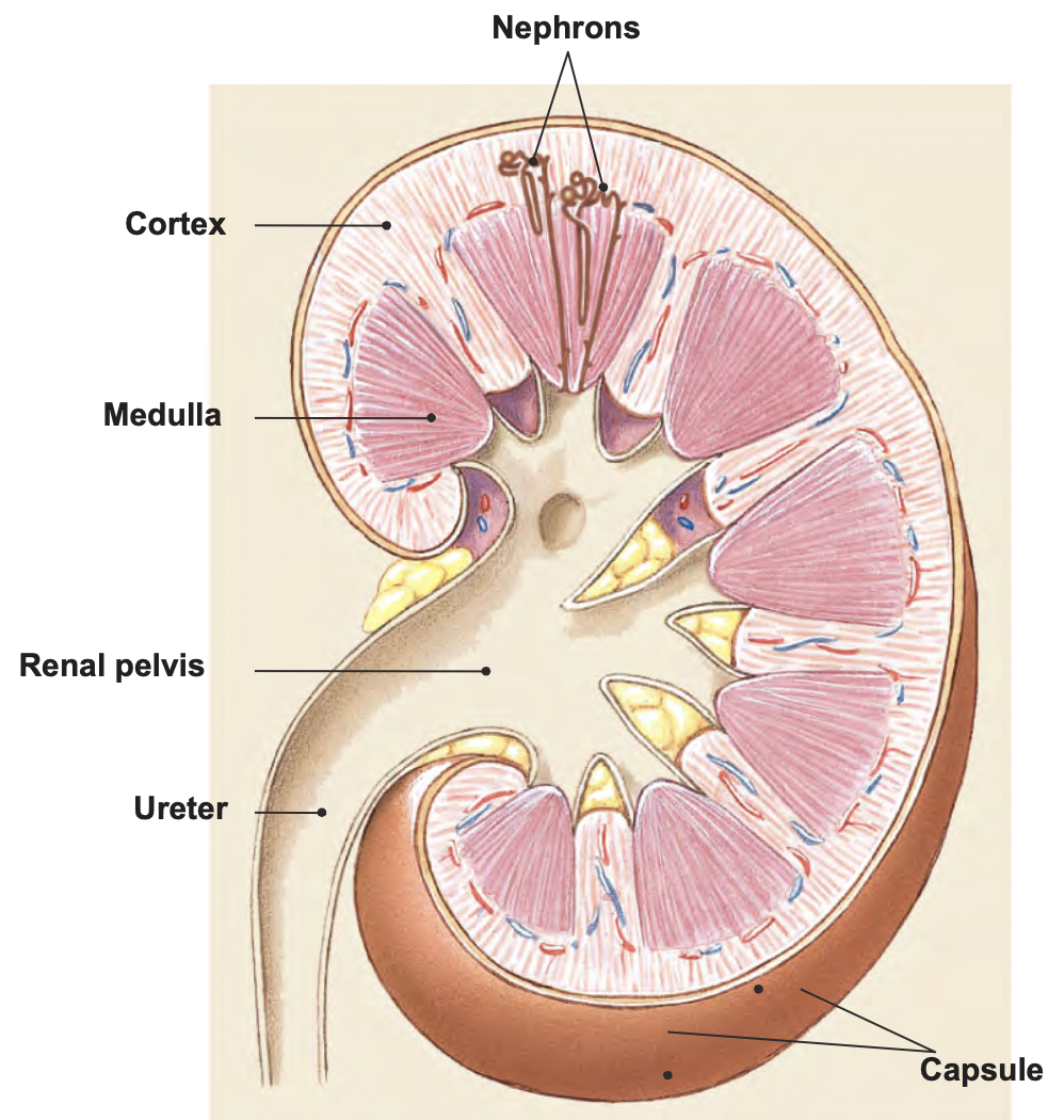
Renal cortex
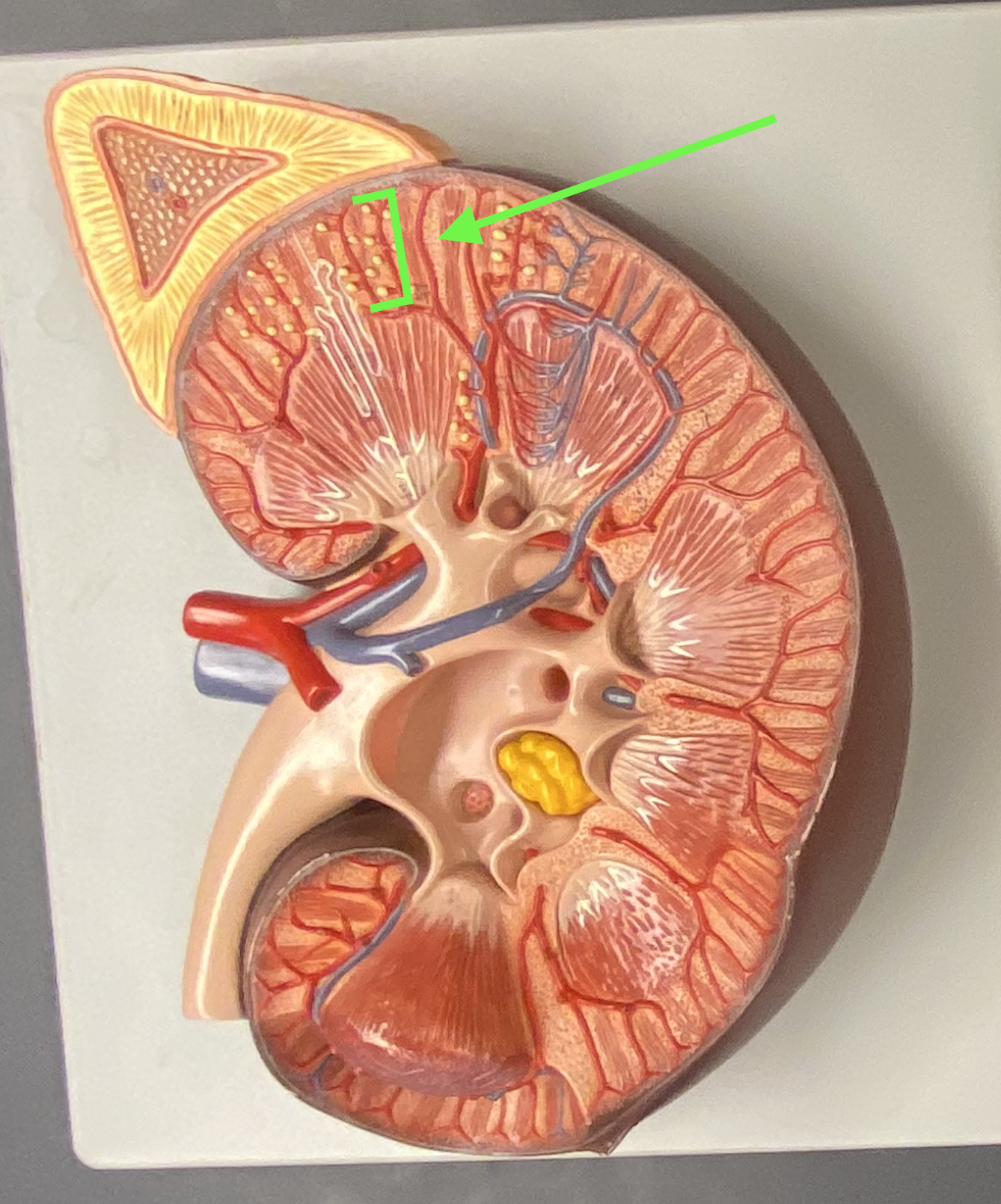
Renal cortex - renal columns
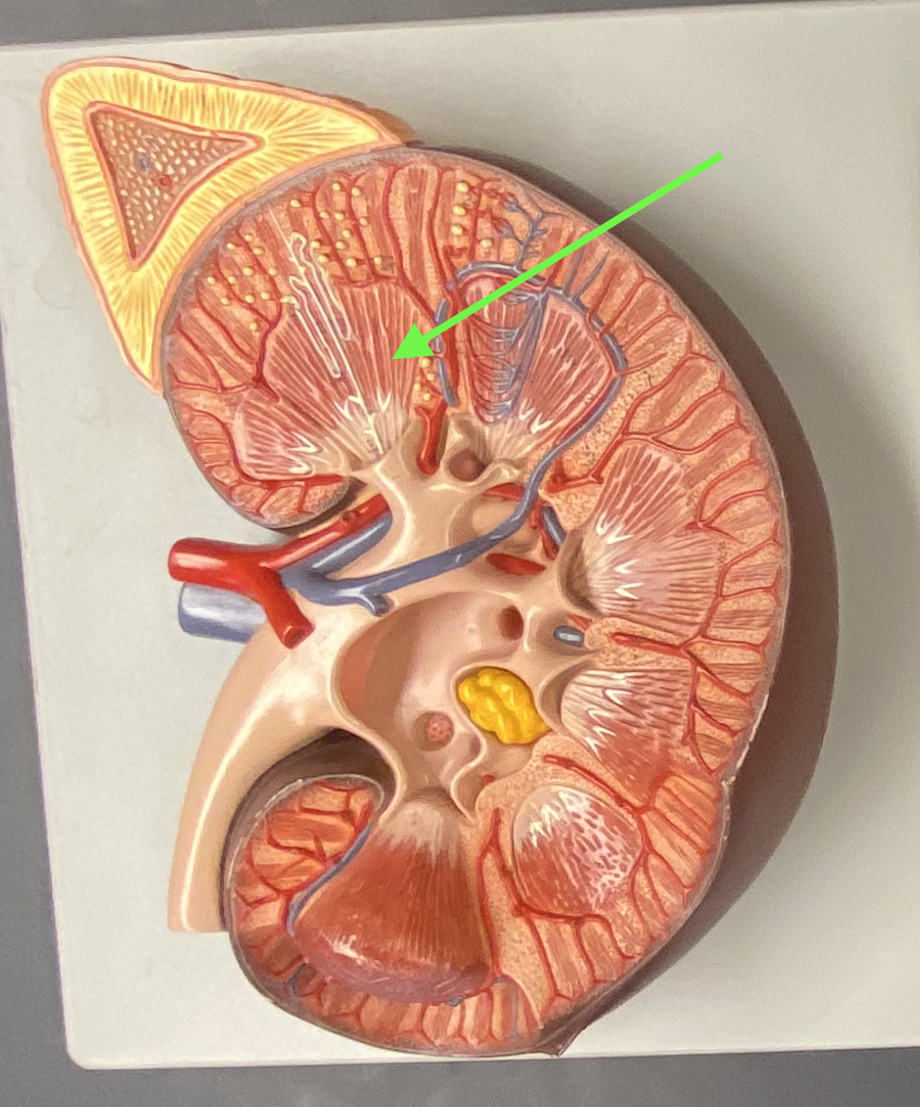
Renal medulla
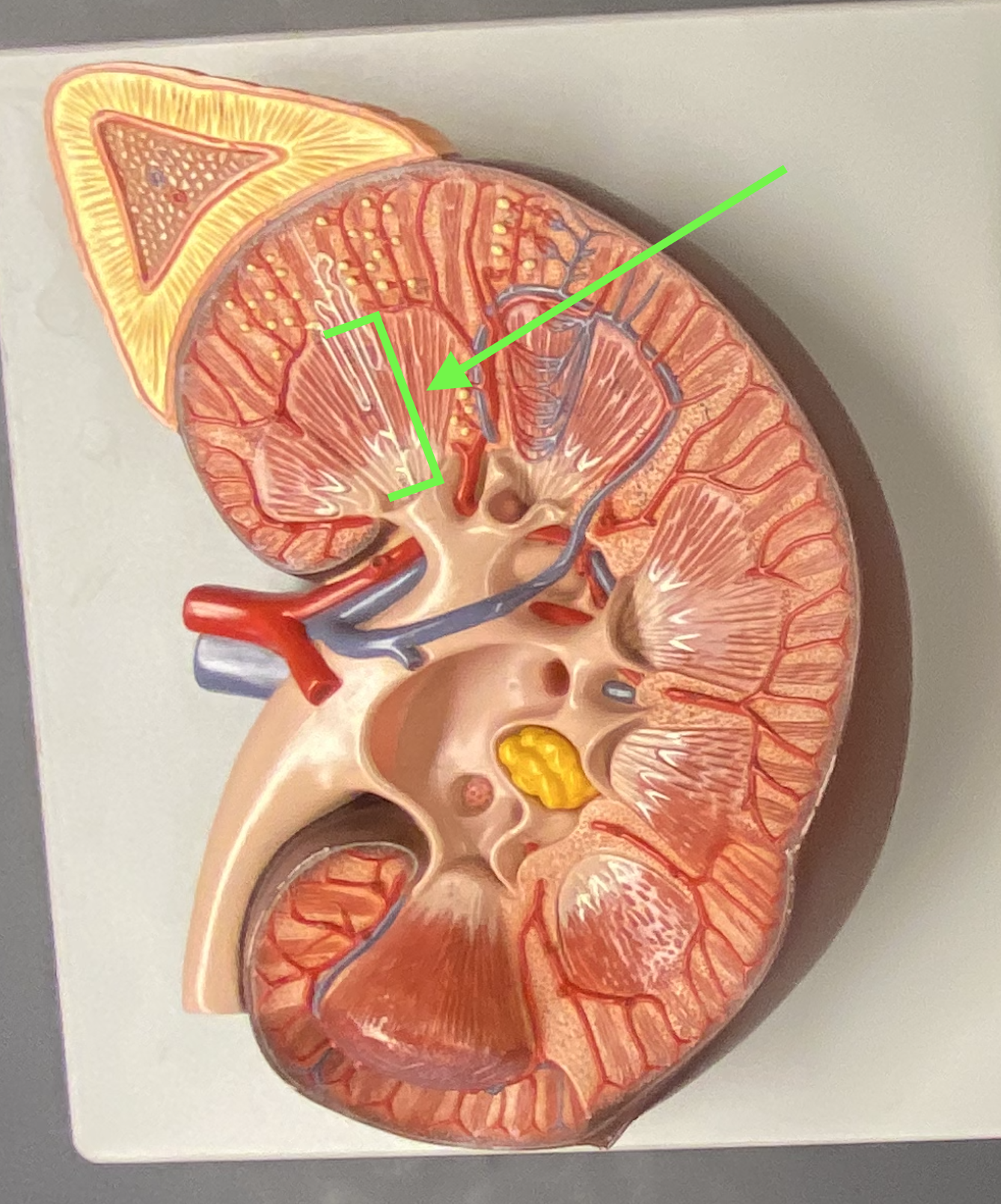
Renal medulla - renal pyramids
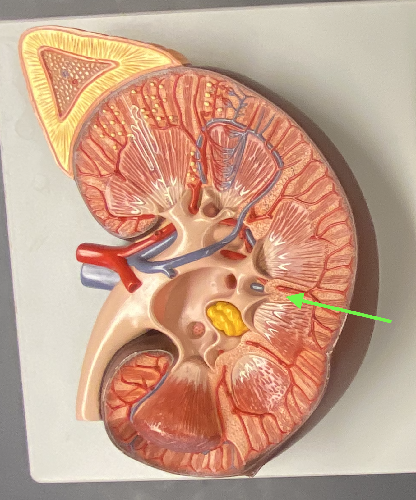
Renal medulla - renal papilla
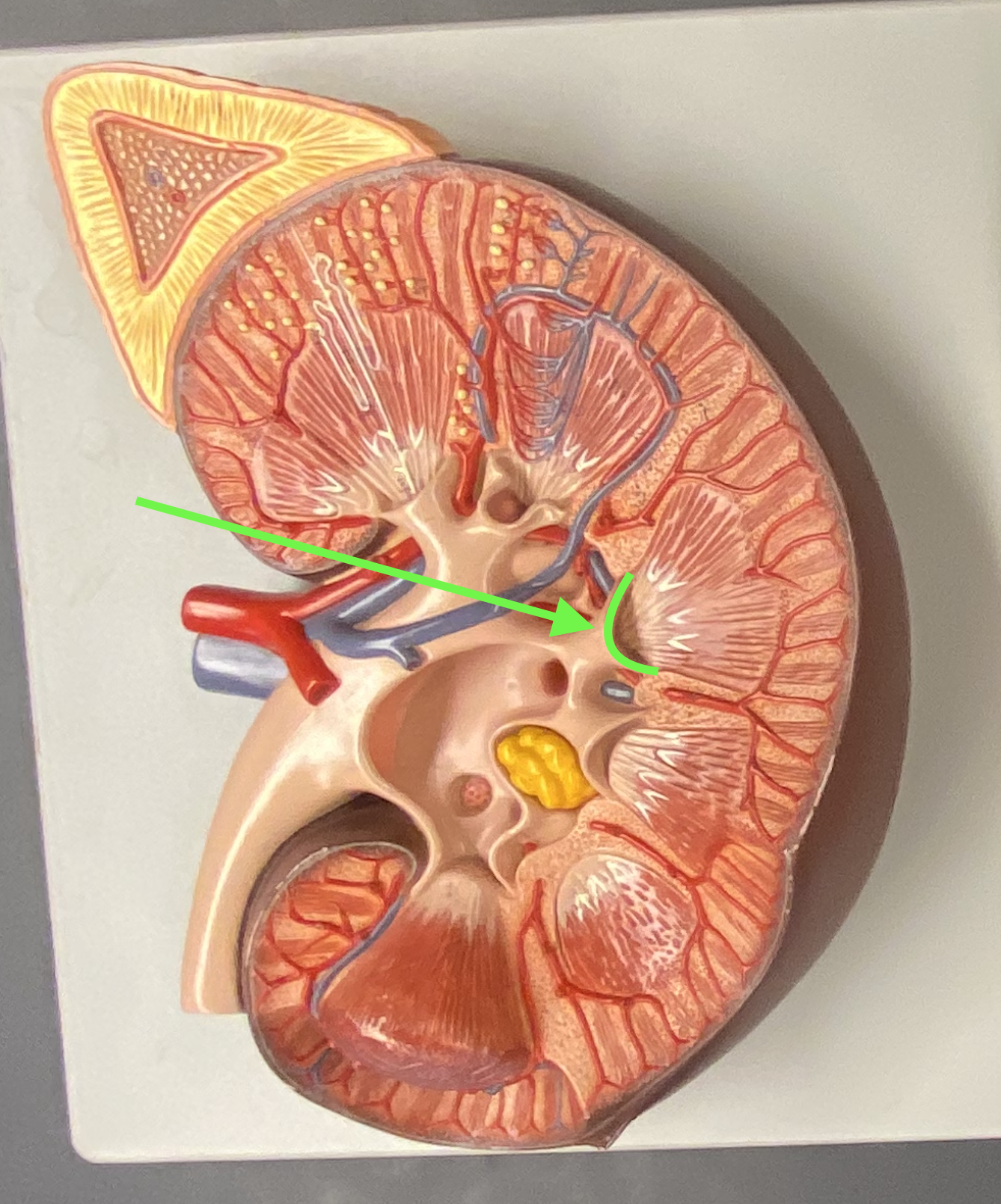
Renal sinus - Major calyx
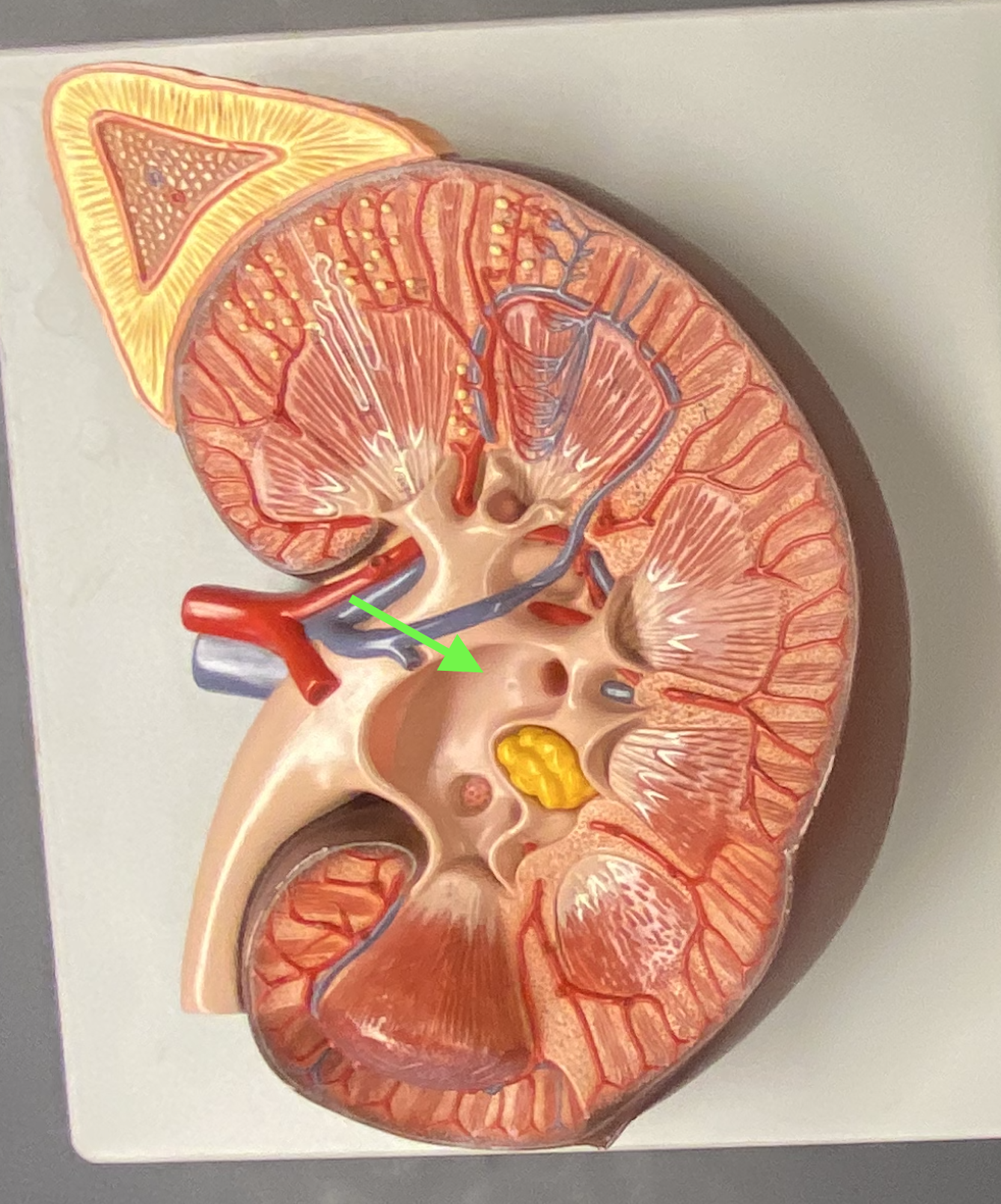
Renal sinus - Minor calyx
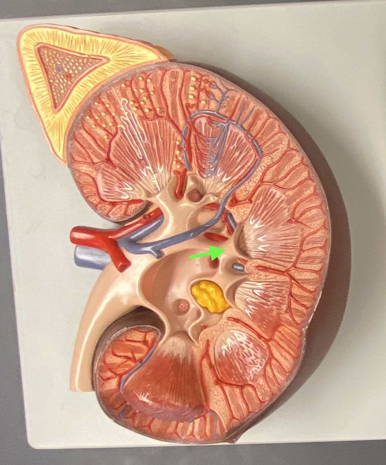
Renal sinus - Renal pelvis
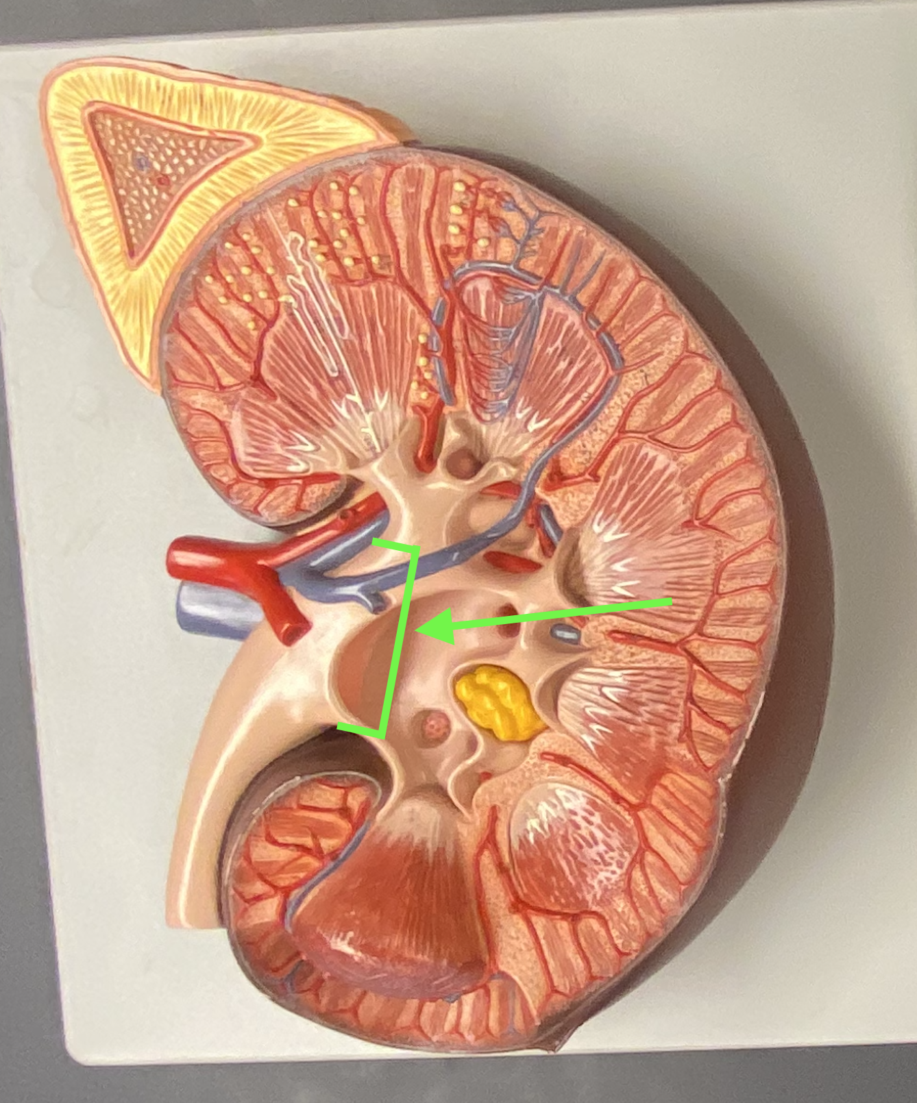
Renal artery and vein
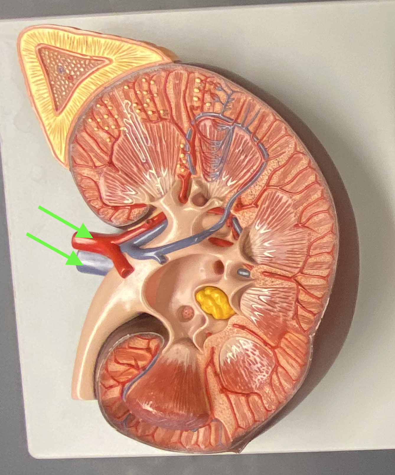
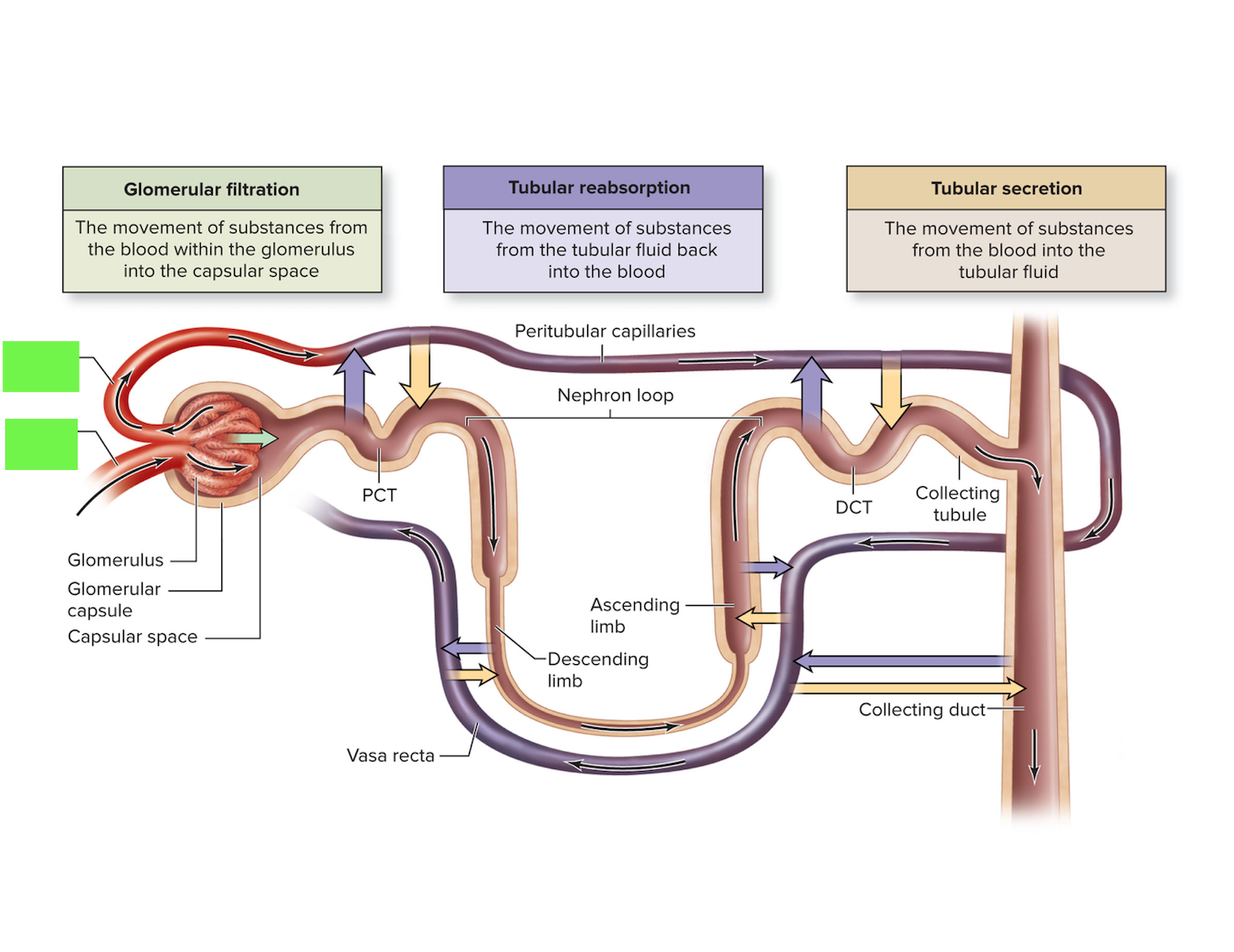
Renal corpuscle (add histology slide)
RC
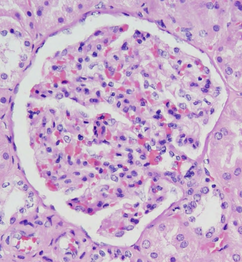
Renal corpuscle
Contains the glomerulus and the glomerular capsule
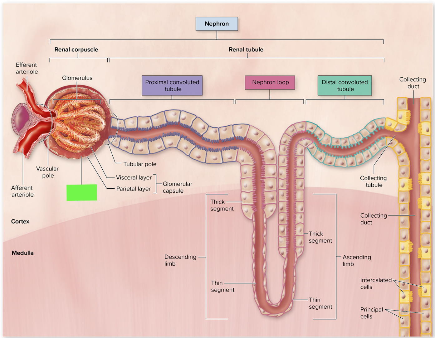
Renal corpuscle - Glomerular capsule
Renal corpuscle - Glomerular capsule (add histology slide)
GC
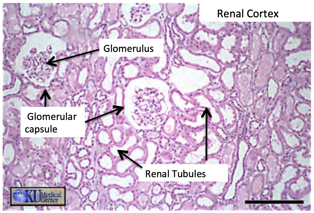
Renal corpuscle - Glomerular space (add histology slide)
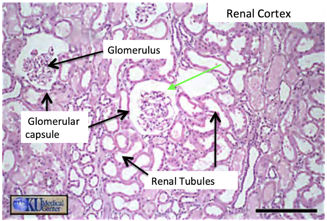
Renal corpuscle - Glomerular space
Renal corpuscle - Glomerulus (add histology slide)
G
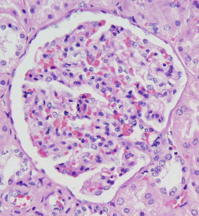
Renal corpuscle - Glomerulus
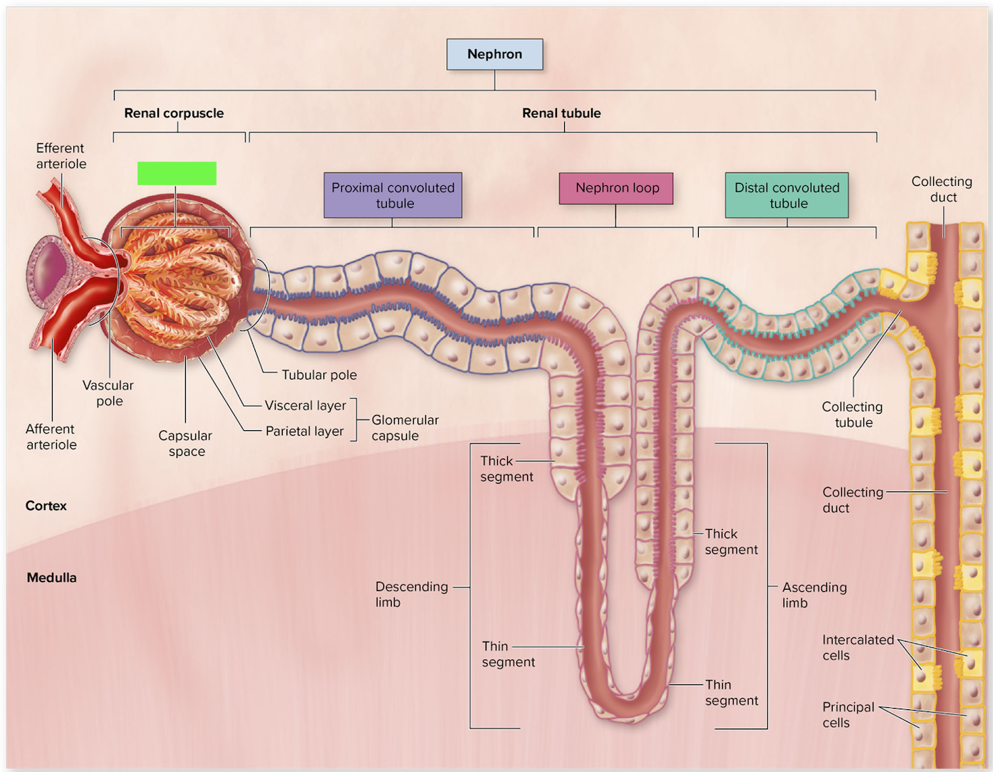
Proximal Convoluted Tubules (add histology slide)
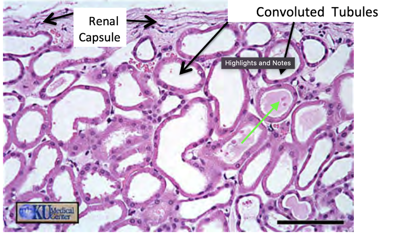
Proximal Convoluted Tubules
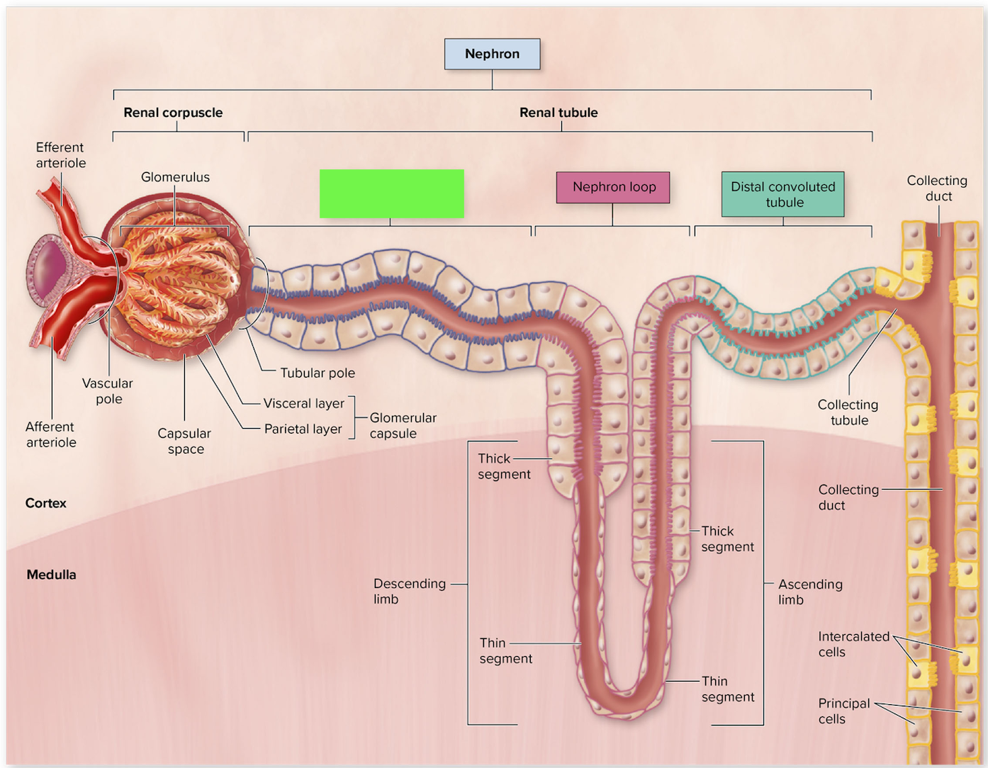
Nephron Loop (Loop of Henle)
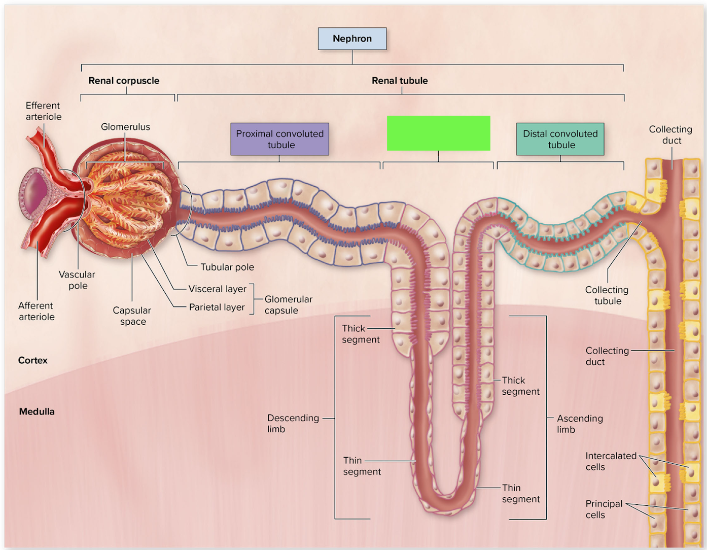
Distal Convoluted Tubules (add histology slide)
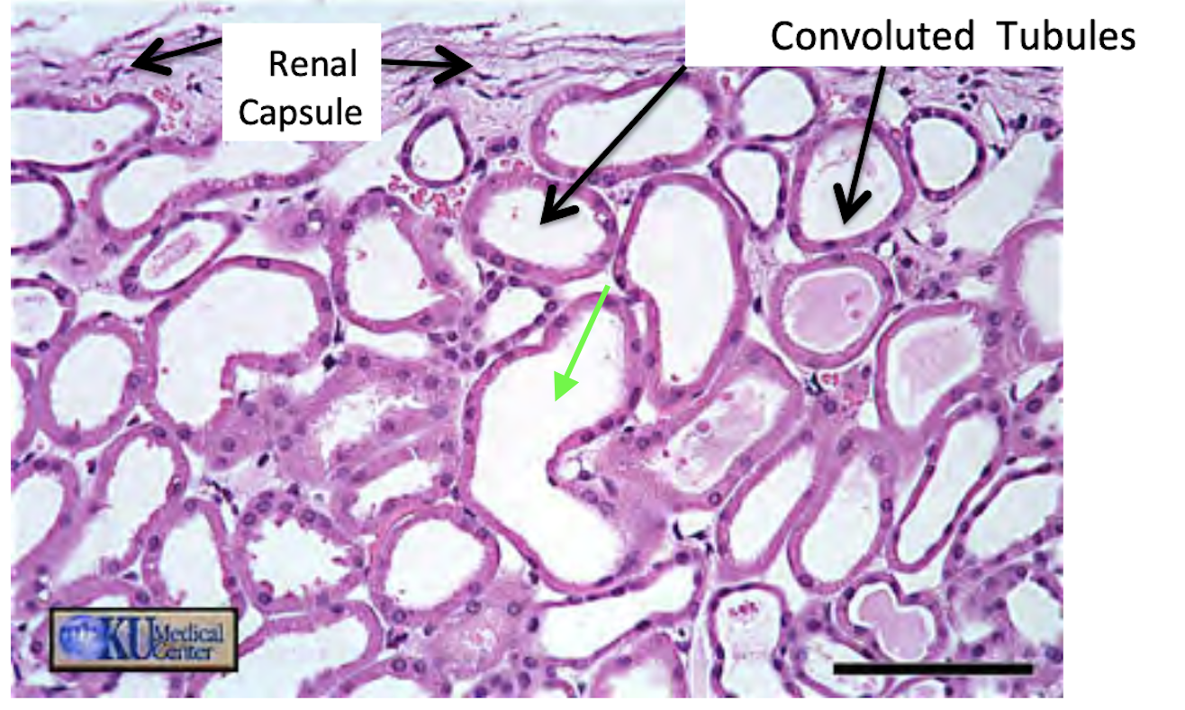
Distal Convoluted Tubules
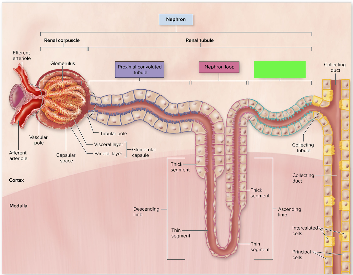
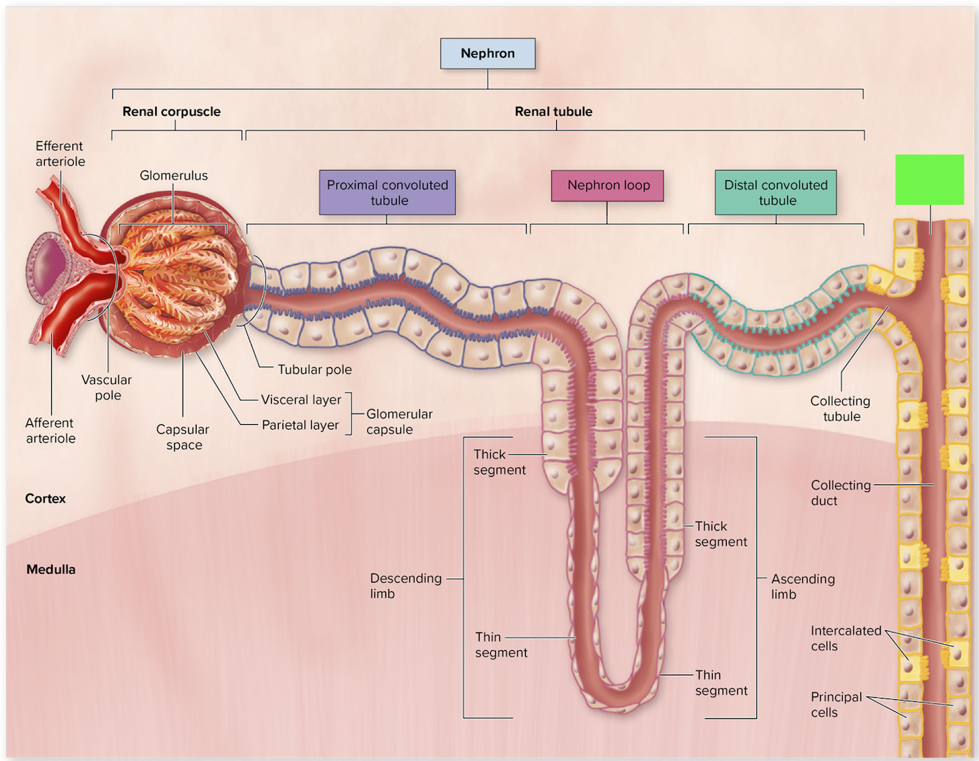
Collecting duct
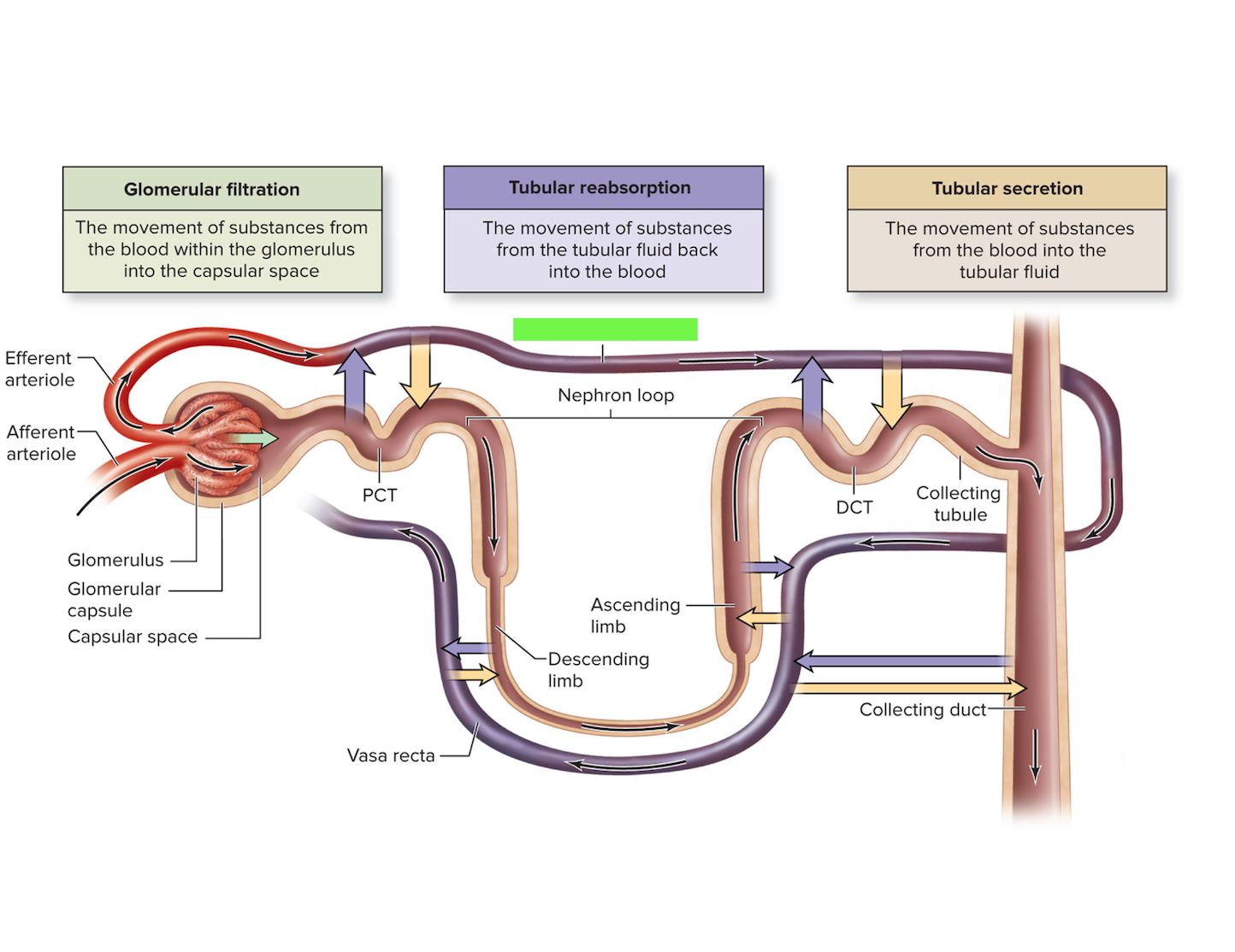
Peritubular capillaries
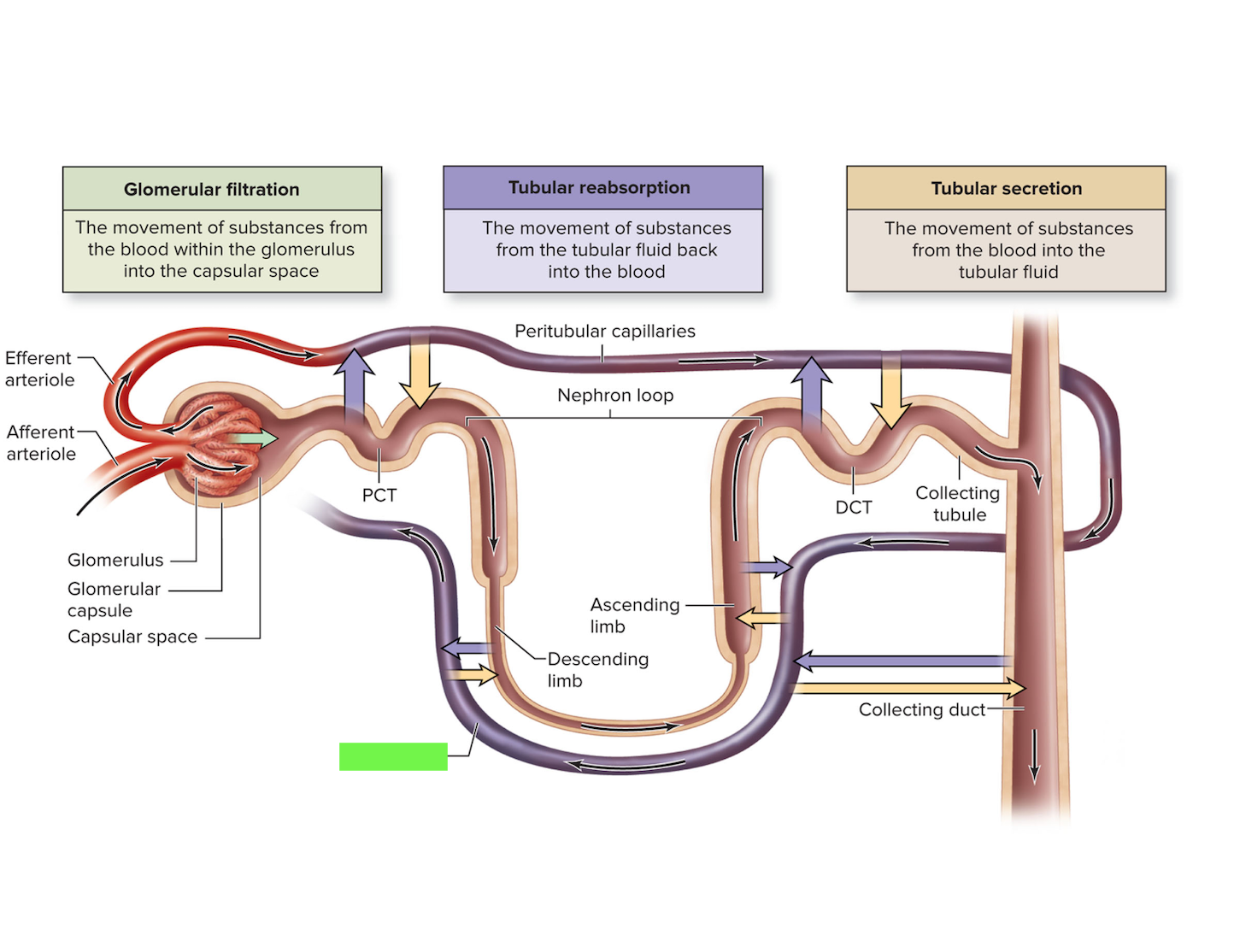
Vasa recta
Afferent and efferent arterioles
List the 5 functions of the urinary system
elimination of metabolic wastes like urea
regulation ion levels of the blood, Na+, Ca++, K+, PO4³
regulate blood pH via H+ and HCO3
regulate blood pressure
eliminate biogically active molecules like hormones and drugs
Identify the 4 major structures that compose the urinary system on the cadaver, models, and images
kidneys
ureters
bladder
urethra
Identify the two distinct regions of the kidney on models and images, and describe the features of each
renal cortex - outer layer, lighter in color, 85% of nephrons
renal column extends to medulla
renal medulla - inner layer, darker in color, 15% of nephrons
renal pyramid
List the 3 components of the renal sinus
minor calyx
major calyx
renal pelvis
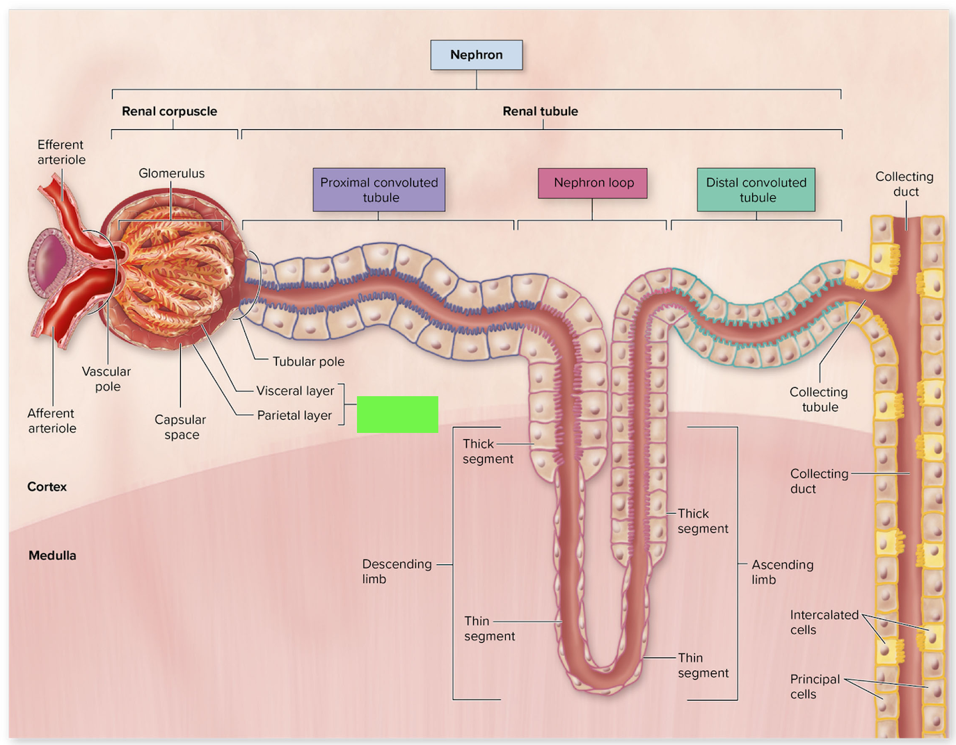
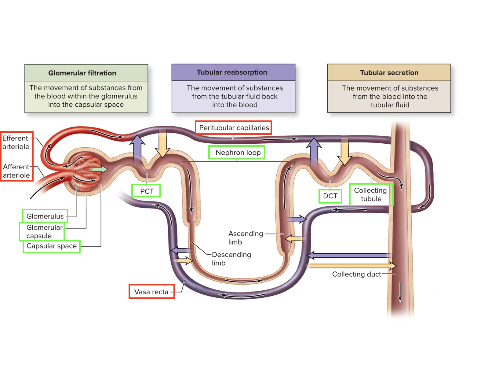
Draw a nephron, labeling the 7 structures from the terms list; include 2 vessels associated with the glomerulus, and 2 vessels associated with the nephron
List the 3 components of the renal corpuscle
glomerulus
glomerular capsule
glomerular space
Identify the glomerulus, glomerular space, glomerular capsule, proximal convoluted tubule, and distal convoluted tubule, on histology slides and images
PCT has brush border and lots of filtrate; DCT clearer; collecting duct cuboidal
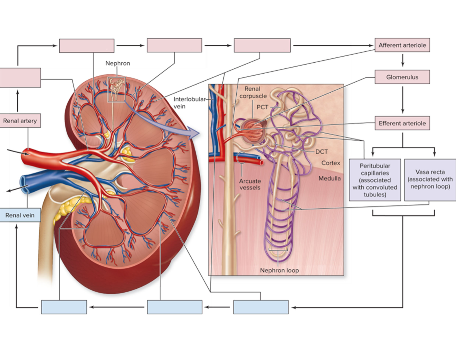
Describe the vasculature of the kidney, including the location and function of afferent and efferent arterioles, peritubular capillaries, and vasa recta.
Afferent arteriole:
supplied blood to a glomerulus
some blood plasma is filtered into the glomerulus
Efferent arteriole:
where blood exits the glomerulus
branches into PERITUBULAR CAPILLARIES or VASA RECTA
Peritubular capillaries:
associated with and intertwined around the proximal and distal convoluted tubules
reside in the cortex of the kidney
Vasa recta:
associated with nephron loop
primarily reside in the medulla
Trace fluid from its formation at the renal corpuscle until it exits the body through the urethra
glomerulus, glomerular space; PCT, loop of Henle, DCT, collecting duct, renal papilla, minor calyx, major calyx, renal pelvis, ureter, bladder, urethra
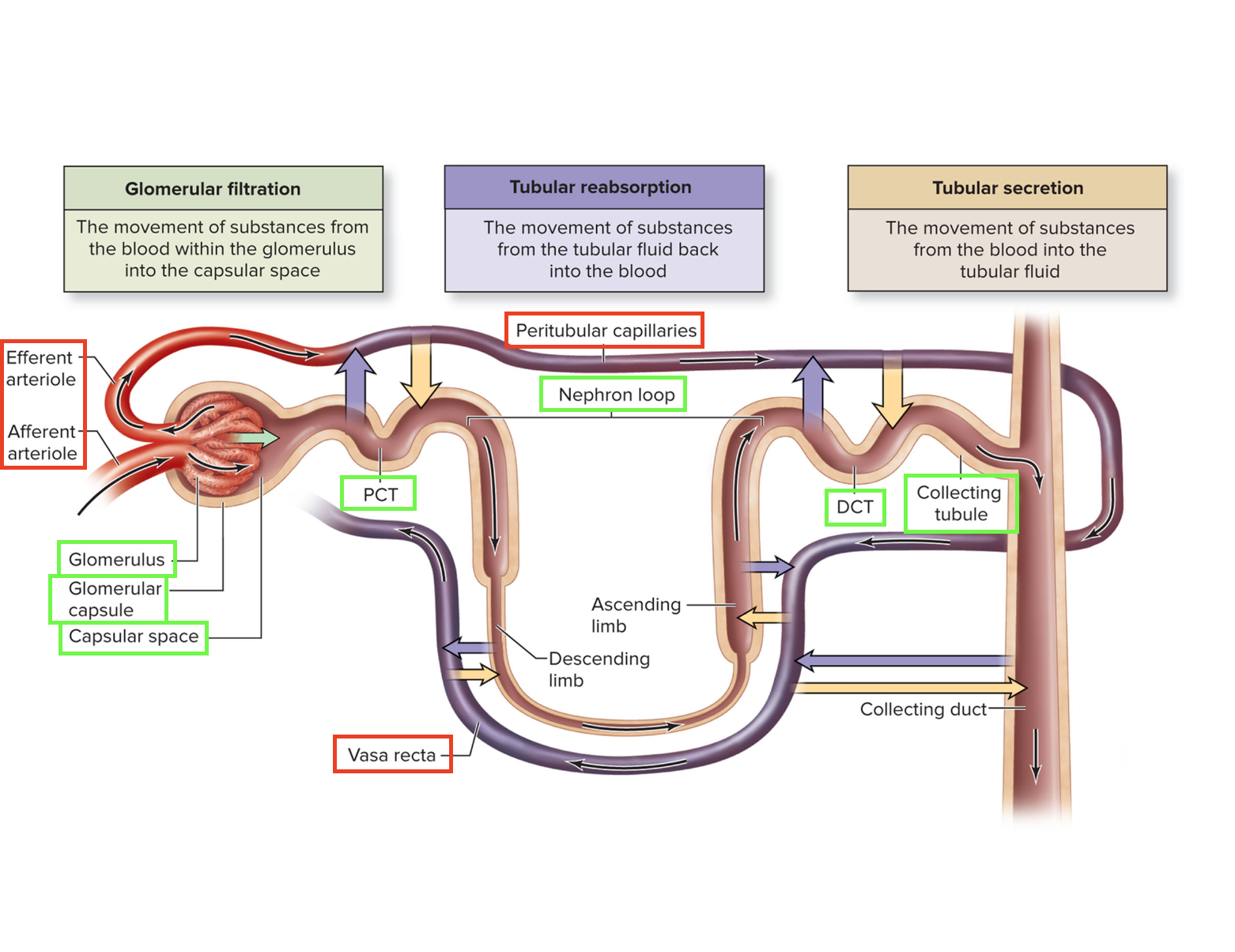
Compare and contrast the processes of filtration, reabsorption, and secretion.
filtration - movement of substances out of the blood into capsule
reabsorption - movement of subtances from tubular fluid back into blood
secretion - movement of substances from blood to tubular fluid
For these substances: water, glucose, proteins, ketones, amino acids, erythrocytes, leukocytes:
Indicate whether they filtered in the nephron,
and if reabsorbed, indicate the location of reabsorption.
Indicate whether they are normally present in urine,
and if not, name a disease or condition that would explain its presence.
Water
Filtered in nephron: Yes
If reabsorbed, where: PCT, Loop of Henle, DCT, Collecting duct
Normally present in urine: Yes
If no, name a disease or condition that would explain its presence:
Glucose
Filtered in nephron: Yes
If reabsorbed, where: PCT
Normally present in urine: No
Name a disease or condition that would explain its presence: Diabetes
Proteins
Filtered in nephron: small = YES, large = NO
If reabsorbed, where: small in PCT
Normally present in urine: No
Name a disease or condition that would explain its presence: Kidney damage
Keytones
Filtered in nephron: Yes
If reabsorbed, where: PCT
Normally present in urine: small amounts = YES, large amounts = NO
Name a disease or condition that would explain its presence: Diabetes
Amino acids
Filtered in nephron: Yes
If reabsorbed, where: PCT
Normally present in urine: No
Name a disease or condition that would explain its presence: …
Erythrocytes
Filtered in nephron: No
If reabsorbed, where: No
Normally present in urine: No
Name a disease or condition that would explain its presence: UTI
Leukocytes
Filtered in nephron: No
If reabsorbed, where: No
Normally present in urine: No
Name a disease or condition that would explain its presence: UTI
Trace the path of glucose from glomerulus to renal vein in a healthy person.
glomerulus - glomerular space - PCT - peritubular capillaries - venules - renal vein
Explain the effects of antidiuretic hormone on urine volume; note the site of production, site of action in the nephron, the direction and movement of water, and the effects on blood pressure.
Effects on urine volume: reduces urine volume
Site of production: posterior pituitary
Site of action: CD (Collecting Duct)
Water movement: reabsorption via aquaporins
Effect on BP: increases
Compare and contrast the female urethra and male urethra.
male 7.5” and surrounded by prostate gland
female 1.6”
State the volume of filtrate and urine produced daily by each kidney.
180L of filtrate, 90L per kidney
Urine volume = 1-2L total, so 0.5-1L per kidney
Urine pH
The pH for urine typiacally ranges between 4.5 and 8.0 - changes with diet