ANA301 Term Test 2
 Call Kai
 Learn
Learn Practice Test
 Spaced Repetition
 Match
Match Flashcards
 Knowt Play
1/129

There's no tags or description
Looks like no tags are added yet.
Name | Mastery | Learn | Test | Matching | Spaced | Call with Kai | Chat |
|---|
No analytics yet
Send a link to your students to track their progress
130 Terms
What does the intraembryonic coelom give rise to?
Pleural, pericardial, and peritoneal cavities
Parts of the respiratory system that are endoderm-derived
Respiratory epithelium
Parts of the respiratory system that are mesoderm derived
Intraembryonic splanchnic mesoderm
Cartilage/CT/SMCs of trachea + bronchi
Visceral pleura
Blood vessels of the lungs
Beginning of respiratory development (1st step)
Primordial foregut (cranial aspect) => pharynx
Paired mesenchymal swellings = pharyngeal arches => face/pharnynx/largyngeal structures
Respiratory outgrowth forms
What gives rise to pharyngeal arches
Mesoderm + neural crest cells
How many pharyngeal arches
6 pairs (5th pair regresses)
Respiratory outgrowth (what/where/when)
Ventral outgrowth that gives rise to the respiratory system
At the level of the 4th-6th pharyngeal arches
Week 4
How does the trachea separate from the esophagus
1: laryngotracheal groove forms
2: evagination ventrally to form laryngotracheal diverticulum (week 4)
3: distal end of diverticulum = respiratory bud
4: elongation + formation of tracheoesophageal folds
5: folds fuse = tracheoesophageal septum (end of week 5)
Tracheoesophageal folds
Growth that separates the trachea from the esophagus
Laryngeal inlet
Cranial opening between the primordial pharynx and developing trachea
Formation of the bronchioles
Respiratory bud bifurcates (= L + R primary bronchial buds) = week 4
Branching = secondary and tertiary bronchial buds = week 6-8
Tracheoesophageal fistula
Tracheoesophageal folds fail to fuse
Abnormal connection between the airway and the esophagus
Adult respiratory system structures
Nasal cavity
Oral cavity
Pharynx
Larynx
Trachea (15-20 cartilage rings + trachealis muscle)
Primary bronchi
Secondary bronchi
Segmental bronchi
Bronchioles
Alveoli
R + L lungs
Diaphragm
Carina
Wedge-shaped cartilage
Located at the bifurcation point of trachea
Superior = cough reflex
Inferior = no cough reflect
Right vs Left primary bronchus in the adult
Right = wider, shorter, vertical
Left = narrow, longer, horizontal
Right vs Left secondary (lobar) bronchi in adults
Right = 3
Left = 2
Right lung structures
3 lobes = superior, middle, inferior
Horizontal fissure (superior/middle)
Oblique fissure (middle/inferior)
Left lung structures
2 lobes = superior + inferior
Cardiac notch + lingula in superior lobe
Oblique fissure (superior/inferior)
Hilum of the lung
Depression on the mediastinal surface of the lungs
Contains the root of the lung (pulmonary arteries, pulmonary veins/primary bronchus/nerves/lymphatics)
State of the body cavities after folding
Embryonic folding = intra-embryonic coelom (U-shaped cavity)
- cranial = primitive pericardial coelom
- more caudal = paired pericardioperitoneal canals
- most caudal = peritoneal cavities (temporary communication with extraembryonic coelom
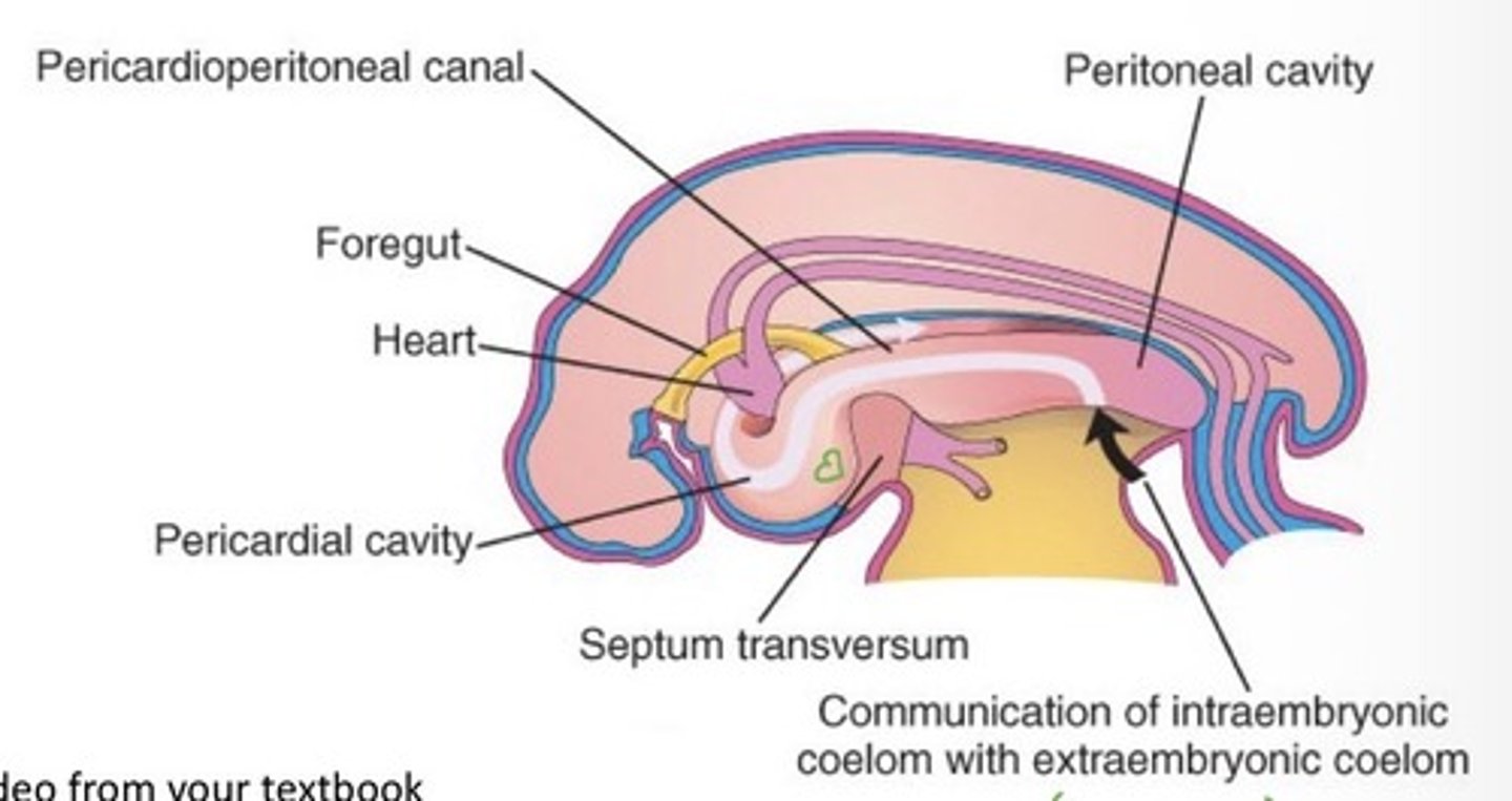
Development of the pleural cavities
Bronchial buds grow into the pericardioperitoneal canals
Expansion of the lungs reduces the cavity size => visceral and parietal pleura come into close apposition = pleural cavities
Pleural cavity in the adult
Lined by visceral pleural lining (adjacent to lung tissue) and parietal pleura (adjacent to ribs)
Space between linings filled with small amount of pleural fluid
Lung maturation stages
Pseudoglandular Stage (5-17 weeks)
Canalicular Stage (16-25 weeks)
Terminal Sac Stage (24 weeks - birth)
Alveolar Stage (late fetal period - 8 years)
Pseudoglandular stage
Rapid growth of conducting airway
Terminal bronchioles = exocrine glands
Gas exchange not possible
- epithelium = cuboidal
- capillaries = not closely associated with bronchioles
Canalicular stage
Development of respiratory elements
Gas exchange possible but limited
- epithelium = largely cuboidal
- capillaries = more & some apposed
Terminal sac stage
Many more terminal sacs bud off
Gas exchange is possible
- epithelium = thin + squamous (type 1 pneumocytes) & type II pneumocytes make surfactant
- capillaries = close apposition
Surfactant (time + purpose)
Begins in weeks 20-22
Functional levels around 26-28 weeks
Reduces surface tension + increases lung compliance
Low levels = neonatal respiratory distress syndrome (NRDS)
Alveolar stage
Terminal sacs mature into alveoli
Gas exchange gets more efficient
Diaphragm anatomy in adult
Attaches to margins of inferior thoracic aperture
Inserts in the midline at the central tendon
Hiatus
1. Caval opening for inferior vena cava
2. Esophageal hiatus
3. Aortic hiatus
Location of each hiatus in the diaphragm
I8 10 Eggs At 12
Inferior vena cava (T8)
Esophagus (T10)
Aorta (T12)
Embryonic components that give rise to the diaphragm
Septum transversum = central tendon
Pleuroperitoneal membranes = close pericardioperitoneal canals
Dorsal mesentery of esophagus = crura
Muscular ingrowth from body wall = peripheral skeletal muscle
Septum transversum for diaphragm development
Mesoderm derived structure
Week 3 = appears cranial to pericardial cavity
Week 4 = cranial folding moves it caudal
Extends dorsally from the ventrolateral body wall
- fuses with the mesenchyme and pleuroperitoneal membranes
Pleuroperitoneal membranes for diaphragm development
Grows ventromedially from dorsolateral body wall
Fuses with septum transversum + dorsal mesentery of esophagus (week 6)
Important in the fetus but minor contribution in the infant (replaced by muscle)
Dorsal mesentery of the esophagus for diaphragm development
Suspends esophagus to dorsal body wall
Located in the midline
Fuses with septum transversum + pleuroperitoneal membranes
Myoblasts from cervical somites (paraxial mesoderm) infiltrate + form crura
Muscular ingrowth from body wall for diaphragm development
Myoblasts from cervical somites (paraxial mesoderm) migrate into the lateral body wall
- form the muscular portion of the diaphragm
Congenital diaphragmatic hernia (CDH)
Defect in the diaphragm = abdominal viscera enter thoracic cavity
- can impair lung development (lack of space)
Most commonly due to incomplete fusion of pleuroperitoneal membranes with other components
When can heart beat be detected in the fetus
4th week (gestational week 6)
Before this point, blood flow = ebb + flow
Germ cell origins of the cardiovascular system
Heart = intraembryonic splanchnic mesoderm
Pericardial cavity = intraembryonic coelom + visceral + parietal pericardium
Aortic arches = neural crest cells
Formation of the heart tube
1. Primary heart field develops in the splanchnic mesoderm (week 3)
- U-shaped, bilateral
2. Primary heart field (mesenchymal cells) = angioblasts = aggregate to form angioblastic chords = canalize to form bilateral endocardial heart tubes
3. Secondary heart field = progenitor cells to elongate the arterial pole
What does lateral folding do for heart development (+ when)
Paired endocardial heart tubes moved to the midline
- Fuses = single primitive heart tube
Paired pericardial coeloms into midline
- fuse = pericardial cavity
Day 20-22
Suspension of the heart tube
Heart tube lies within the pericardial cavity (attached dorsally by a mesentery = dorsal mesocardium)
Mid-portion often the dorsal mesocardium degenerates = transverse pericardial sinus (day 28)
Anchoring points of the primitive heart tube (day 22-28)
Cranial = truncus arteriosus (arterial outflow)
Caudal = sinus venosus (venous inflow)
Layers of the embryonic heart wall
Endocardium = endothelial lining (derived from endocardial tube)
Myocardium = middle muscular layer (derived from splanchnic mesoderm), separated by cardiac jelly from the endocardium
Epicardium = outer layer (derived from the secondary heart field), coronary vessels
Constrictions + dilations of the heart tube
Day 23 - 28
Truncus arteriosus (TA) = arterial outflow, continuous with pharyngeal arch arteries
Pharyngeal arch arteries (PAA)
Bulbous Cordis (BC) = future R ventricle
Primitive ventricle (PV) = largest dilation = future L ventricle
Primitive Atrium (PA) = future L+R atria
Sinus Venosus (SV) = venous inflow segment
Septa in the adult heart
Atrioventricular septum
Interatrial septum
Interventricular septum
Heart looping
Day 23 (linear) - 28 (S-shaped)
Caused by differential growth in a confined space
- Bulbus cordis and primitive ventricle grow faster than the rest
Cranial end buckles ventrally, caudally, slightly to the right
Caudal end buckles dorsally, cranially, and slightly to the left
Trabeculation
Late week 4 - early week 5
Smooth myocardial surface => muscular ridges (Trabeculae)
Dextrocardia
Looping occurs in the opposite direction
Chambers on the wrong side of the
General mechanisms of septum formation
Tissue growth
- opposing ridges proliferate + extend across the lumen (from opposite sides or from a single wall)
Differential remodelling
- septal ridge present but fixed
- adjacent chambers expand/dilate
Formation of septa in the heart
1. Dorsal + ventral endocardial cushions form in the AV canal (end of week 4) + left + right cushions narrow the AV canals
2. Active growth of cushions + fusion = separation into R and L atroventricular canals (week 5)
3. Septum primum (thin membrane) extends from the roof of the atrium towards the cushions
- gap is left called ostium primum
4. Septum primum fuses with the cushions but cell death makes a new opening (ostium secundum)
5. Septum secundum forms (crescent-shaped with a gap (foramen ovale)
Function of the ostium secundum
R to L shunting during fetal life (no need for pulmonary circulation
Fossa ovale
Depression in the R atrial wall
Location where the septum primum and septum secundum fused after birth
Patent foramen ovale
Septa (primum + secundum) fail to fuse
- stay closed under normal pressure conditions
How do the atria develop
Primitive right atrium enlarges - pectinate
Encorporates right horn of the sinus venosus (into posterior wall) = sinus venarum - smooth-walled
Primordial pulmonary vein sprouts from the dorsal wall of the primitive atrium (week 4) + connects to vascular plexus
- bifurcates into 4 pulmonary veins
Left atrium enlarges and incorporates the pulmonary veins (until week 8)
Interventricular septum anatomy in the adult
Membranous part = small, superior, posterior
- results from the fusion of the right conotruncal (bulbar) ridge, left conotruncal (bulbar ridge), fused dorsal + ventral endocardial cushions
Muscular part = large, inferior, anterior
- leaves a gap called interventricular foramen
Timeline for heart development
Mostly between day 23 and day 28
Role of the membranous part of the interventricular septum
closes up the interventricular foramen
Formation of the aorticopulmonary septum
Swellings develop in the walls of the truncus arteriosus and bulbus cordis
- L + R truncal ridges in TA
- L + R bulbar ridges in BC
Neural crest cells migrate into the ridges
Ridges align, spiral, and merge = aorticopulmonary septum
Outflow tract partitioned into ascending aorta and pulmonary trunk
What happens to the interventricular foramen
Fusion of L + R (conotruncal) bulbar ridges
Fusion of dorsal + ventral endocardial cushions
Fusion to the superior margin of the muscular interventricular septum
Lateral expansions of the sinus venosus
L + R sinus horns (bilateral by day 26)
Concomitant with heart looping
Cardinal vein (anterior/posterior),
Umbilical vein (placenta)
Vitelline vein (UV)
Flow of blood in the embryo (day 26)
L+R head => L+R anterior cardinal vein =>
+
L+R body => L+R posterior cardinal vein =>
common cardinal vein => sinus venosus
Anatomy of the adult right atrium
Inflow vessels = superior + inferior vena cava
Pectinate muscles (Trabeculated part of the wall)
Sinus venarium (smooth part of atrial wall)
Interatrial septum (wall to L atrium)
Fossa Ovalis (depression with rim from septum secundum and floor from septum primum
Right Auricle (ear-like flap)
Right AV valve (tricuspid valve)
Adult anatomy of the right ventricle
AV valve (tricuspid)
Trabecular carnae (muscular ridges on wall of ventricle)
Interventricular septum
Pulmonary valve (semilunar)
Outflow vessels (pulmonary trunk + R and L pulmonary artery)
Adult anatomy of the left atrium
Inflow vessels (pulmonary veins (superior + inferior right + left))
Pectinate muscles (rough part of anterior wall)
Smooth posterior wall
Interatrial septum
Valve of foramen ovale (thinning in septum)
Left auricle (ear-like flap)
AV valve (bicuspid/mitral)
Adult anatomy of the left ventricle
AV valve (bicuspid/mitral)
Chordate tendinae (close valve)
Papillary muscles (close valve)
Trabecular carnae (muscular ridges)
Interventricular septum
Aortic semilunar valve
Outflow vessels (ascending aorta, aortic arch, brachiocephalic, left common carotid, left subclavian arteries)
Where is the heart located
Middle mediastinum
Between 2 pleural cavities
Enclosed in the pericardium (outer fibrous + inner serous layer)
Superior to diaphragm
Inner serous layer of pericardium
Parietal layer (somatic mesoderm)
Visceral layer (splanchnic mesoderm) = epicardium
Pericardial cavity contains pericardial fluid
Fate of the sinus venosus in the embryo
R atrium enlarges + incorporates right horn into posterior wall (sinus venarium in adult) = week 6
Venous return shifts to the R, so left horn of sinus venosus shrinks (persists as coronary sinus in adult) = week 8
Coronary sinus
Blood vessel on the posterior side that drains blood from coronary circulation into the right atrium
Coronary arteries that supply the heart tissue
Right Coronary A
- branch of ascending aorta
- in atrioventricular sulcus
- R marginal branch (R ventricle)
- Posterior interventricular artery
- supplies RA, RV, some LV
Left Coronary A
- branch of ascending aorta
- posterior to pulmonary trunk + bifurcates
- Anterior interventricular artery (anastomosis with posterior interventricular artery at apex)
- circumflex a (AV sulcus) (anastomosis with R coronary artery)
- left marginal a (branch of circumflex a)
- supplies LA and LV)
Fate of Anterior Cardinal veins
Day 26 = bilateral venous drainage from head
Week 7-8 = anastomosis between L+R anterior cardinal vein = shift blood flow from Left to Right
Caudal L anterior cardinal vein + L common cardinal vein regress
Anastomosis = L brachiocephalic vein
Caudal R anterior cardinal vein + R common cardinal vein = superior vena cava
Cranial R anterior cardinal vein = R brachiocephalic vein
Fate of Posterior Cardinal Veins
Day 26 = bilateral venous drainage
Posterior cardinal veins regress
- except contribute to the root of the Azygos vein
Flow of blood in week 8
L upper body => L brachiocephalic vein => SVC => right atrium
R upper body => R brachiocephalic vein => SVC => right atrium
Fate of the umbilical veins
R umbilical vein disappears (7 weeks)
L umbilical vein persists
- cranial aspect (heart - liver) degenerates
- caudal aspect (placenta - liver) persists as umbilical vein
- ductus venosus between L umbilical vein and IVC (bypass the liver)
Blood flow from placenta
Placenta => umbilical vein => ductus venosus => IVC => right atrium
Fate of the vitelline veins
Bilateral at Day 26
Week 7 = left vitelline vein regresses
Right vitelline vein = IVC + hepatic portal system (return nutrient-rich blood from abdominal region to heart)
Blood flow from the vitelline veins
Abdominal venous blood =>
portal vein =>
hepatic veins =>
IVC =>
right atrium
Outflow of blood pathway in embryo (day 26)
Primordial atrium
Aortic Sac (distal Truncus Arteriosus)
Pharyngeal Arch Arteries
Dorsal Aorta
- dorsal segmental arteries => intercostal + lumbar arteries in adult
- vitelline artery (to UV via omphaloenteric duct)
- umbilical artery
Fate of the vitelline arteries
Multiple arise from the dorsal aorta (day 26)
Most regress during weeks 5-6
3 unpaired ventral branches present at the end of week 6
- celiac artery (foregut)
- superior mesenteric artery (midgut)
- inferior mesentery artery (hindgut)
Adult anatomy of outflow vessels
Thoracic aorta (between esophagus + vertebral bodies)
Paired posterior intercostal arteries
Passes through the diaphragm at the aortic hiatus (T12) = Abdominal aorta
Branches
- paired lateral branches (ex. Renal)
- unpaired: celiac, superior mesenteric, inferior mesenteric arteries
Terminates at L4, bifurcates:
- L+R common iliac arteries, branch:
- internal illiac artery (bladder/rectum/reproductive organs)
- superior vesicle artery
- medial umbilical ligaments
- external iliac artery (lower limb)
Fate of umbilical arteries
Initially = travel through connecting stalk
Remodelled in week 5-6 as placental circulation is established
In adult:
- Distal portions regress (=medial umbilical ligaments
- Proximal portions persist (internal iliac arteries + superior vesical arteries)
Fate of dorsal aortae
Cranial portions remain bilateral
- left = primordial aorta
- right = regress + remnants = proximal part of right subclavian artery + distal internal carotid arteries
Caudal portions fuse to single thoracic/abdominal aorta (by day 37)
- 30 intersegmental branches = intercostal arteries/lumber arteries/common iliac artery
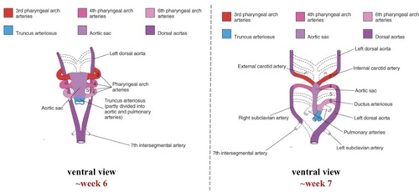
Pharyngeal arch arteries
6 pairs, (1 per pharyngeal arch)
Form during heart looping (4th week)
Blood flow from distal aspect of the truncus arteriosus (aortic sac) into pharyngeal arch arteries (6th => 1st)
5th pharyngeal arch artery regresses early (in 50%)
Fate of the pharyngeal arch arteries (week 7)
1st + 2nd also mostly regress by 6 weeks
1st = maxillary arteries (ears, teeth, muscles of face)
2nd = dorsal remnants form stems of the small stapedial arteries (middle ear)
dorsal aorta degenerates between 3rd & 4th = physical separation between 3rd and 4th outflows
3rd = internal carotid artery (both sides)
4th = bilateral (parallel to 3rd PAA)
6th = ductus arteriosus
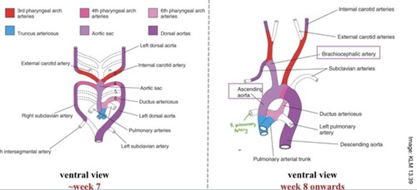
Fate of the pharyngeal arch arteries (week 8 onwards)
3rd = proximal aspect of internal carotid artery + common carotid artery (both sides)
4th =
- R = proximal segment of right subclavian artery
- L = arch of aorta between L common carotid and L subclavian
6th =
- R = degenerates & proximal = R pulmonary artery
- L = proximal = L pulmonary artery + distal = ductus arteriosus
ductus arteriosus
Shunt between pulmonary artery and aorta
Allows blood to flow from pulmonary trunk to L dorsal aorta (future descending aorta)
Fate of the aortic sac
Terminates at the 3rd pharyngeal arch
- 6th pharyngeal arch branches from the caudal aspect
Elongates => sac (week 6) => tube (week 7) => ascending aorta (week 8)
Contributes to the ascending aorta, part of the arch of the aorta, brachiocephalic artery
Right subclavian artery derivatives
Proximal segment = 4th pharyngeal arch artery
Distal = R dorsal aorta, 7th intersegmental artery
Adult vasculature coming off the arch of aorta
Brachiocephalic artery
- R subclavian
- R Common carotid (R external carotid + R internal carotid)
L Common Carotid (L internal carotid + L external carotid)
L subclavian artery
Changes in blood flow between the fetus and neonate
Umbilical vein = ligamentum teres that attaches inferior liver to umbilicus
Umbilical arteries = distal parts medial umbilical ligament on either side of the bladder. Proximal part = superior vesical arteries of the bladder
Oval foramen = closed by valve of septum primum (fuses at 3m of age)
Ductus venosus (bypasses liver) = ligamentum venosum
Ductus arteriosus (bypasses lungs) = ligamentum arteriosum
Adult derivatives of the foregut
Pharynx
Esophagus
Stomach
Proximal duodenum
Liver
Gallbladder
Cystic duct
Pancreas
Respiratory system
Adult derivatives of the midgut
Distal duodenum (starting after major duodenal papilla)
Jejenum
Ileum
Cecum
Appendix
Ascending colon
Proximal 2/3 of transverse colon
Adult derivatives of the hindgut
Distal 1/3 of transverse colon
Descending colon
Sigmoid colon
Rectum
Proximal anal canal
Epithelium of urinary bladder + urethra
Allantois
Omphaloenteric duct/vitelline duct/yolk stalk
Connects midgut to umbilical vesicle
Classification of mesoderm surrounding gut tube
Developing tube is invested in intraembryonic splanchnic mesoderm
Enclosed by the intraembryonic coelom (peritoneal cavity)
Intraembryonic somatic mesoderm surrounds overlying ectoderm
Tissues derived from the Endoderm in the digestive system
Epithelial lining of gastrointestinal tract
Epithelial cells of the accessory digestive organs
Blood supply of the digestive system in the adult
Foregut = celiac trunk
Midgut = superior mesenteric artery
Hindgut = inferior mesenteric artery
Name of the somatic and splanchnic mesoderm in the adult
Somatic mesoderm = parietal peritoneum
Splanchnic mesoderm = visceral peritoneum
Serous cavity
Cavity between the parietal and visceral membranes
Mesentery
Site of attachment
Tether the gut tube to abdominal wall
Contain neurovascular components that supply the gut tube