Di Imaging 2 Exam #1 Images
 Call Kai
 Learn
Learn Practice Test
 Spaced Repetition
 Match
Match Flashcards
 Knowt Play
1/45

There's no tags or description
Looks like no tags are added yet.
Name | Mastery | Learn | Test | Matching | Spaced | Call with Kai | Chat |
|---|
No analytics yet
Send a link to your students to track their progress
46 Terms
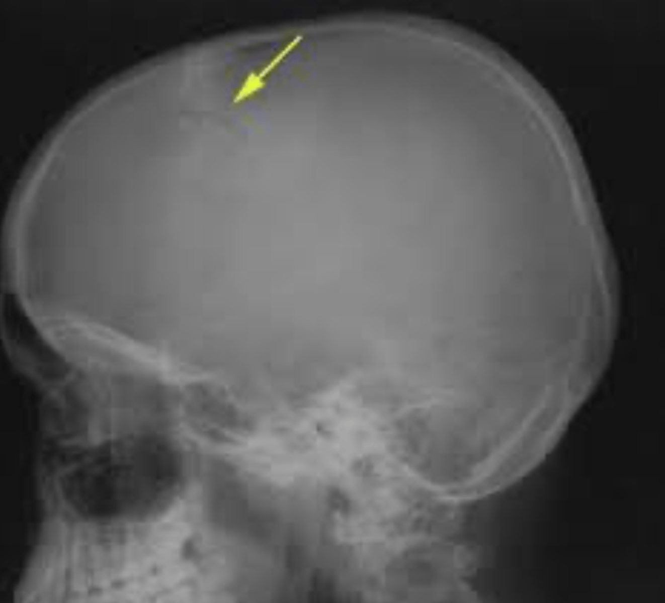
Depressed skull fracture
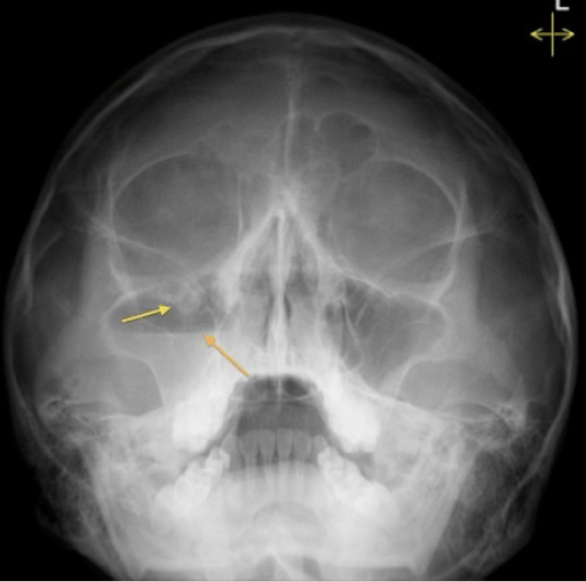
Blowout fracture
- fracture at floor of orbit
- MC due to direct trauma
- inferior rectus muscle may project into maxillary sinus
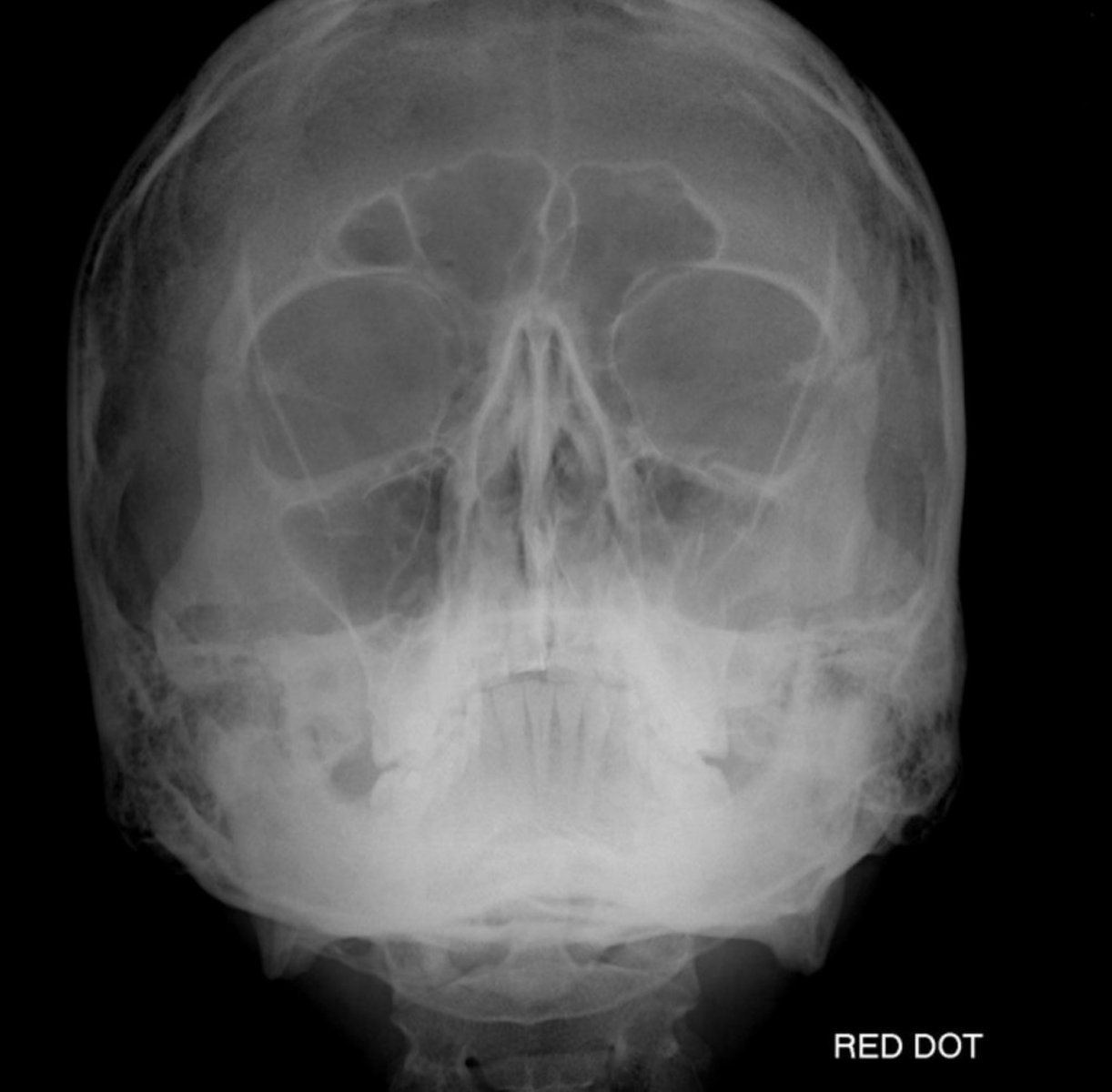
Tripod fracture
- Separation of all three major attachments of the zygoma to the rest of the face
Linear skull fracture
- most common type of skull fracture
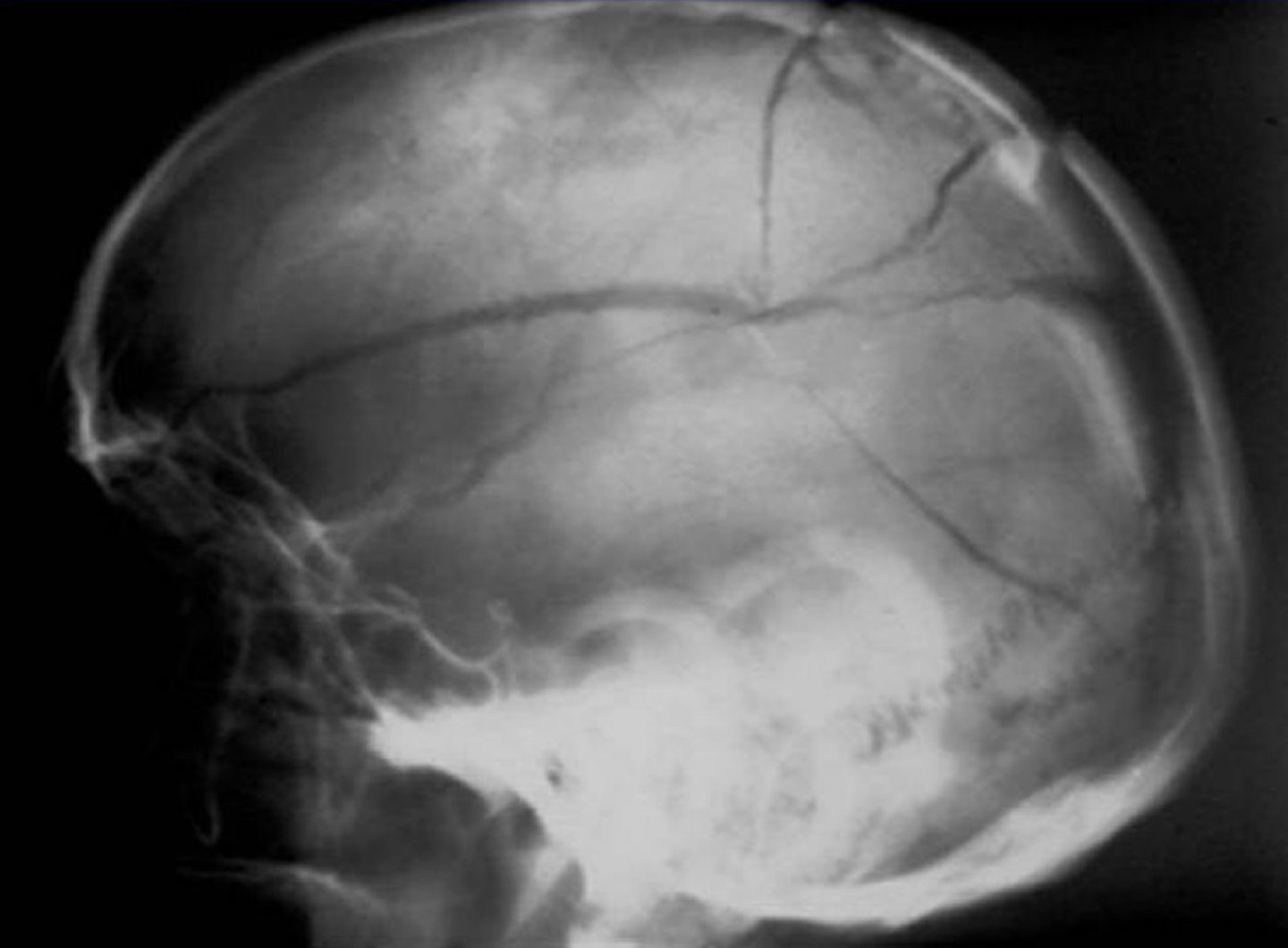
Subdural hematoma
- blood gathers between the dura mater and the brain
- convex in shape
- venous involvement
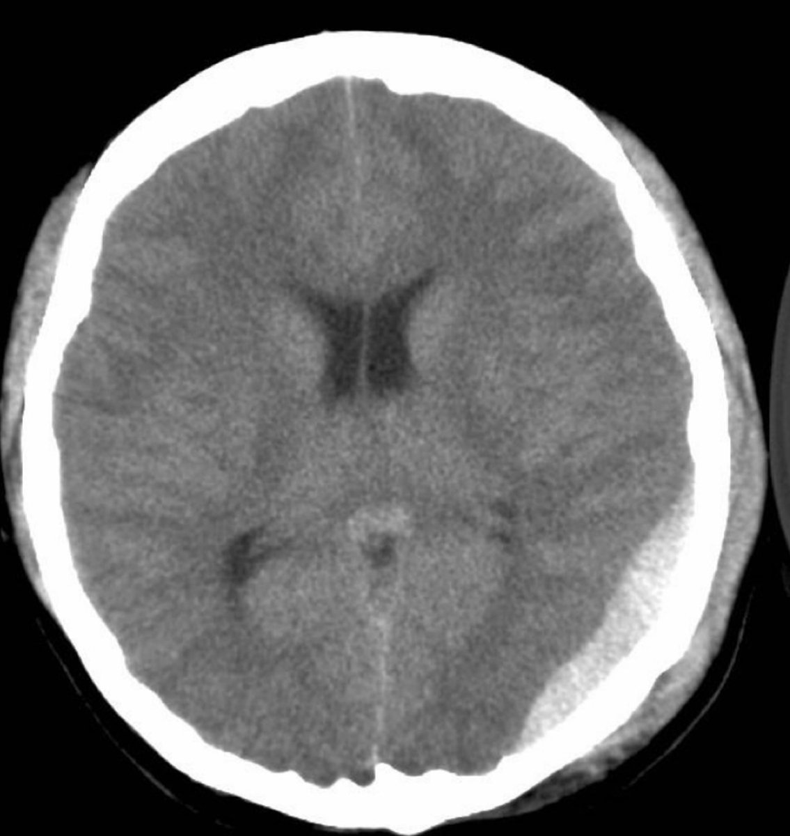
Epidural hematoma
- blood gathers between the dura mater and the skull
- 15-20% are fatal
- concave (crescent) in shape
- arterial involvement
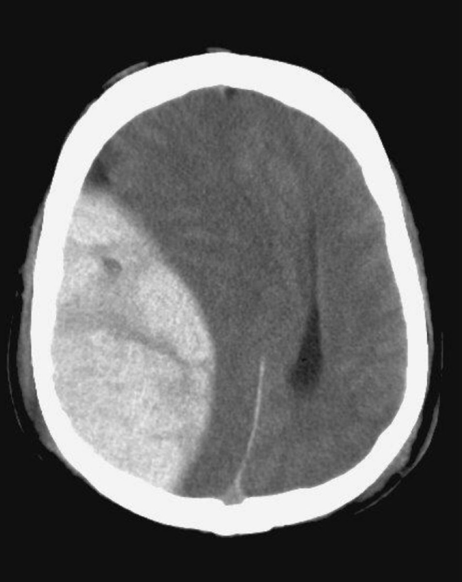
Posterior arch fracture
- MC fracture of atlas
- typically bilateral and vertical
- hyperextension mechanism
- differentiate from agenesis of posterior arch of atlas
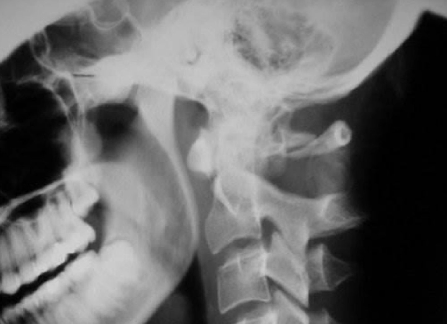
Agenesis of posterior arch of C1 (with enlarged C2 spinous)
- differentiate from posterior arch fracture
- developmental anomaly
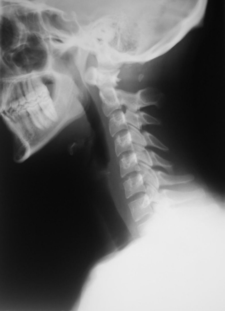
Jefferson's Fracture (Bursting fracture of C1)
- 2nd MC injury to atlas
- axial compression injury
- bilateral fracture of anterior and posterior arches
- overhang sign
- If the transverse atlantal ligament is torn, you'll see >7mm of offset
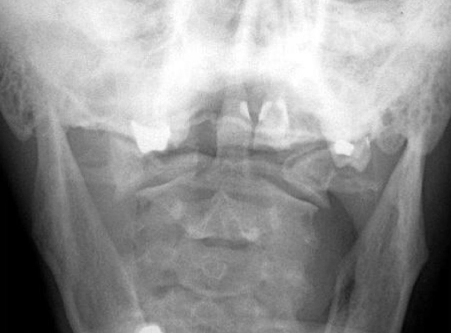
Type 1 odontoid fracture
- fracture of the tip of the dens
- avulsion of dens from alar or apical dental ligaments
- very rare
- stable and heals well
- differentiate from os terminale
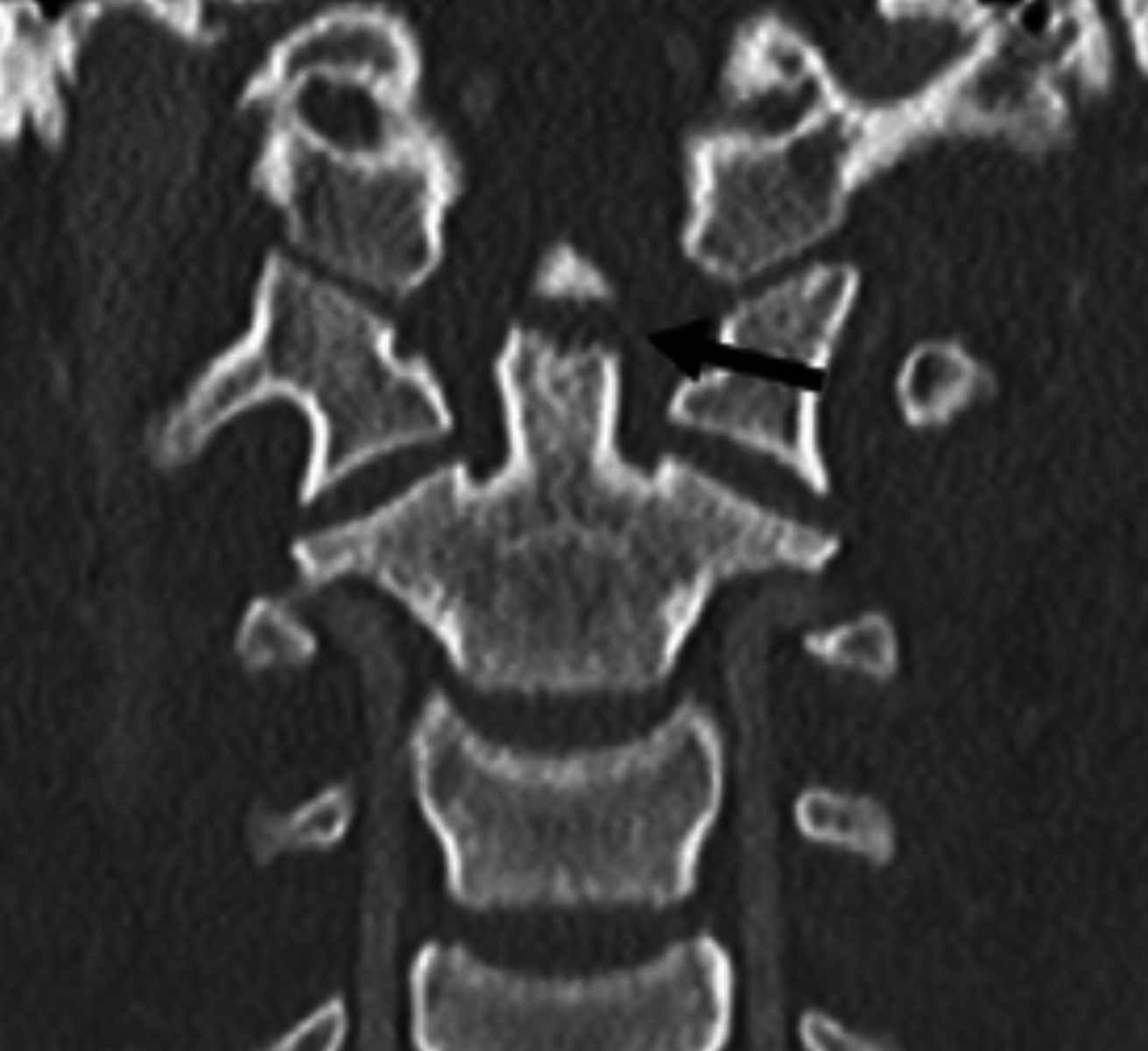
Type 2 odontoid fracture
- MC fracture of dens
- unstable
- nonunion very common (64%)
- if displaced >5mm = 100% nonunion
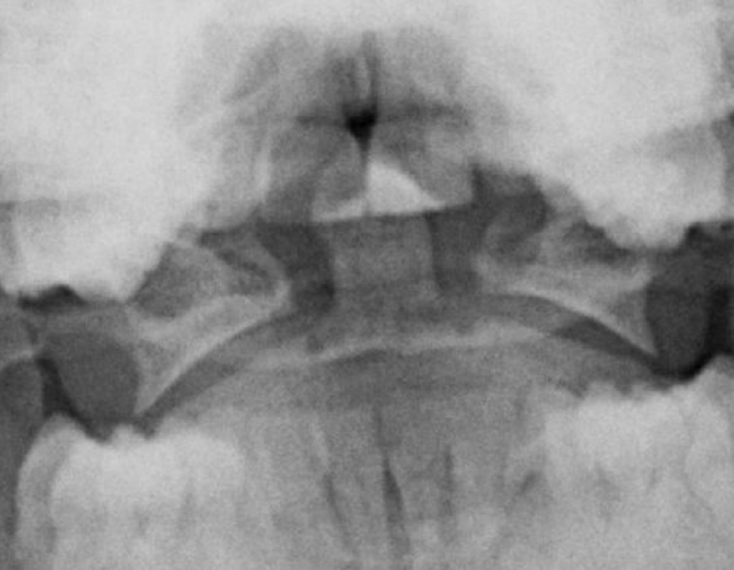
Type 3 odontoid fracture
- fracture through body of C2
- unstable
- May disrupt Harris' ring
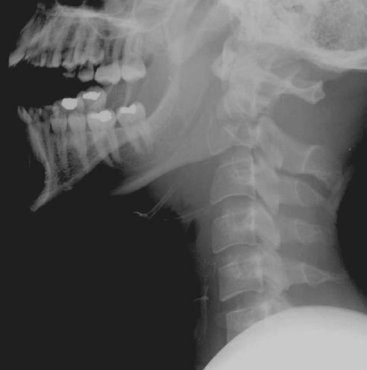
Hangman's fracture
- traumatic spondylolisthesis of C2
- 2nd MC fracture of C2
- bilateral pedicle fracture of C2
- hyperextension mechanism
- Send to ER
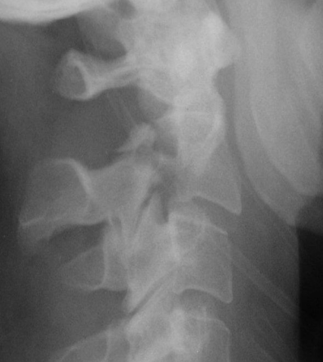
Compression fracture
- flexion mechanism
- Decreased anterior body height, preservation of posterior body height
- Superior endplates are affected
- stable
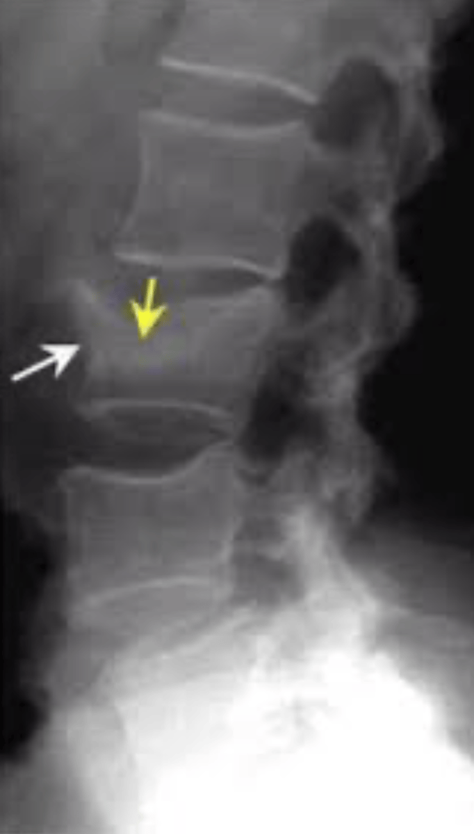
Burst fracture (of C3)
- vertebral body "explodes"
- possible cord compression
- unstable
- interpeduncular widening (seen on AP film)
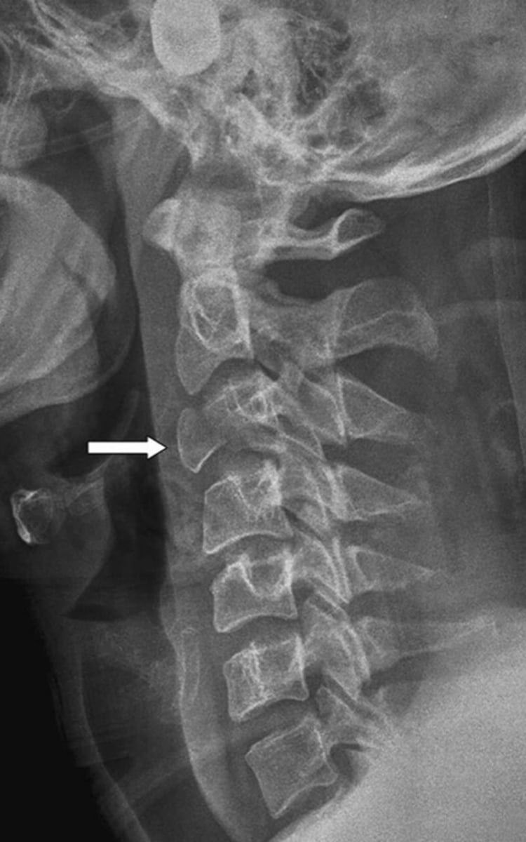
Unilateral facet dislocation (of C4)
- flexion, rotation mechanism
- inferior articular process dislocates into the IVF
- anterior body displacement (25%)
- mechanically stable (relatively)
- decreased laminar length
- bow tie sign
- inverted hamburger sign on CT
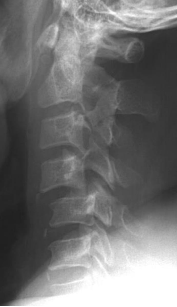
Bilateral facet dislocation
- hyperflexion mechanism
- MC C4-C7
- severe soft tissue injury
- unstable, high incidence of cord injuries
- anterior displacement (typically >50%)
- Two types: (interlocking = complete, perched = incomplete)
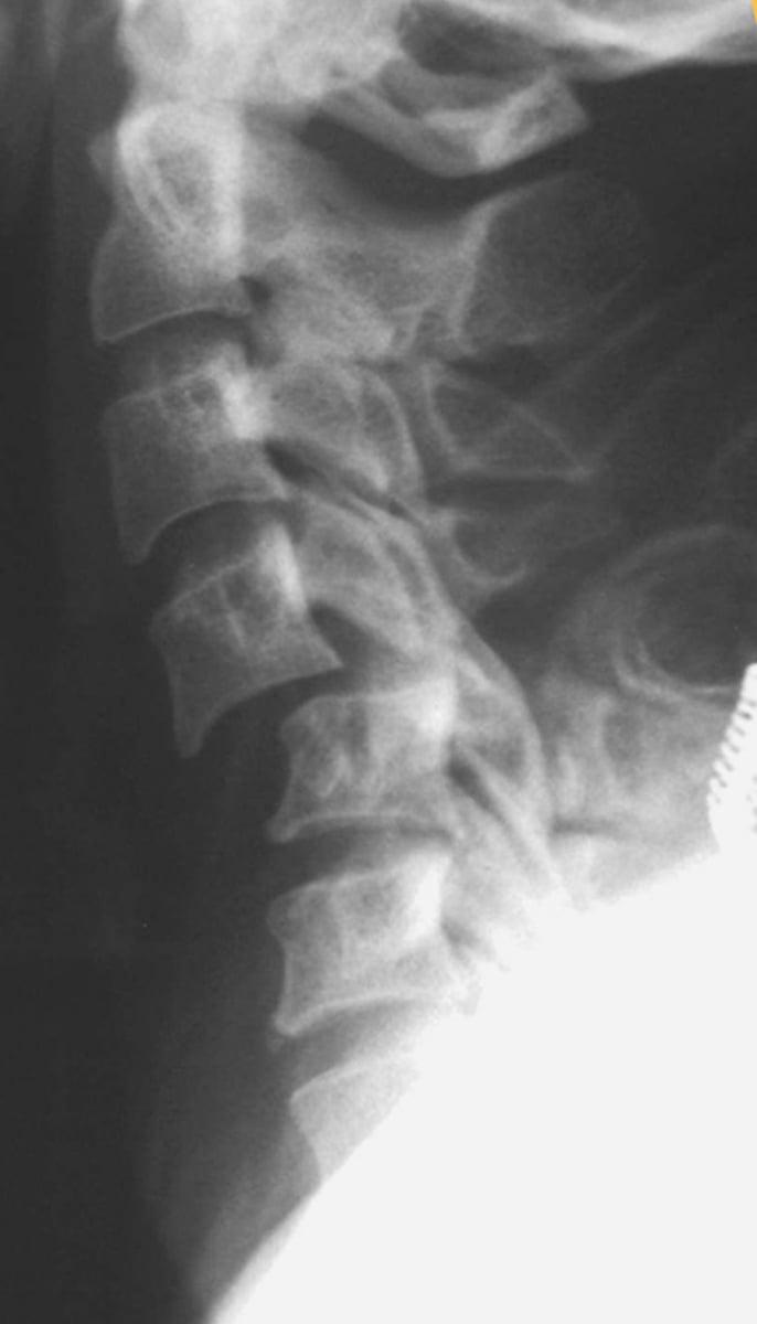
Flexion tear trop fracture
- aka bursting tear trop fracture
- flexion mechanism
- most severe injury of cervical spine
- unstable (anterior cord syndrome)
- facets are "subluxated" or dislocated
- usually C4-C6
- triangular fragment at the anterior inferior vertebral body
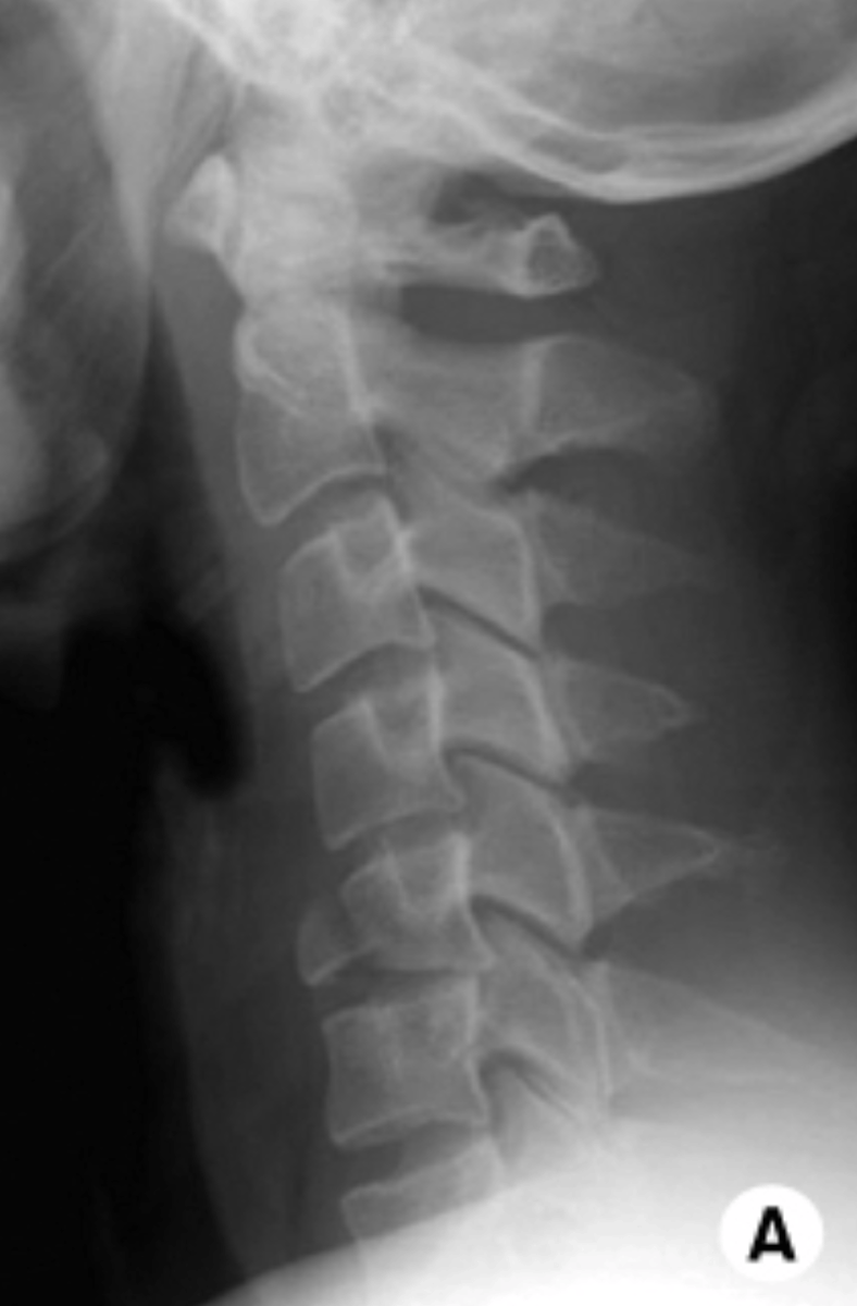
Extension teardrop fracture
- aka avulsion fracture
- extension mechanism
- triangular fragment on anterior inferior vertebral body
- usually C2
- unstable
- buckling of ligamentum flavum
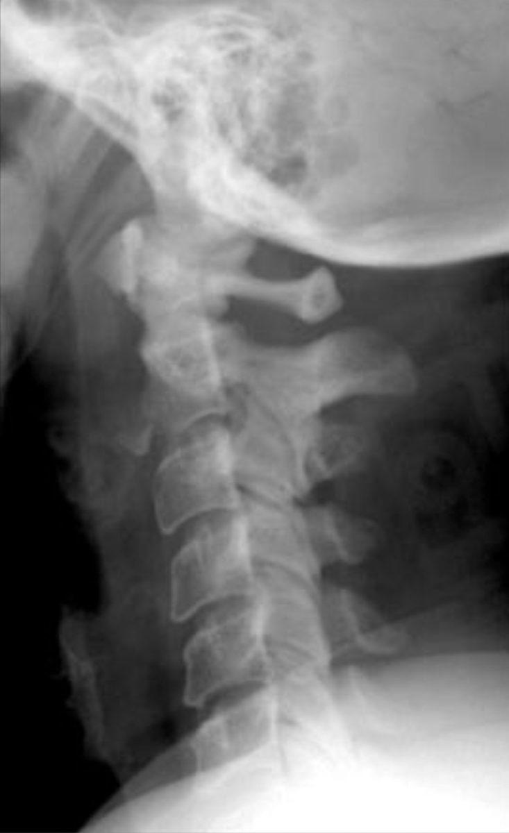
Clay shoveler's fracture
- aka root puller's fracture
- flexion mechanism
- C6-T1, (mc C7)
- avulsion fracture
- stable
- "double spinous sign" on AP film
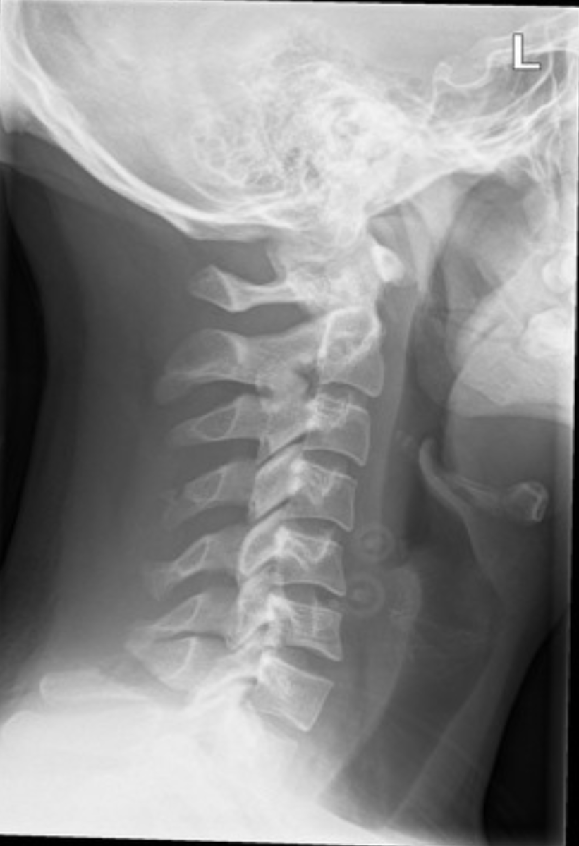
Laminar fracture (of C6)
- uncommon
- mechanically stable
- hyperextension mechanism
- lower cervicals
- may need CT
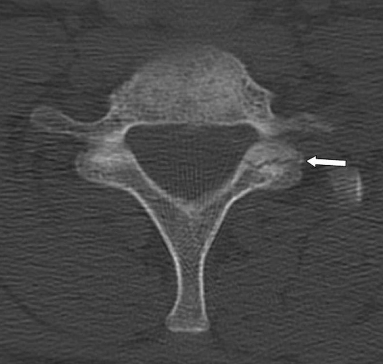
Pillar fracture
- uncommon
- commonly missed on x-ray
- C4-C7
- extension/compression mechanism
- may need CT
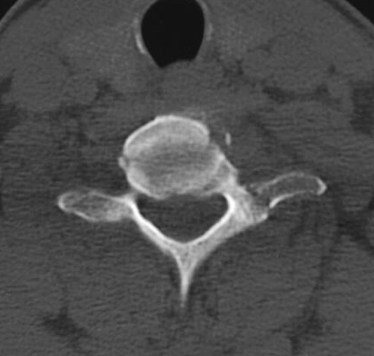
Uncinate fracture
- rare
- stable
- lateral flexion mechanism
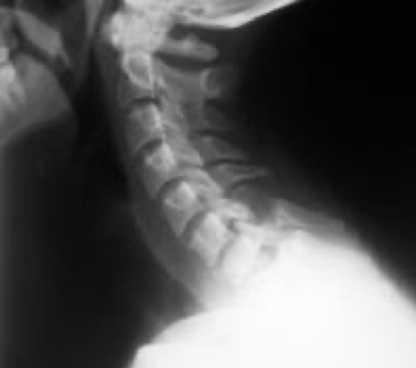
Whiplash
- hyperflexion/hyperextension mechanism
- soft tissue injury (flexion component)
-widened interspinous space (of C5/C6)
- subluxated widened facets
- disruption of posterior ligament complex
- flexion/extension views
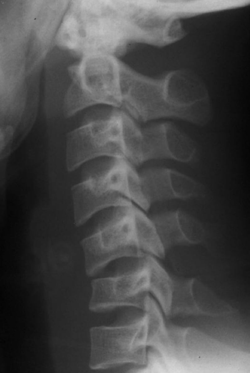
Lumbar compression fracture of L1
- 90% occur between T11-L2
- MC fracture of thoracics and lumbars is compression
- flexion mechanism
- osteoporotic patients could have compression fractures with no trauma
- decreased anterior height
- posterior body height maintained
- may see step defect and zone of impaction (condensation)
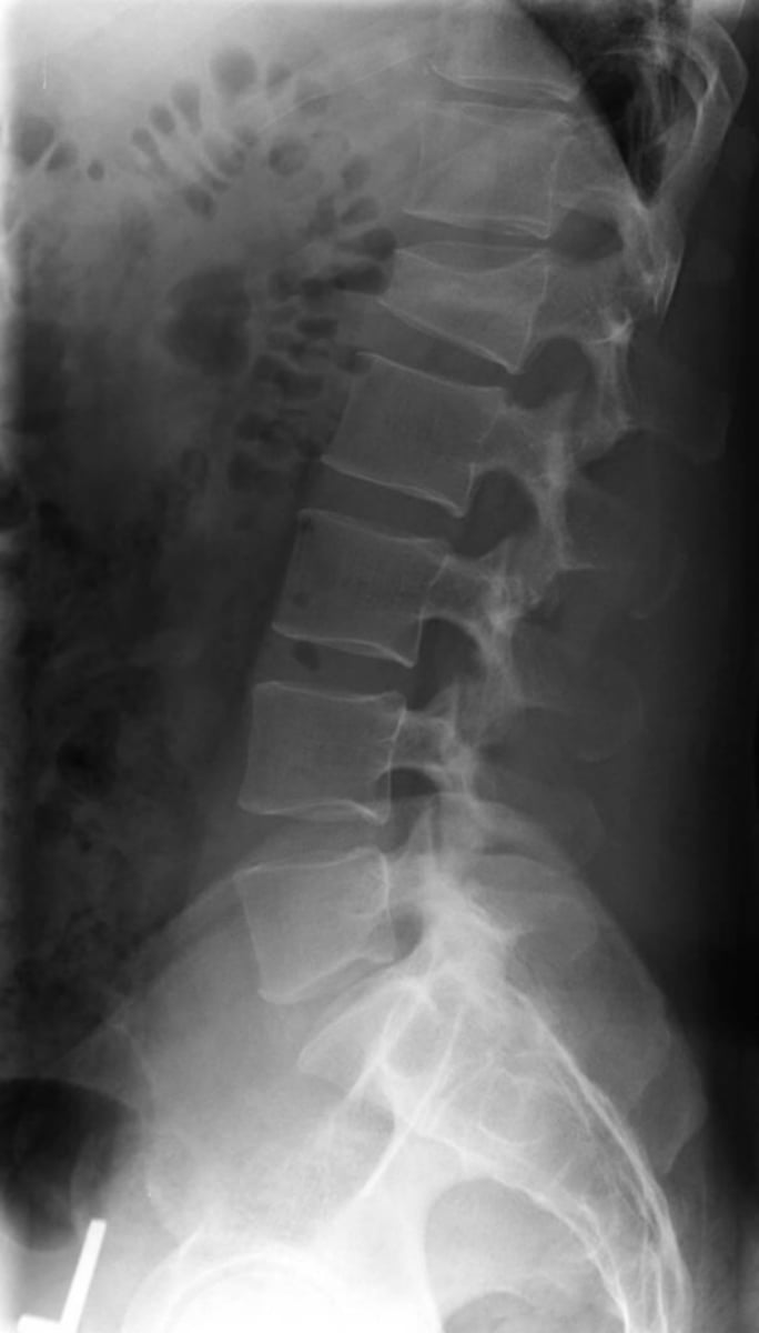
Osteoporotic compression fractures
- More common in females >50 years old
- increased kyphosis (dowager's hump)
- accentuated vertical striations
- usually multiple levels
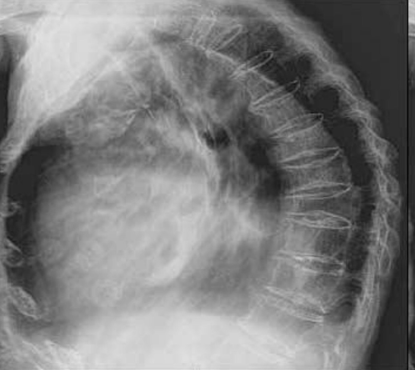
Pathological fracture
- decreased height of the entire body (implies pathology)
- Osteoporosis, Metastasis, or Multiple Myeloma
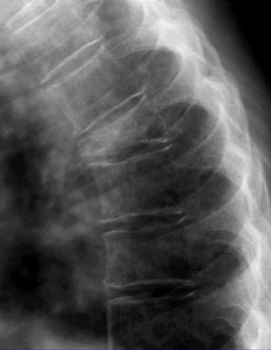
Vertebroplasty & Kyphoplasty
- minimally invasive procedures for treatment of compression fractures
- physicians inject a cement mixture into the fractured bone
- in kyphoplasty, a balloon is first inserted to create space
Green arrow
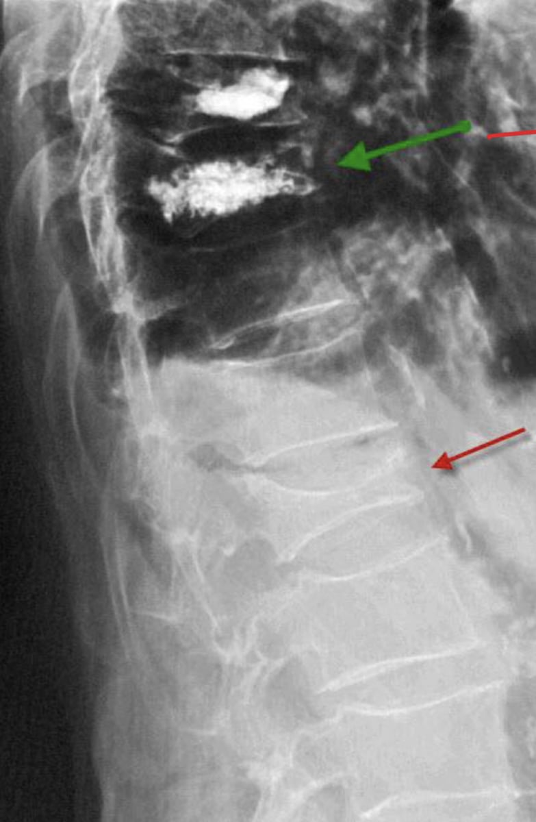
Burst fracture
- bursting "compression" fracture
- posterior body convexity
- retropulsion of the posterior fragments
- widened intrapedicular distance
- CT needed
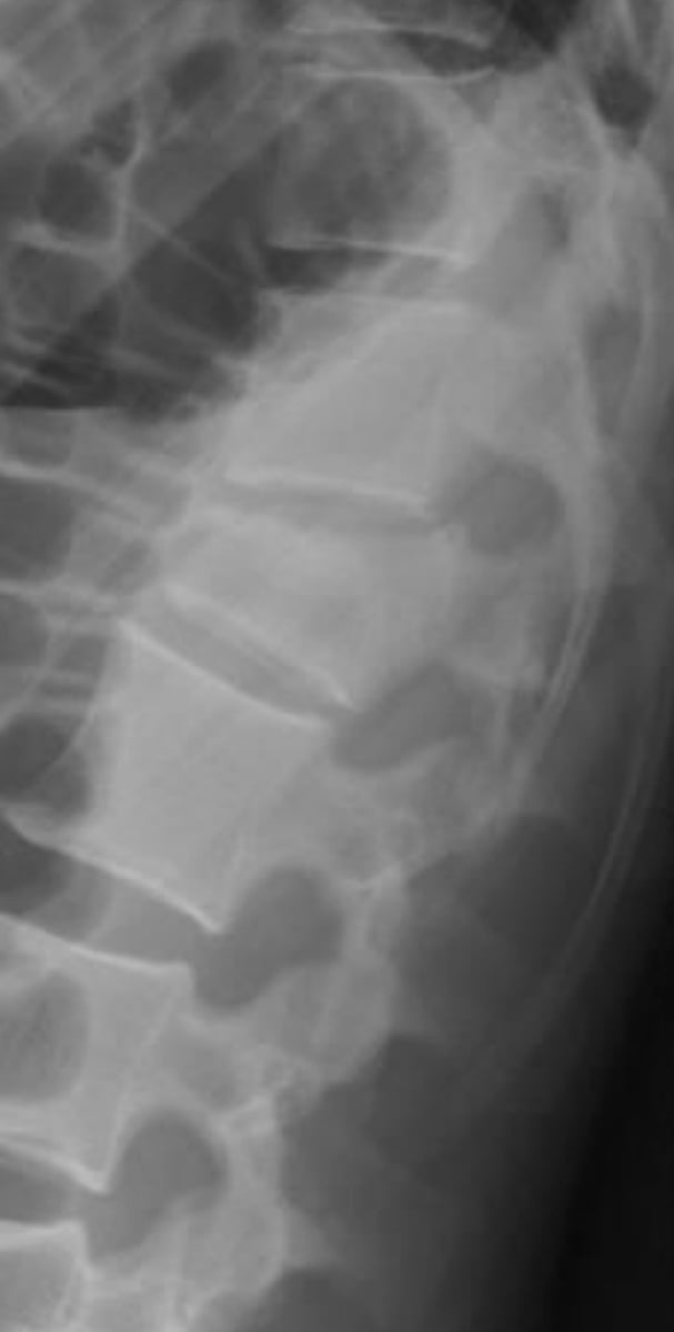
Chance fracture
- "Lap belt" fracture
- flexion/distraction mechanism
- horizontal splitting of the arch and body
- MC L1-L3
- "Empty vertebra" sign
- commonly associated with compression fracture
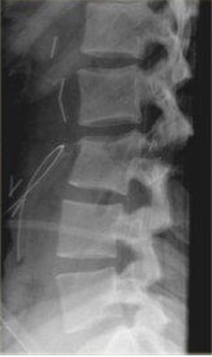
Chance fracture (at L3)
- "Lap belt" fracture
- flexion/distraction mechanism
- horizontal splitting of the arch and body
- MC L1-L3
- "Empty vertebra" sign
- commonly associated with compression fracture
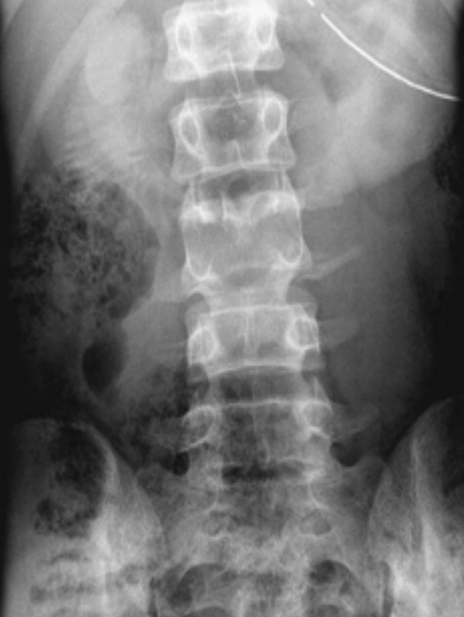
Transverse process fracture
- 2nd most common lumbar fracture
- MC L2-L3
- Vertical to oblique in orientation
- often unilateral and multiple levels
- often obscured by gas and fecal material
- potential renal damage (need urinalysis for potential hematuria)
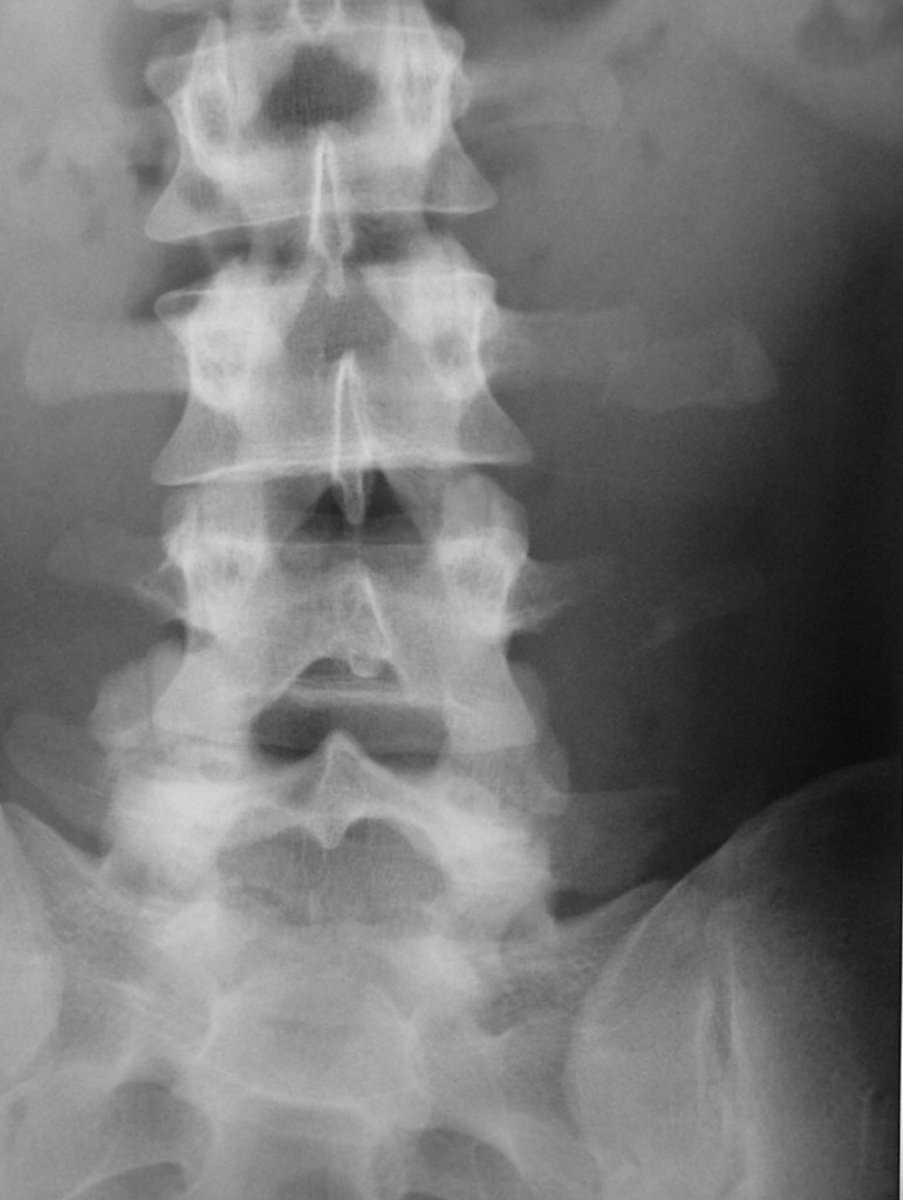
Type 2 spondylolisthesis
- aka spondylolytic or isthmic
- stress fx, elongated pars, or acute fx
- biomechanical stress
- MC type for younger age
- MC at L5
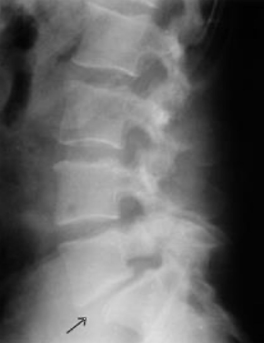
Type 3 spondylolisthesis
- aka degenerative
- due to degeneration of posterior joint
- pars is intact
- MC at L4
- MC in females
- Older age onset
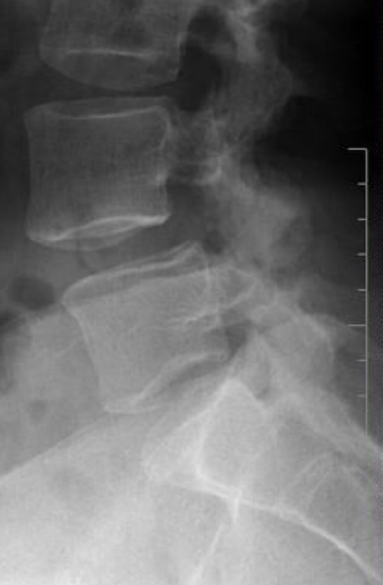
Type 4 spondylolisthesis
- aka traumatic
- acute one time trauma is rare
- MC type is a Hangman's fracture of C2
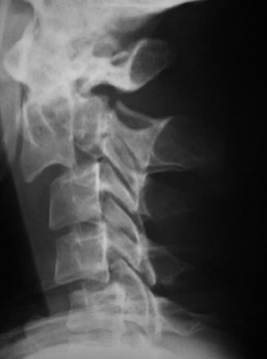
Spondylolisthesis (Radiographic)
- Inverted Napoleon hat
- Detected with George's line
- Graded by Meyerding's method
- May or may not have pars defects
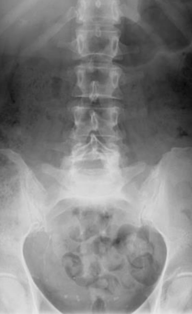
Sacral fracture
- Direct trauma or fall on buttocks
- associated with other pelvic fractures
- most are horizontal near 3rd or 4th segments
- examine sacral foramen carefully
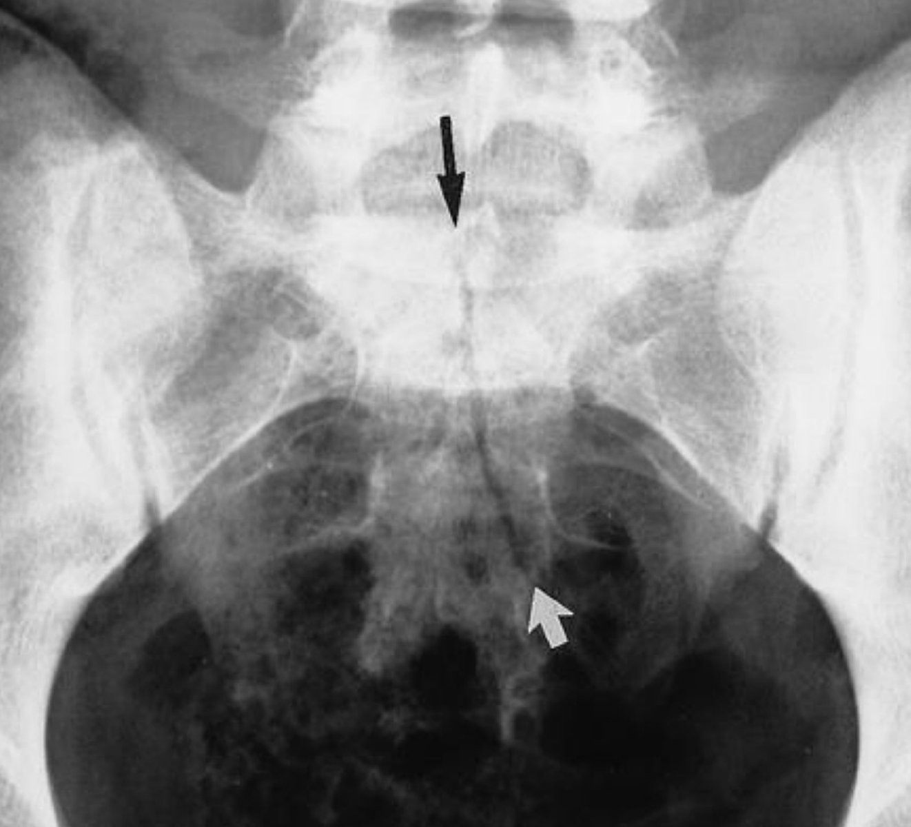
Duverney Fracture
- iliac wing fracture
- splitting of the iliac wing
- direct lateral force
- stable
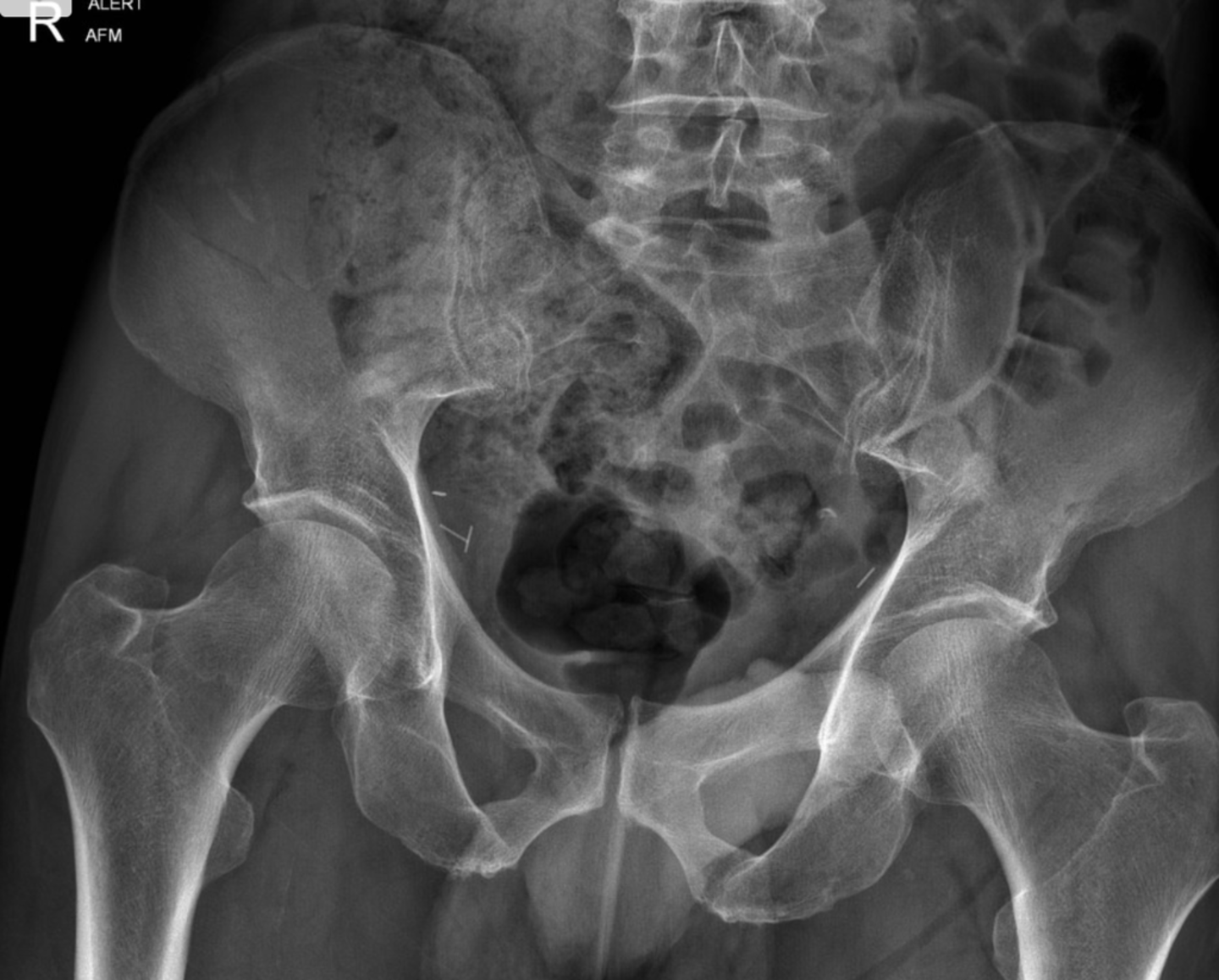
Ischiopubic Rami Fracture
- MC STABLE fracture of the pelvis (40%)
- stable
- Inferior rami more common
- stress fracture may occur
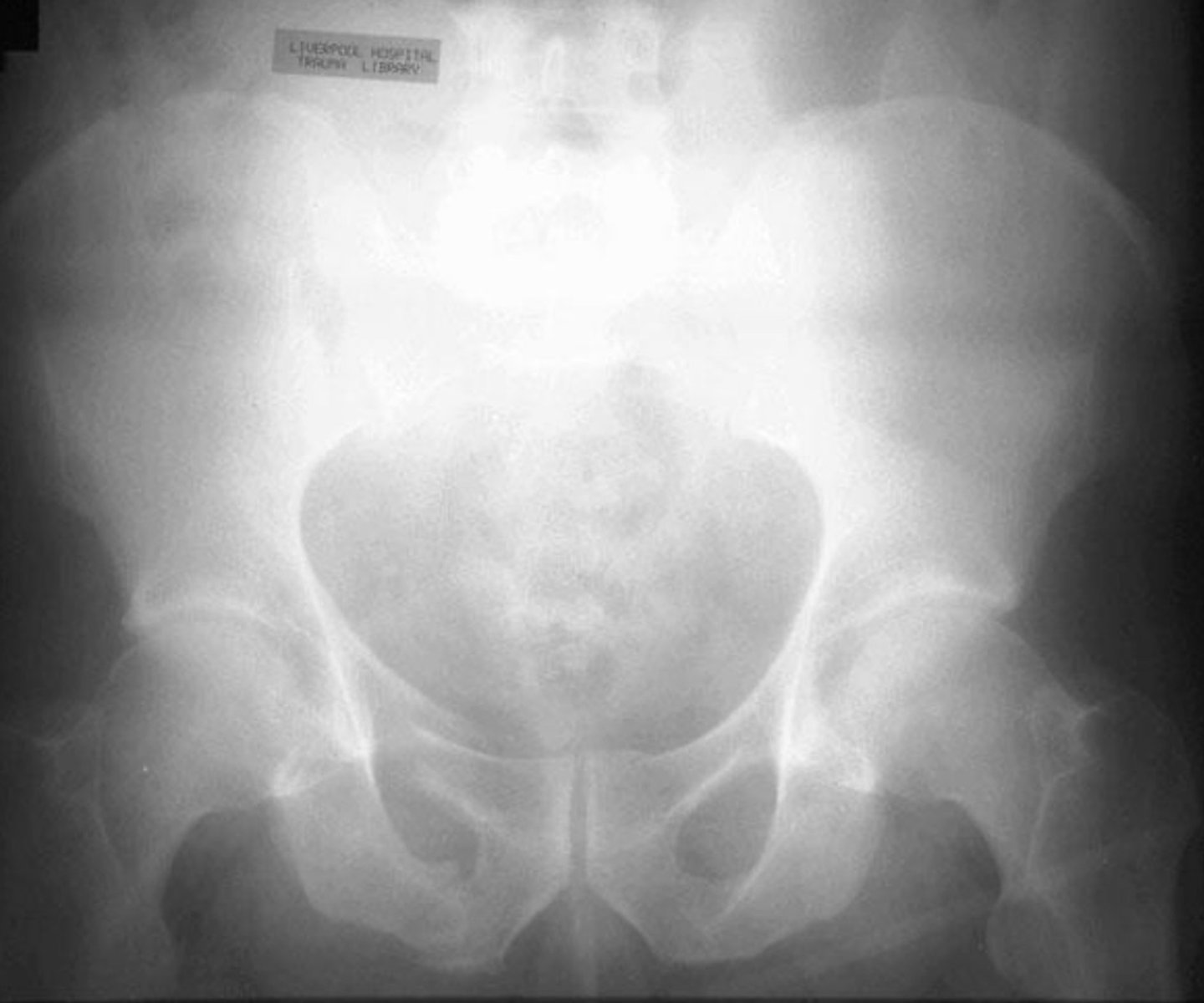
ASIS avulsion fracture
- muscular traction of sartorius muscle leads to avulsion
- seen as curvilinear density
- may unite or remain ununited
- MC is ischial tuberosity avulsion
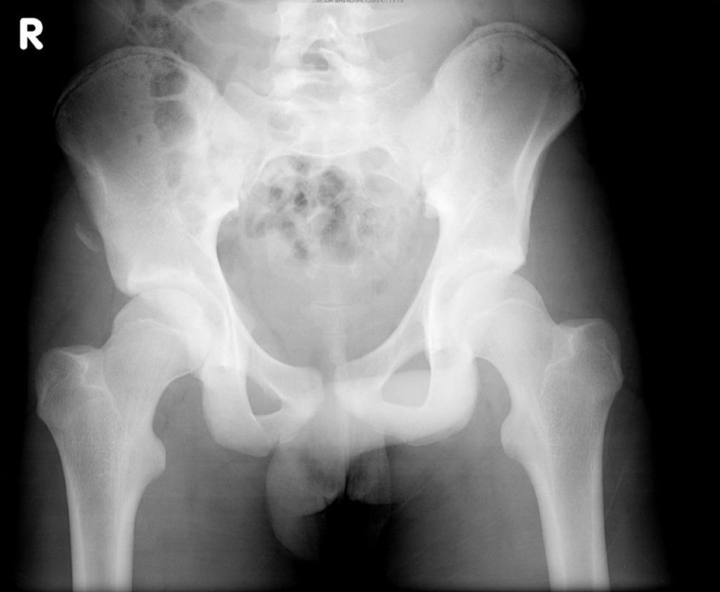
Ischial tuberosity avulsion fracture
- muscular traction of hamstrings leads to avulsion
- seen as curvilinear density
- may unite or remain ununited
- MC type of avulsion fracture
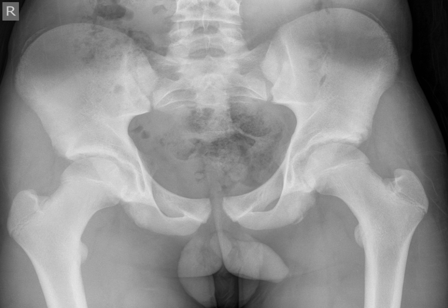
Malgaigne fracture
- ipsilateral double vertical shearing fracture of superior and inferior pubic rami with fx or dislocation about ipsilateral SI joint
- MC UNSTABLE pelvic fx (33%)
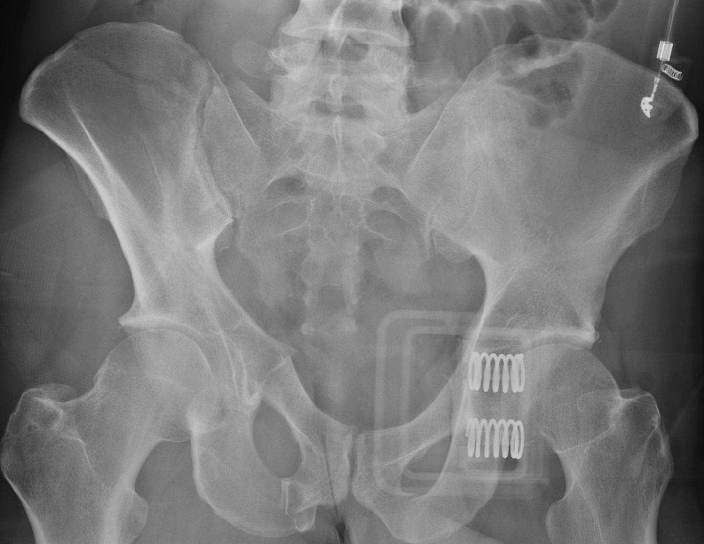
Bucket-Handle fracture
- contralateral double vertical fx
- superior and inferior pubic rami fx
- fx or dislocation about contralateral SI
- unstable
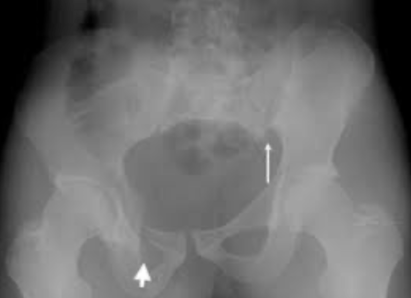
Straddle fracture
- bilateral superior and inferior rami fx
- substantial soft tissue injury
- unstable
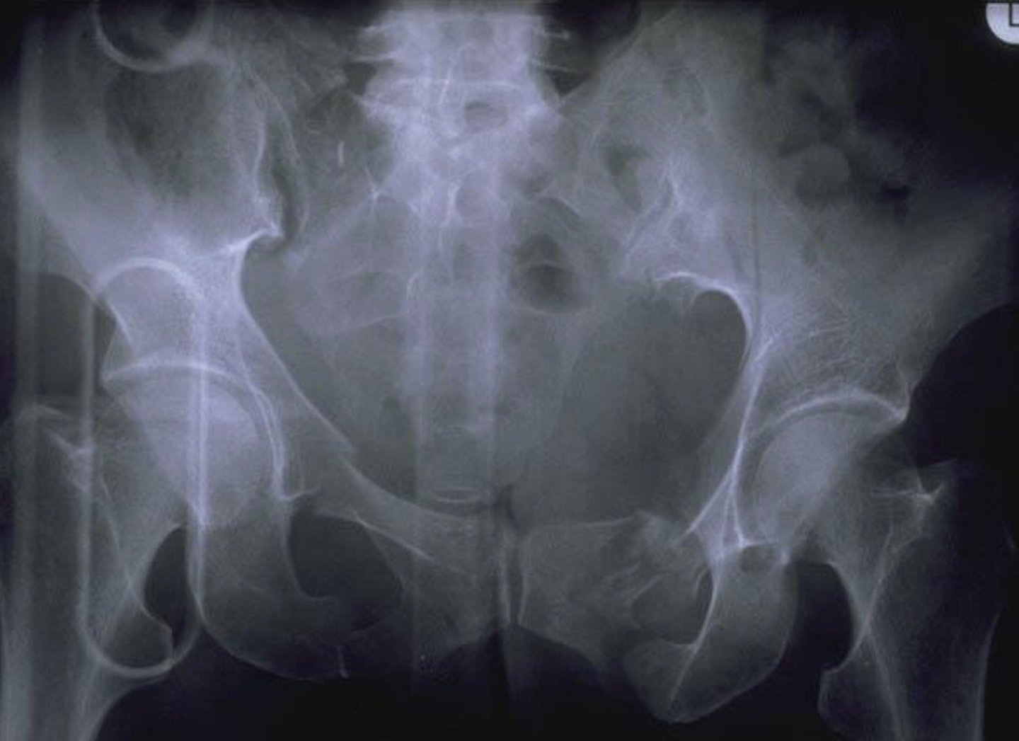
Sprung pelvis
- "Open book" fracture
- diastasis of pubic symphysis
- diastasis of one or both SI joints
- unstable
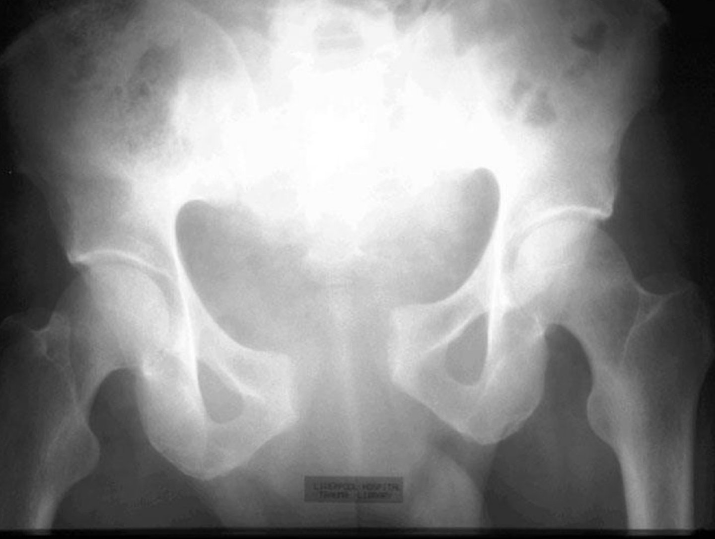
Complex pelvic fracture
- unstable
- severe injury
- difficult to classify
- multiple fractures
- comminution of the pelvis