Unit 2 Antomy and Physiology MasterSet Practical 1
 Call Kai
 Learn
Learn Practice Test
 Spaced Repetition
 Match
Match Flashcards
 Knowt Play
1/126

Earn XP
Description and Tags
lacrimal
Name | Mastery | Learn | Test | Matching | Spaced | Call with Kai | Chat |
|---|
No analytics yet
Send a link to your students to track their progress
127 Terms
frontal bone

mandible
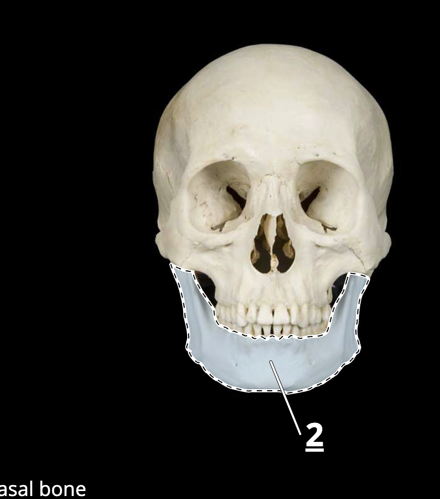
inferior nasal concha

temporal bone

temporal bone
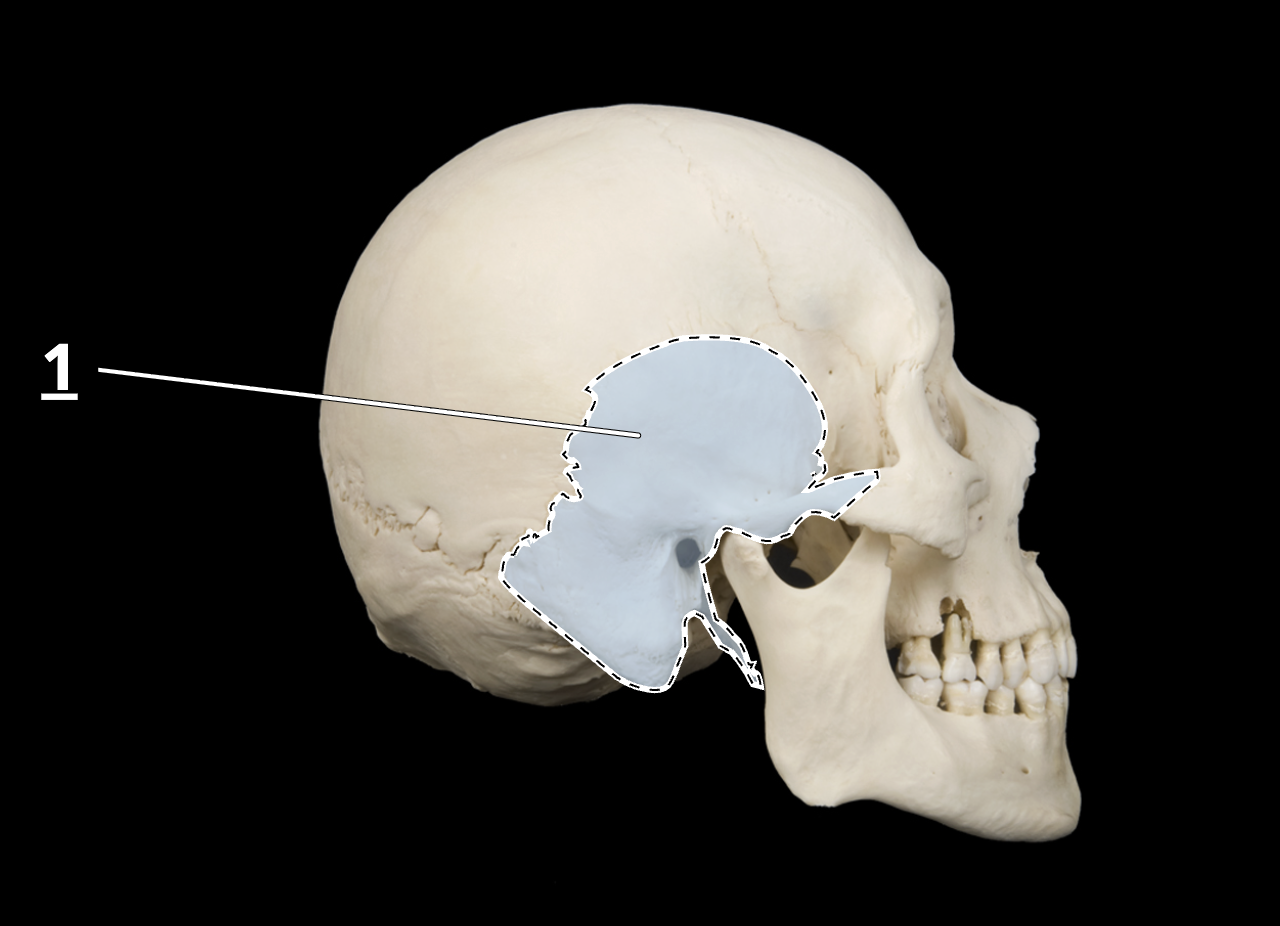
occipital bone

parietal bone
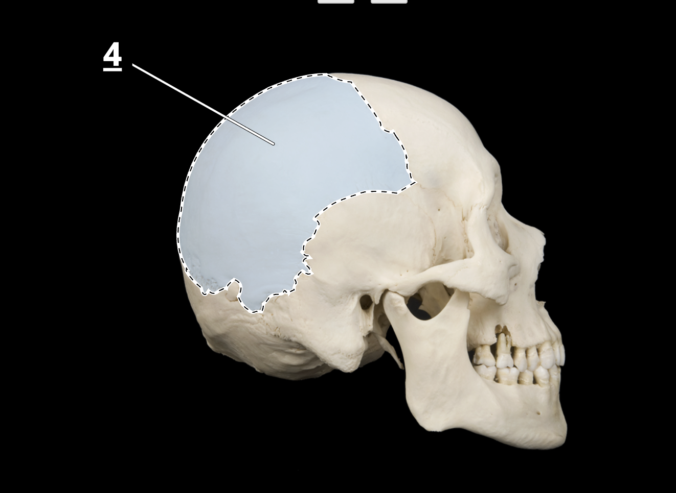
frontal bone
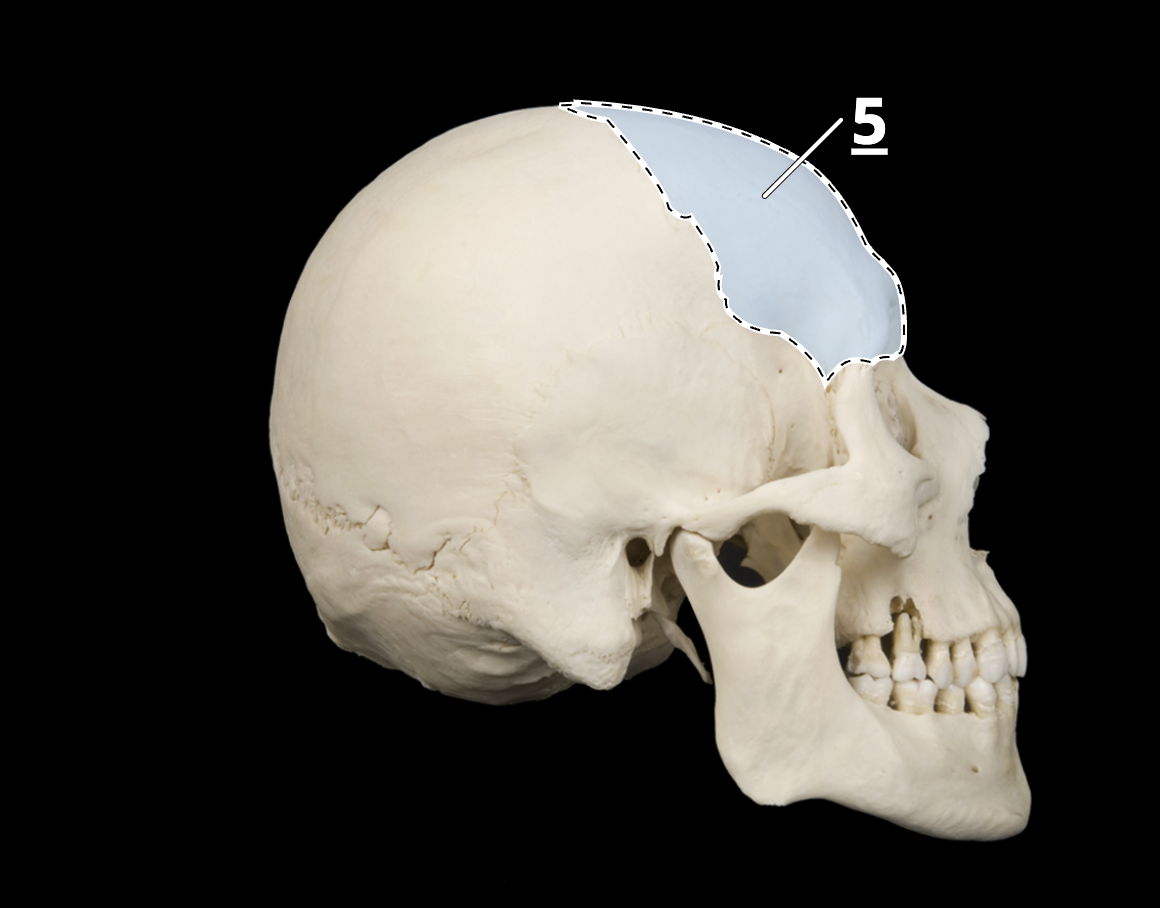
sphenoid bone

mandible

squamous suture

coronal suture
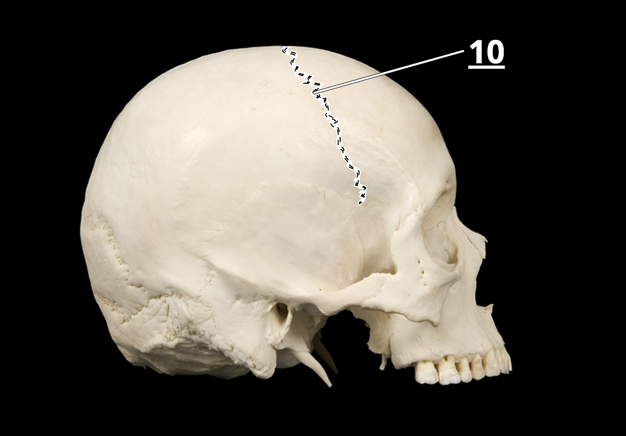
lambdoid suture
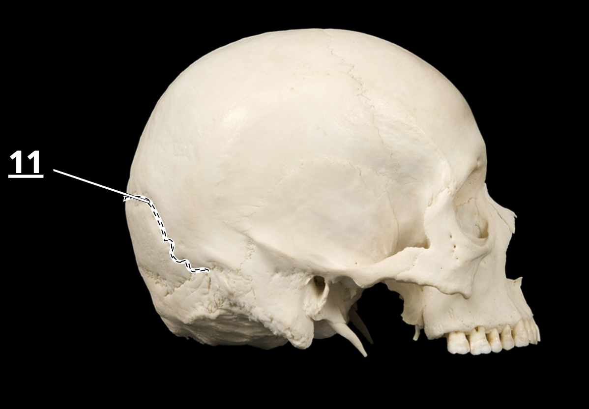
sagital suture

anterior frontal

frontal suture

lambdoid suture

external acoustic meatus

mastoid region/mastoid process of temporal bone

sphenoid bone

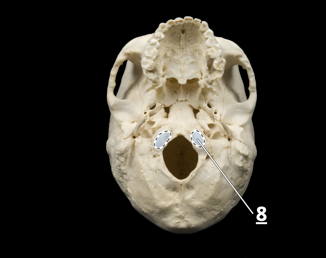
occipital condyle
cervical vertebrae

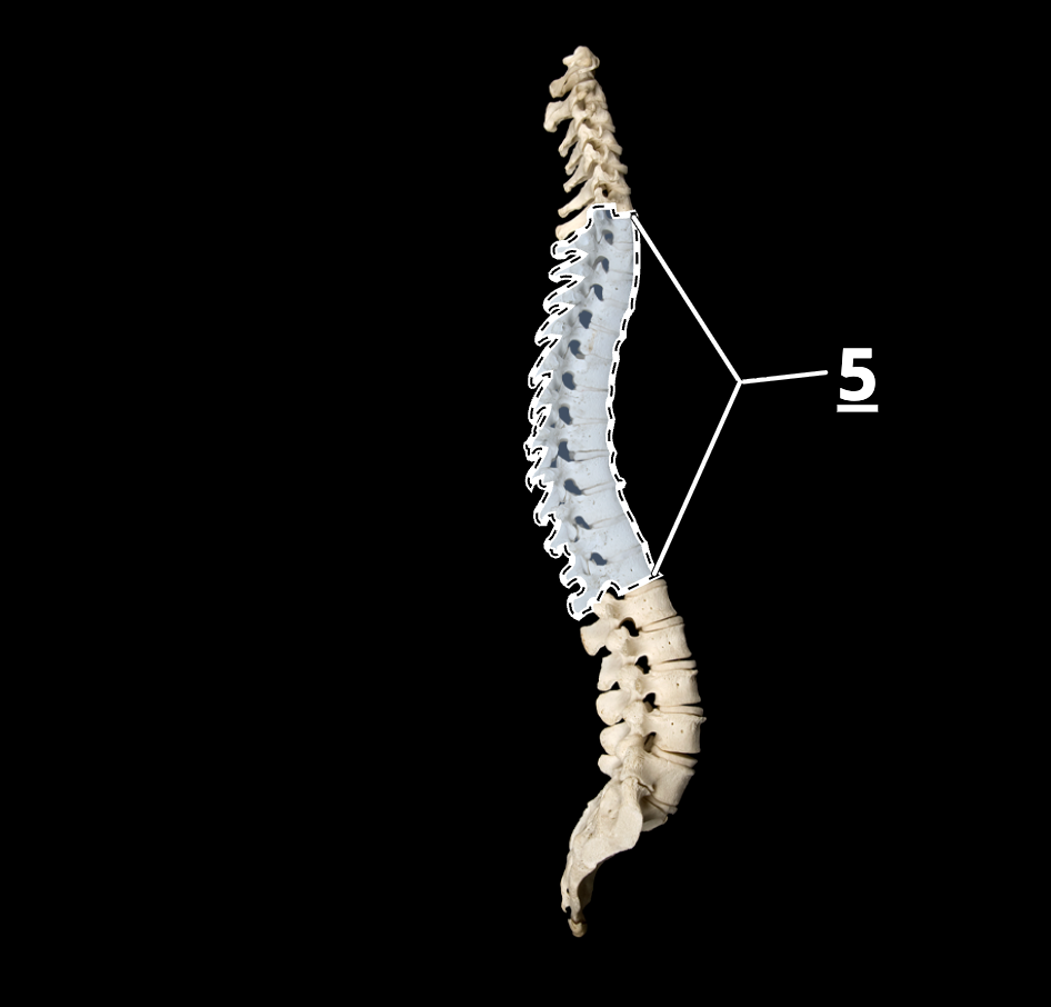
thoracic vertebrae
lumbar vertebrae

intervertebral discs

sacrum and sacral curvature/pelvic vertebrae

spinous process on cervical vertebra
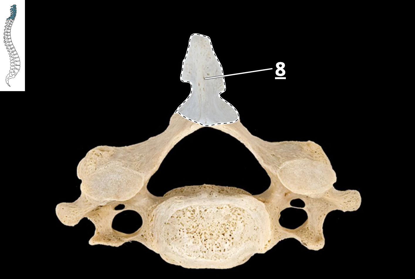
body on cervical vertebra

body of thoracic vertebra

transverse process of thoracic vertebrae

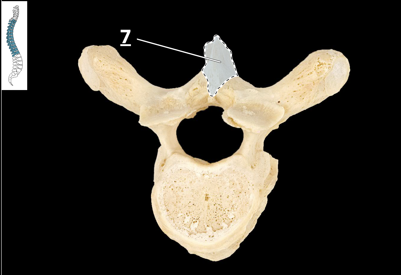
spinous vertebra
body of lumbar vertebra

transverse process of lumbar vertebra

spinous process of lumbar vertebra

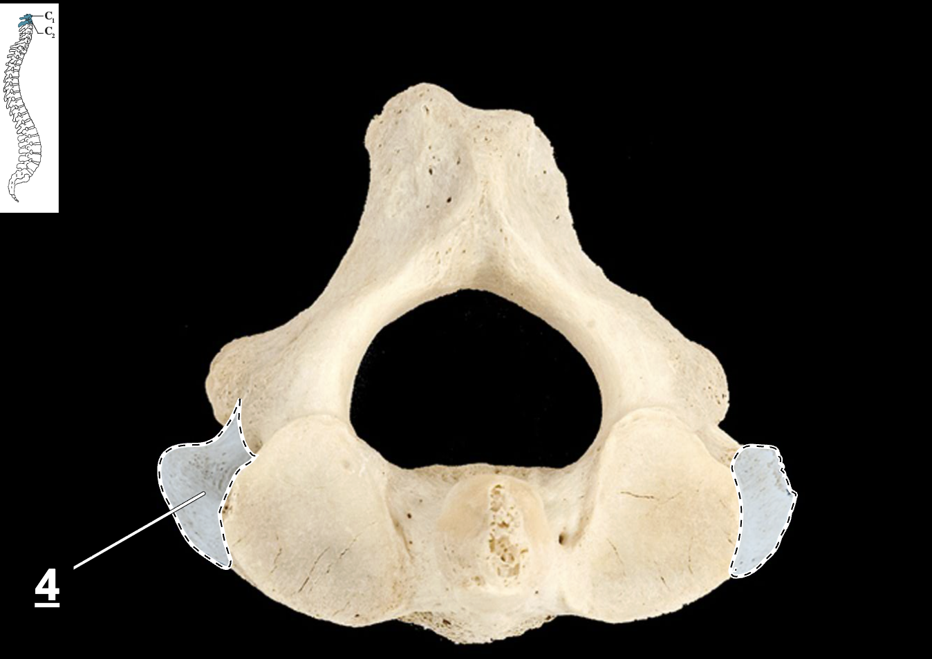
transverse process of atlas, superior view

spinous process of axis

transverse process of axis
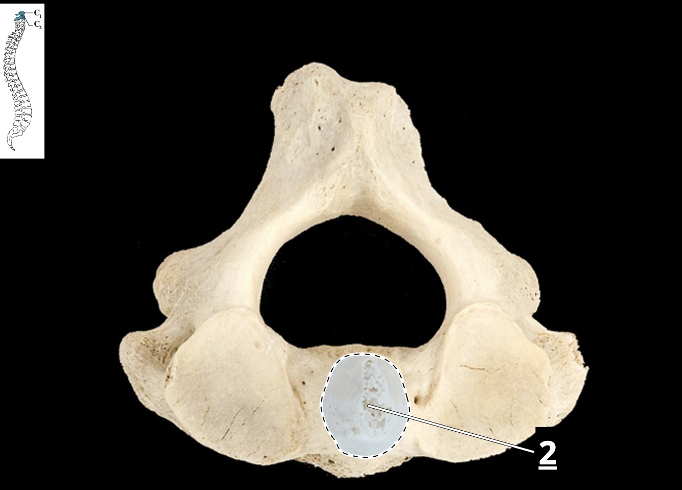
dens/odontoid process of atlas
spinous processes of thoracic vertebrae and curvature

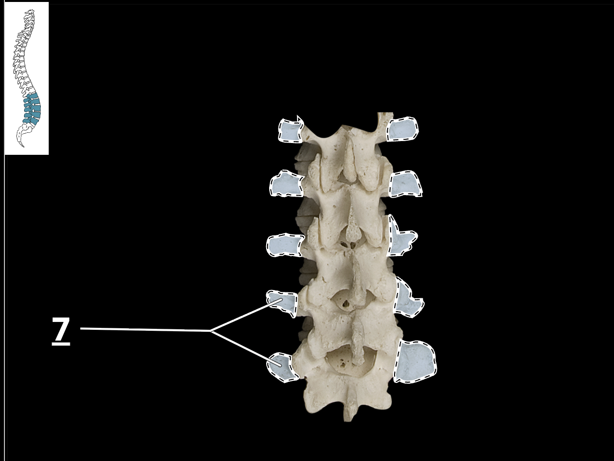
transverse processes of lumbar vertebrae
clavicle

sternum

scapula
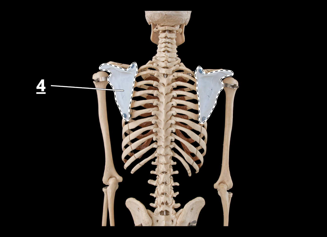
floating ribs
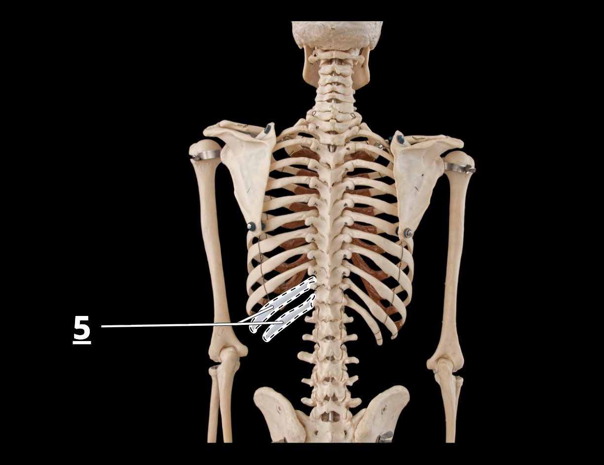
false ribs

true ribs
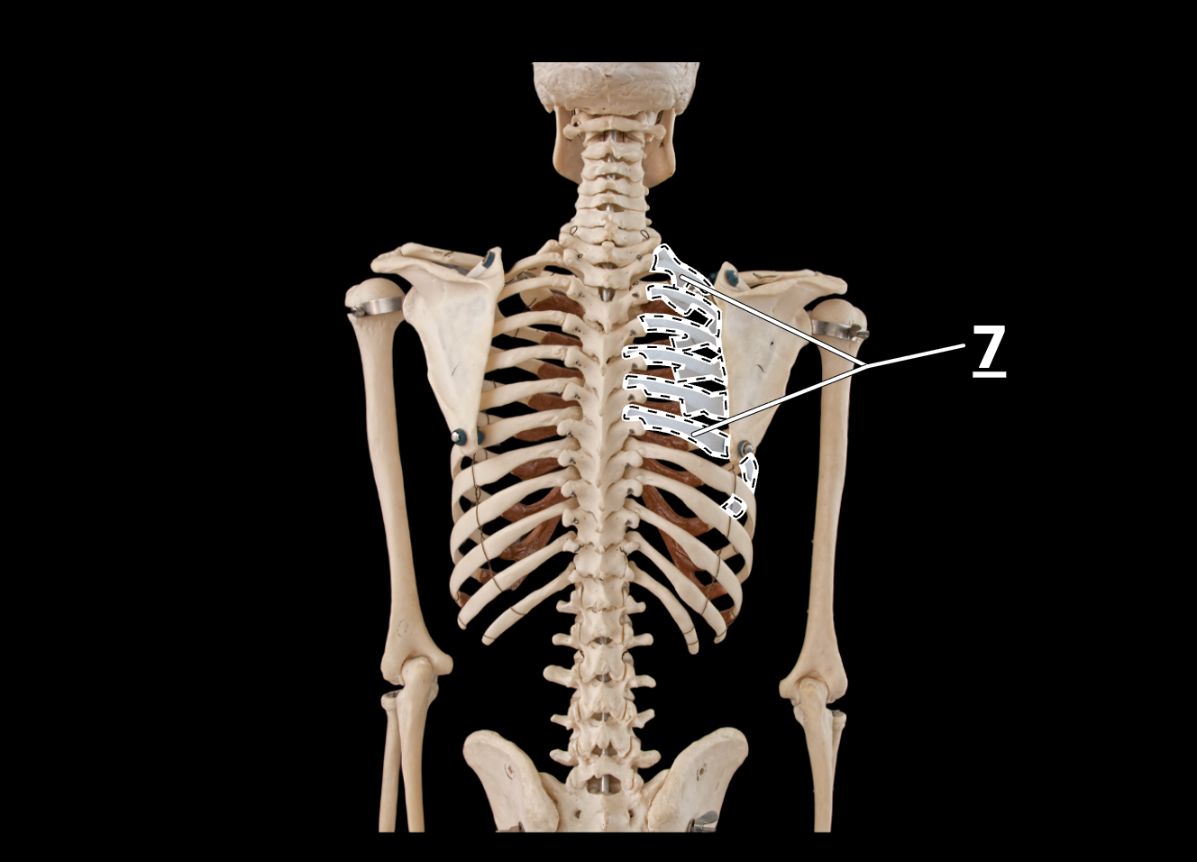
xiphoid process
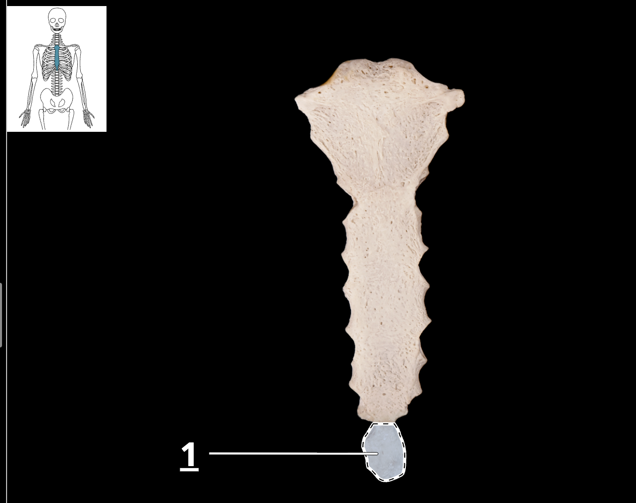
manubrium

costal cartilage of true ribs
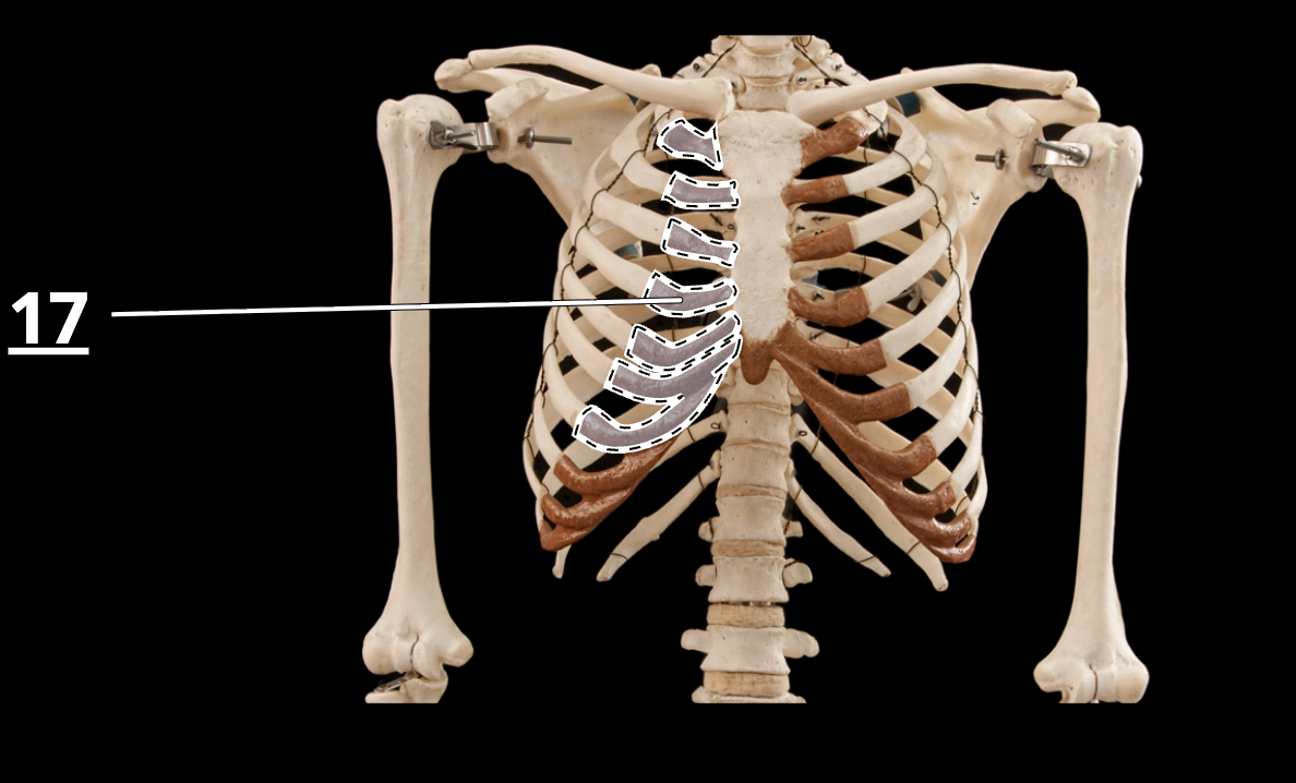
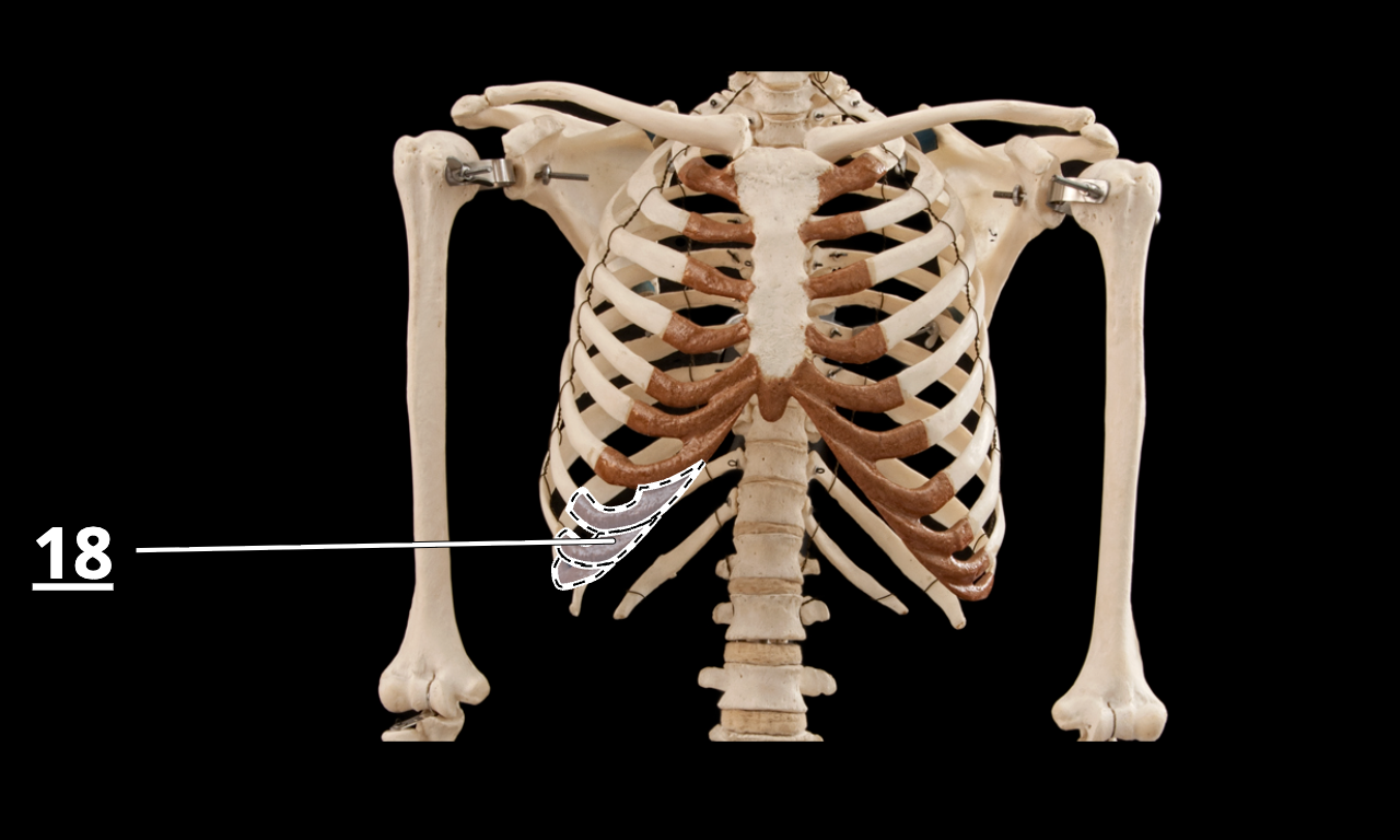
costal cartilage of false ribs
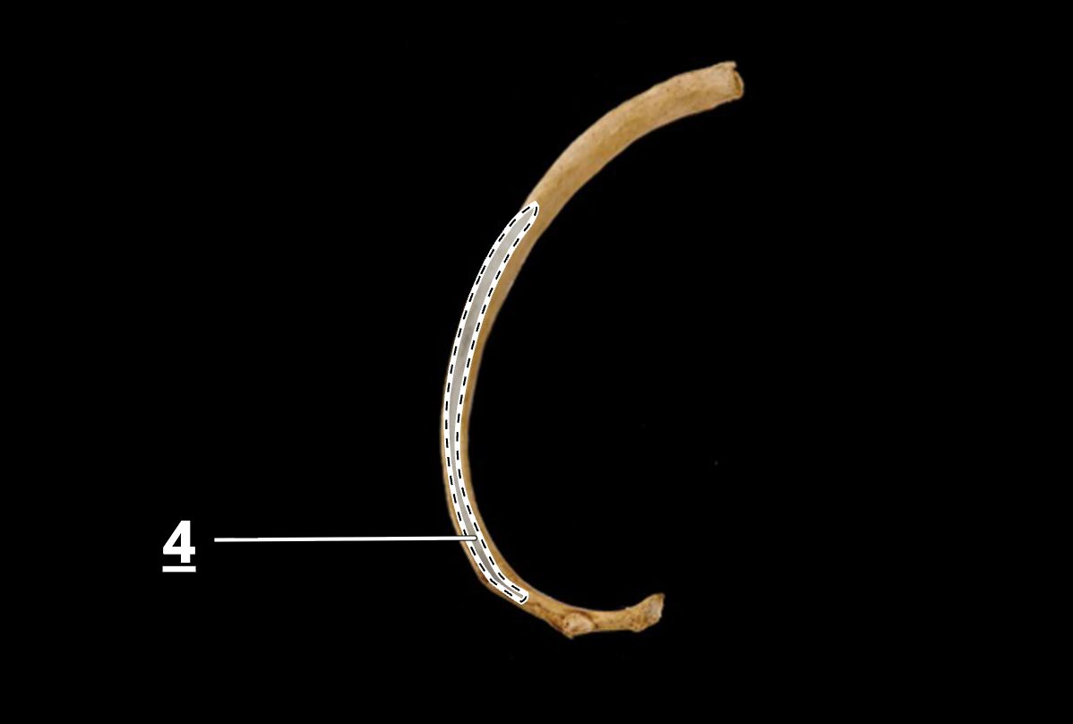
costal groove of rib
greater tubercle of humerous

head of humerus, anterior view
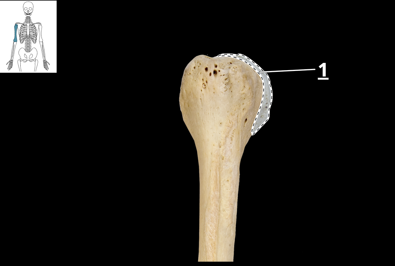
trochlea of humerus
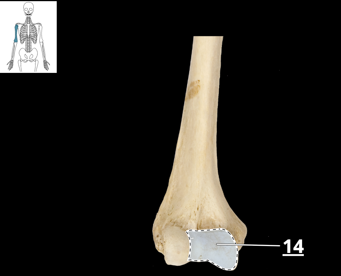
ulna

radius
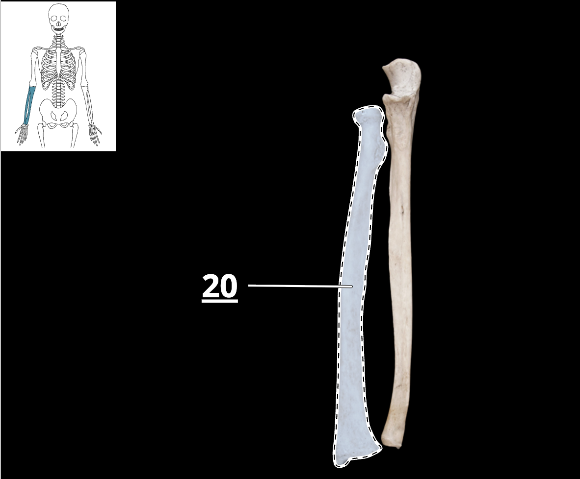
trochlear notch of ulna
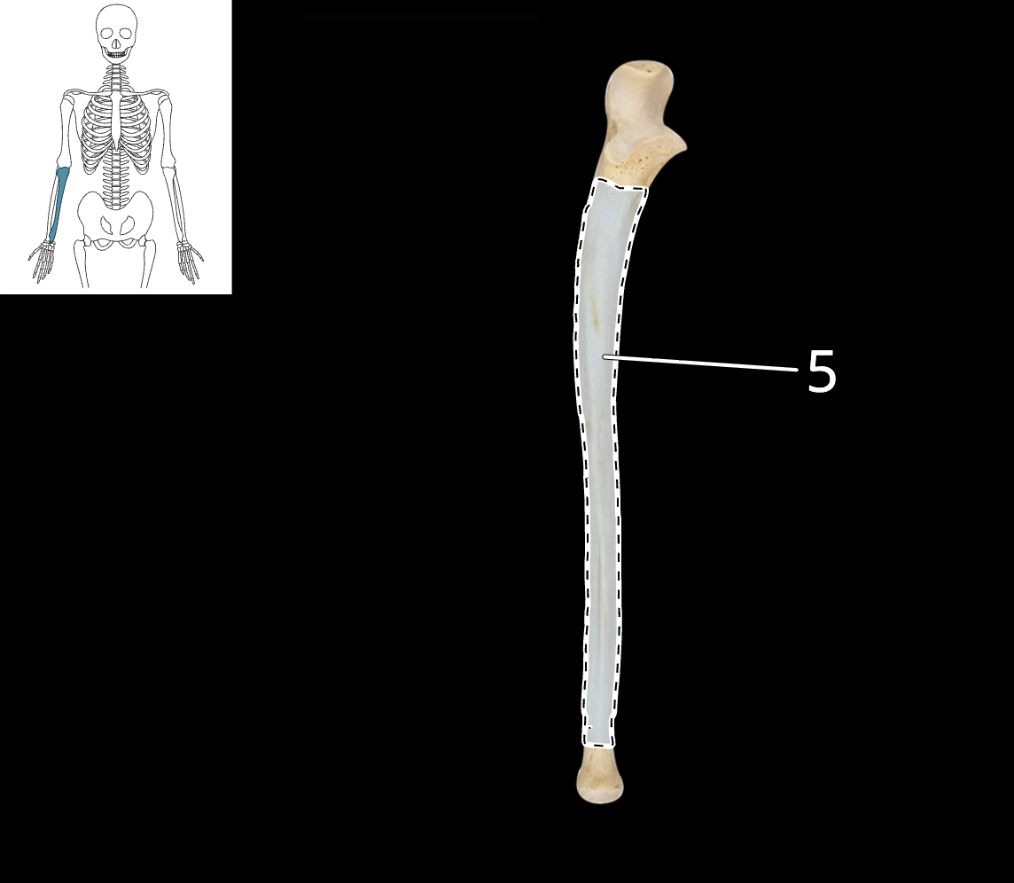
olecranon process of ulna

head of ulna
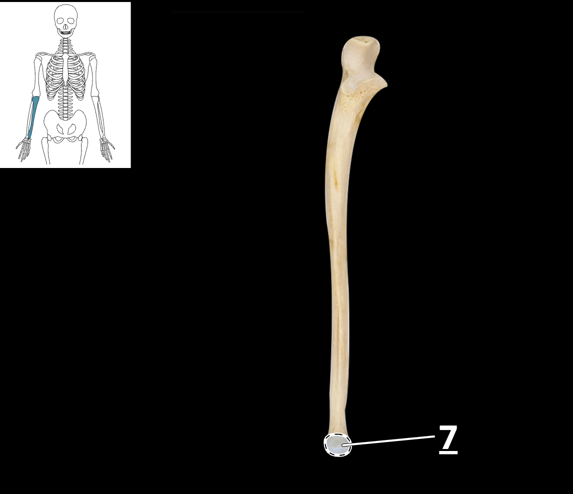
head of radius

neck of radius
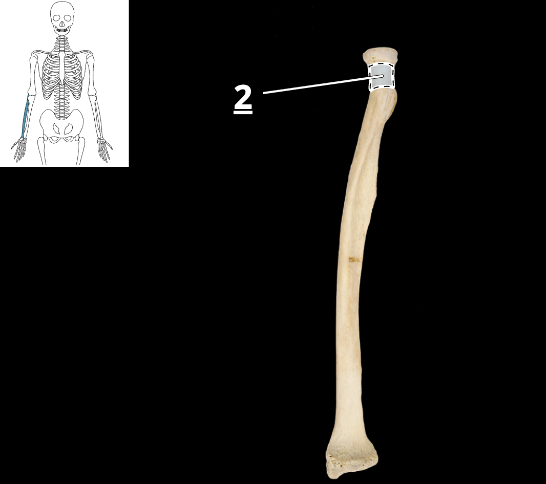
styloid process of radius

sacrum

coccyx
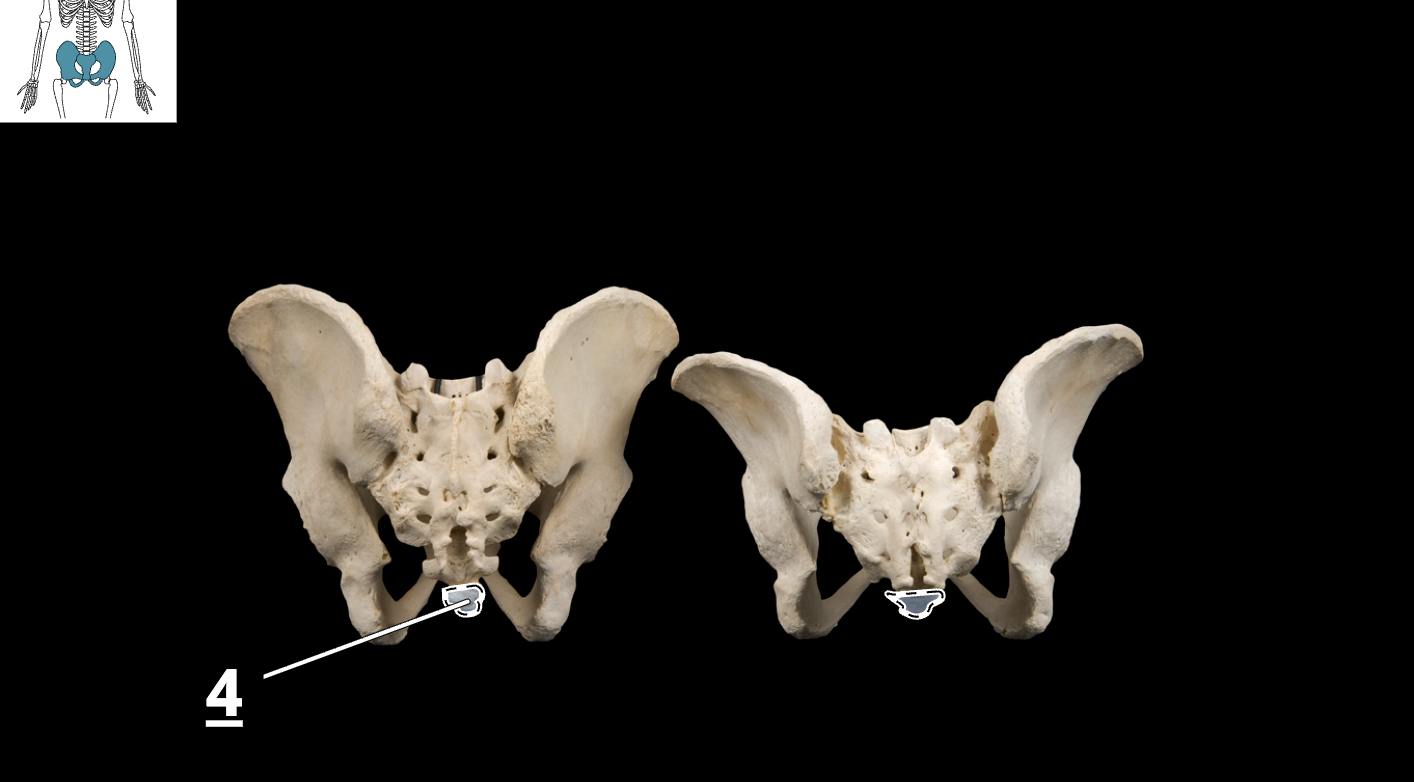
pubic symphysis

obturator foramen
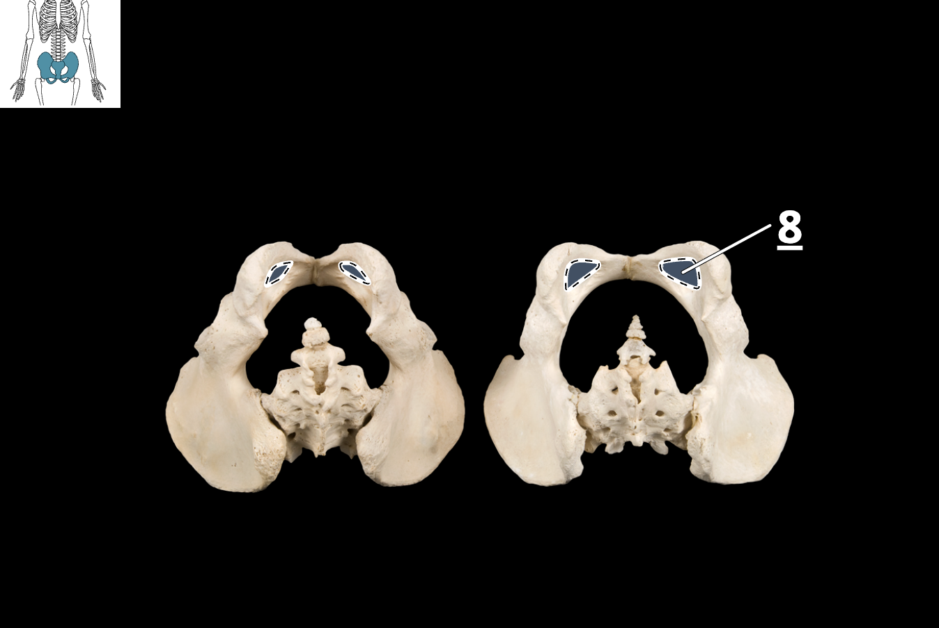
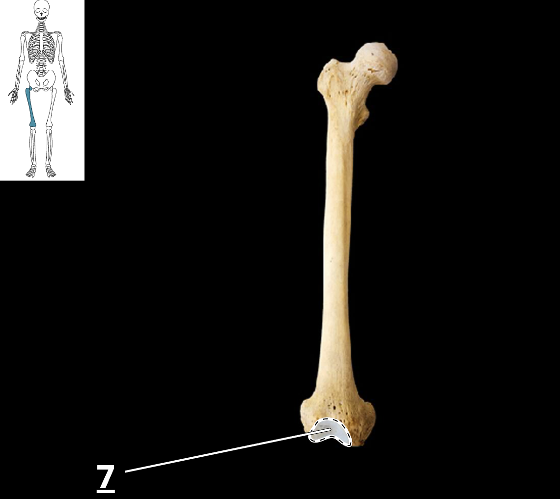
patellar surface of femur
head of femur

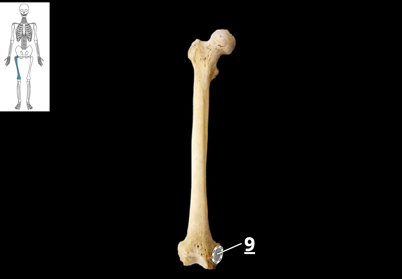
medial epicondyle of femur
greater trochanter of femur

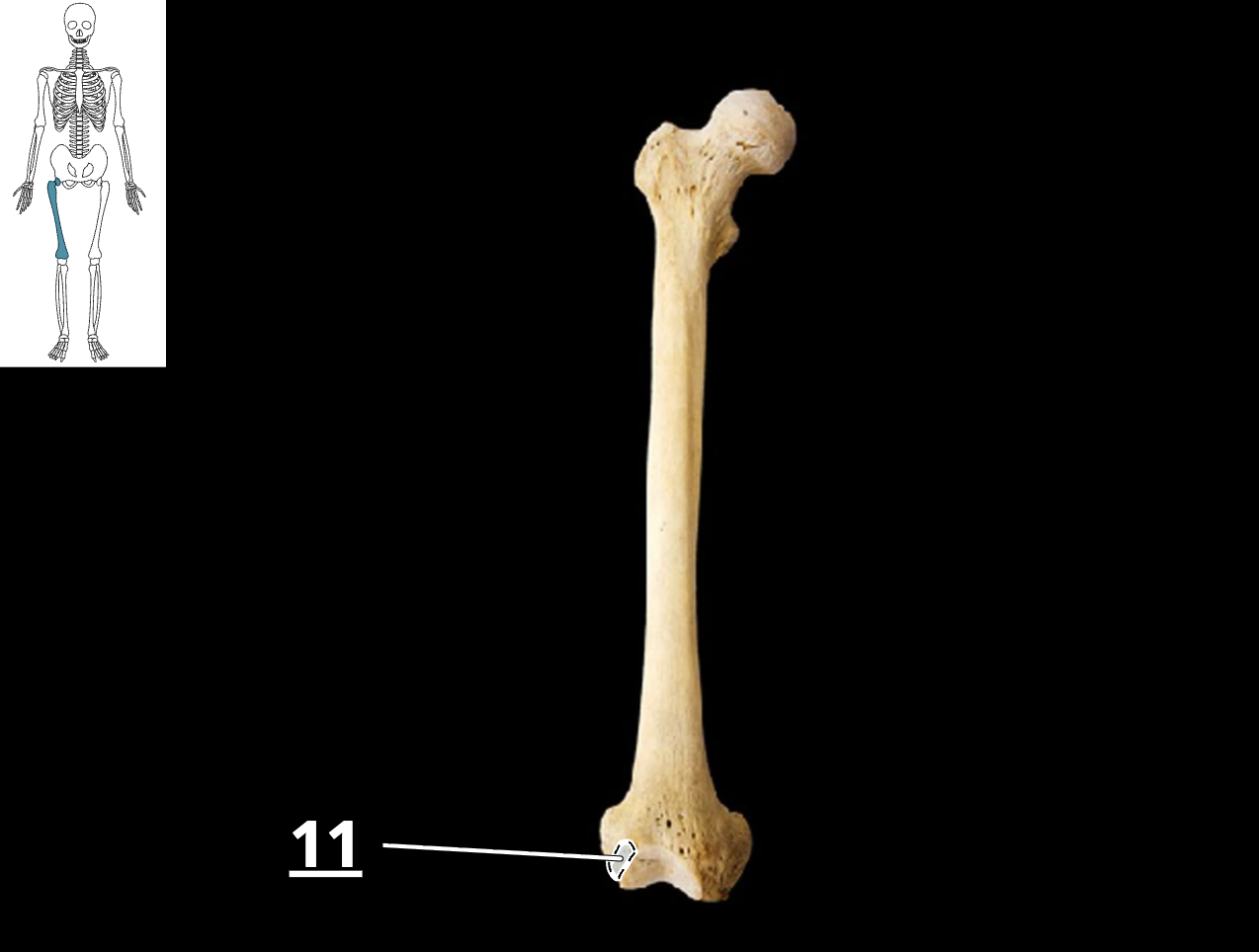
lateral condyle of femur
tibia

fibula

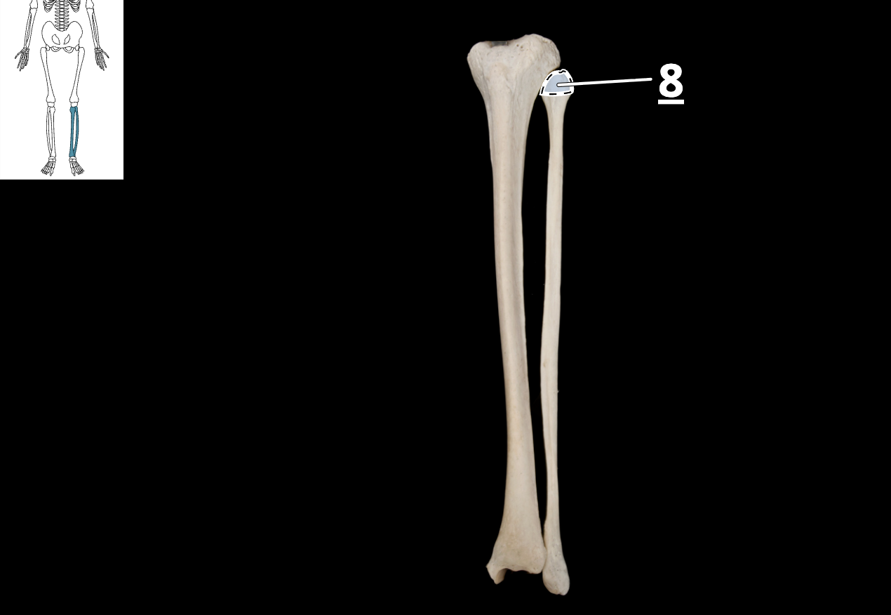
head of fibula
lateral malleolus

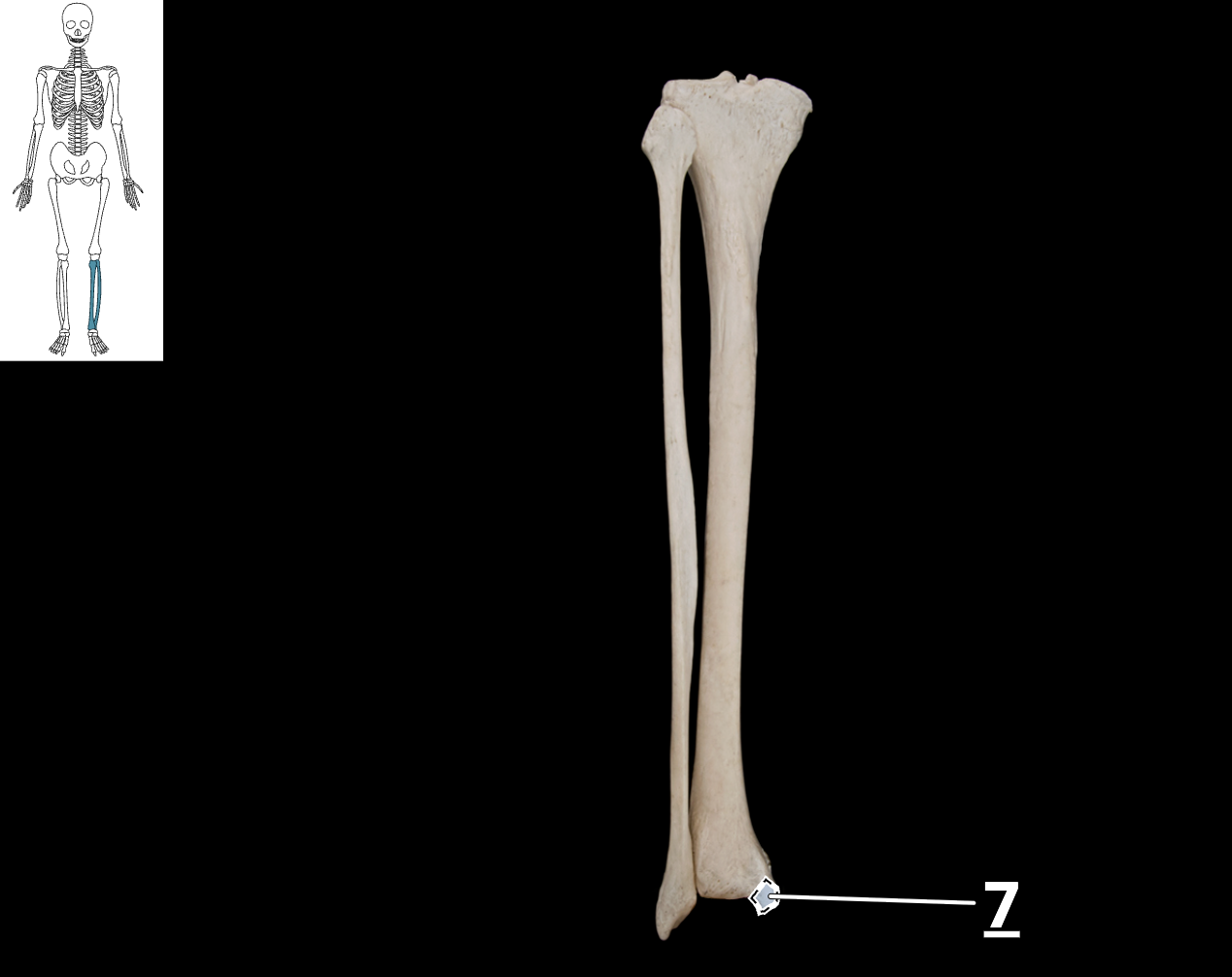
medial malleolus
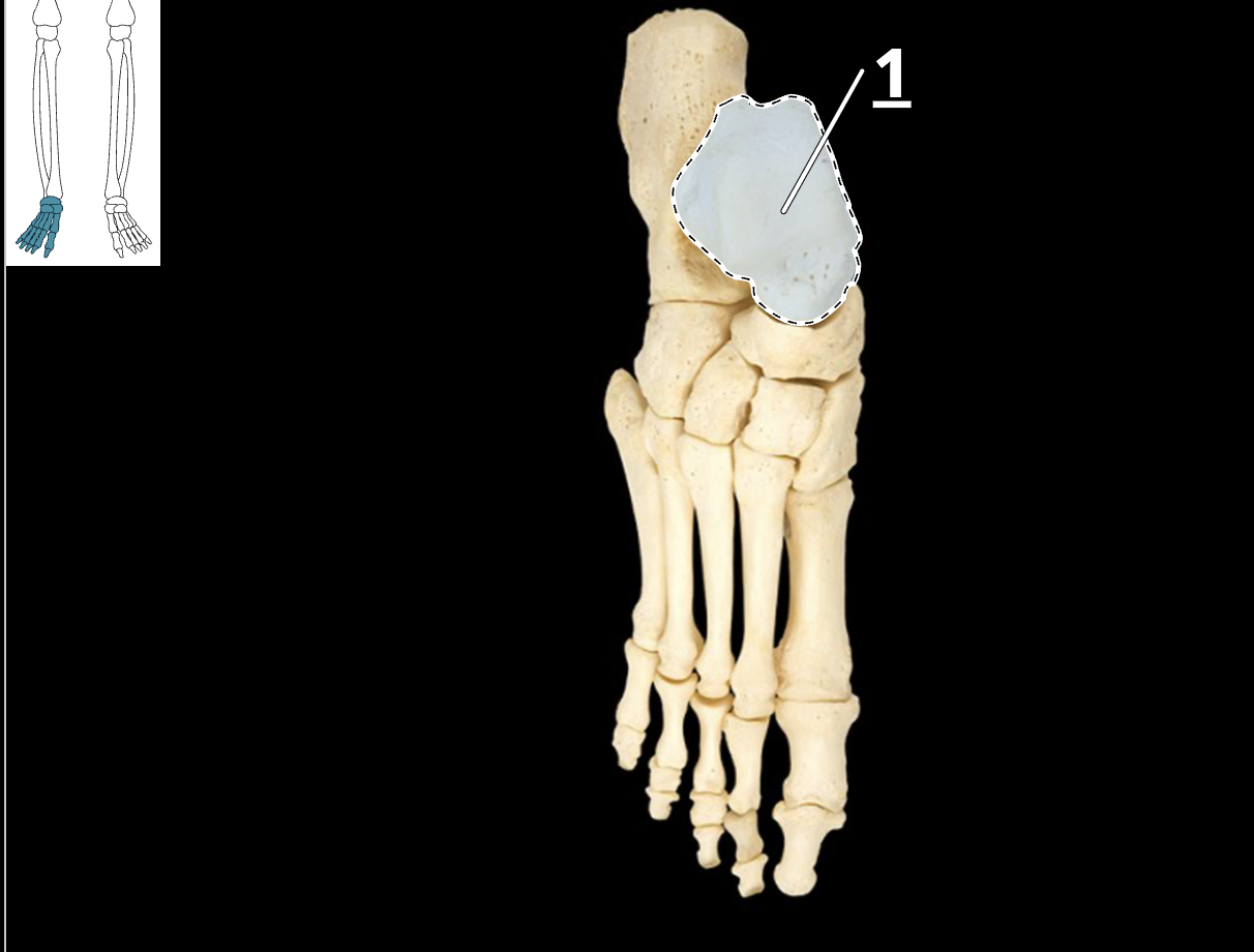
talus
calcaneus bone

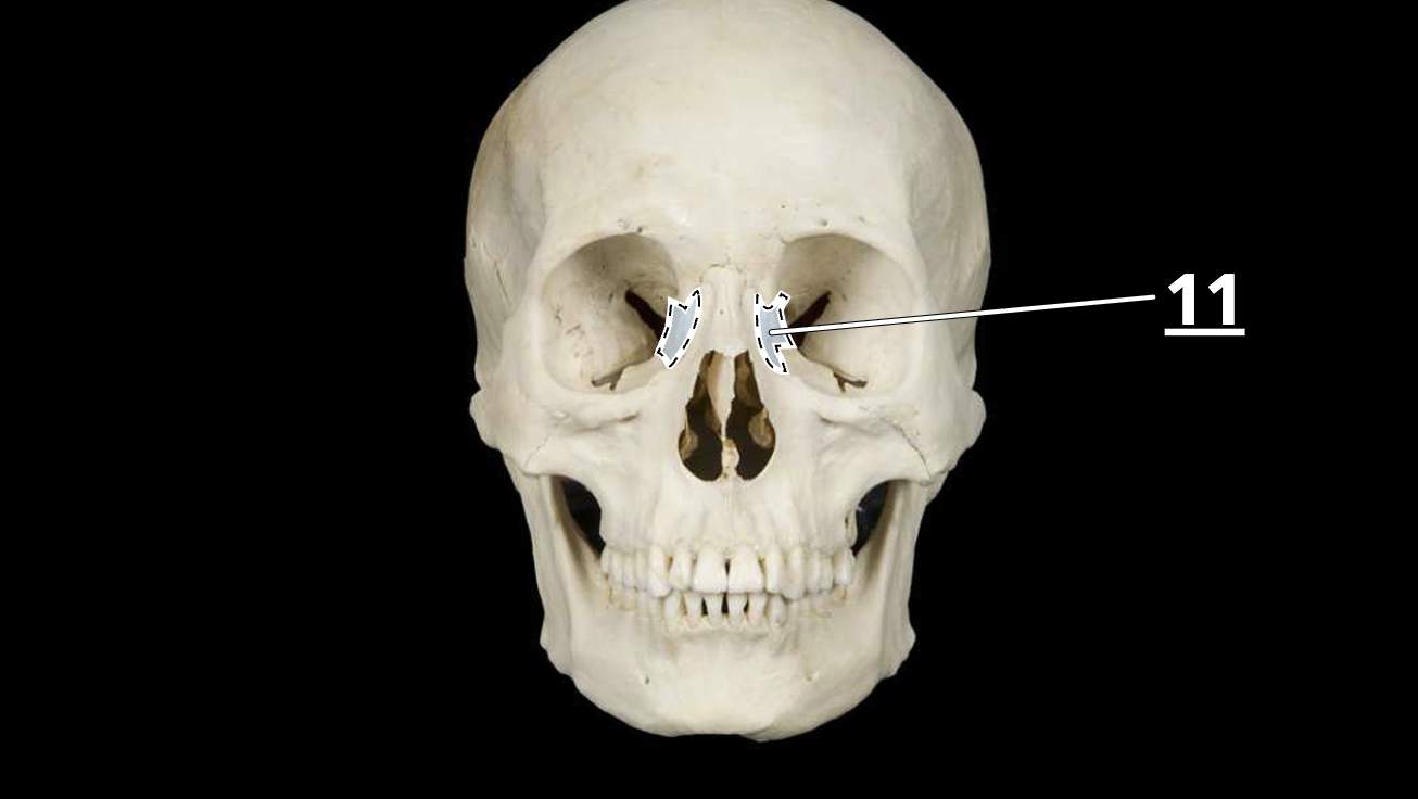
lacrimal bone
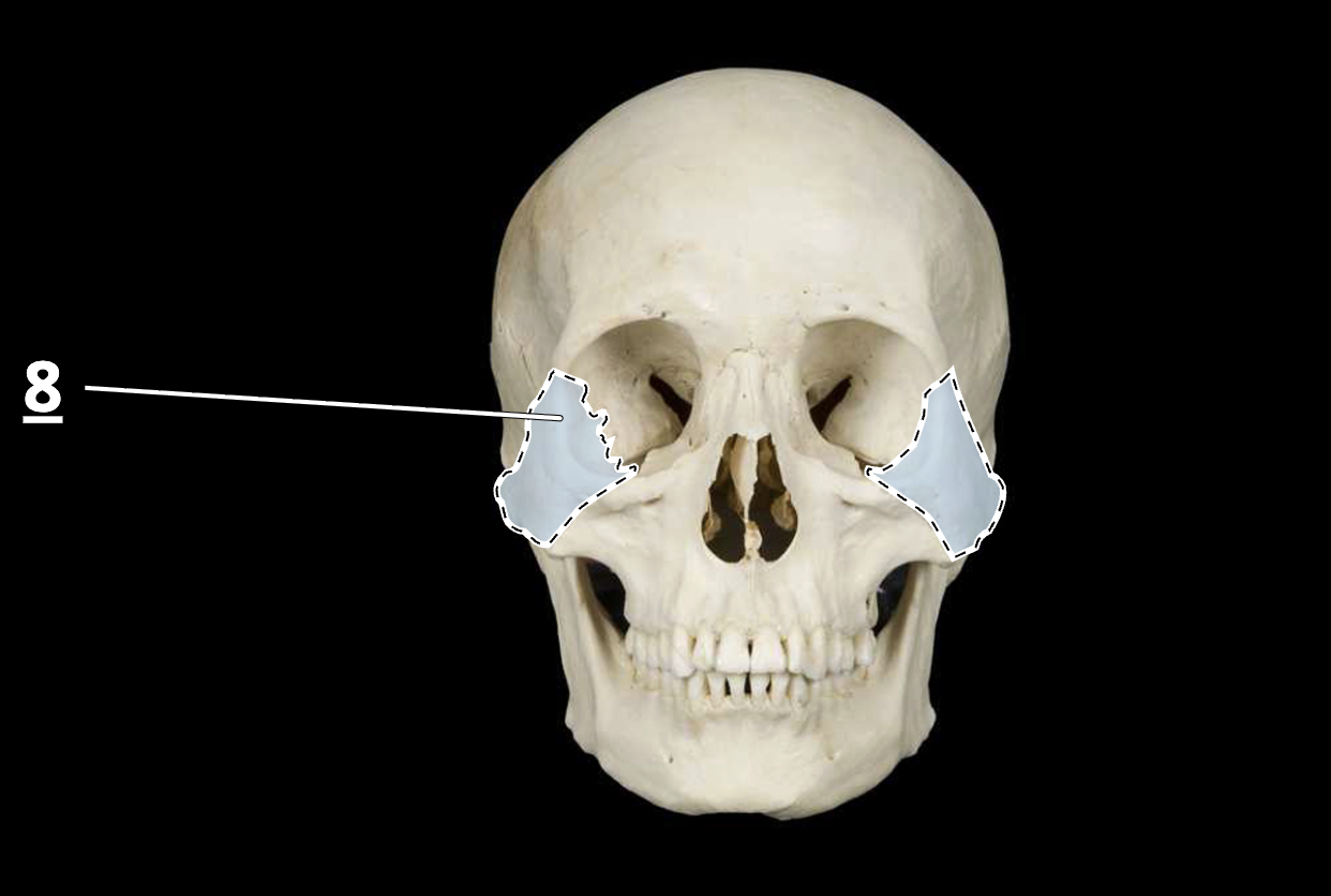
zygomatic
maxilla

mandible

palatine
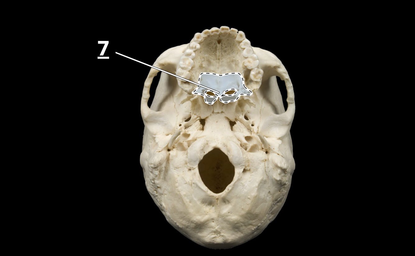
vomer
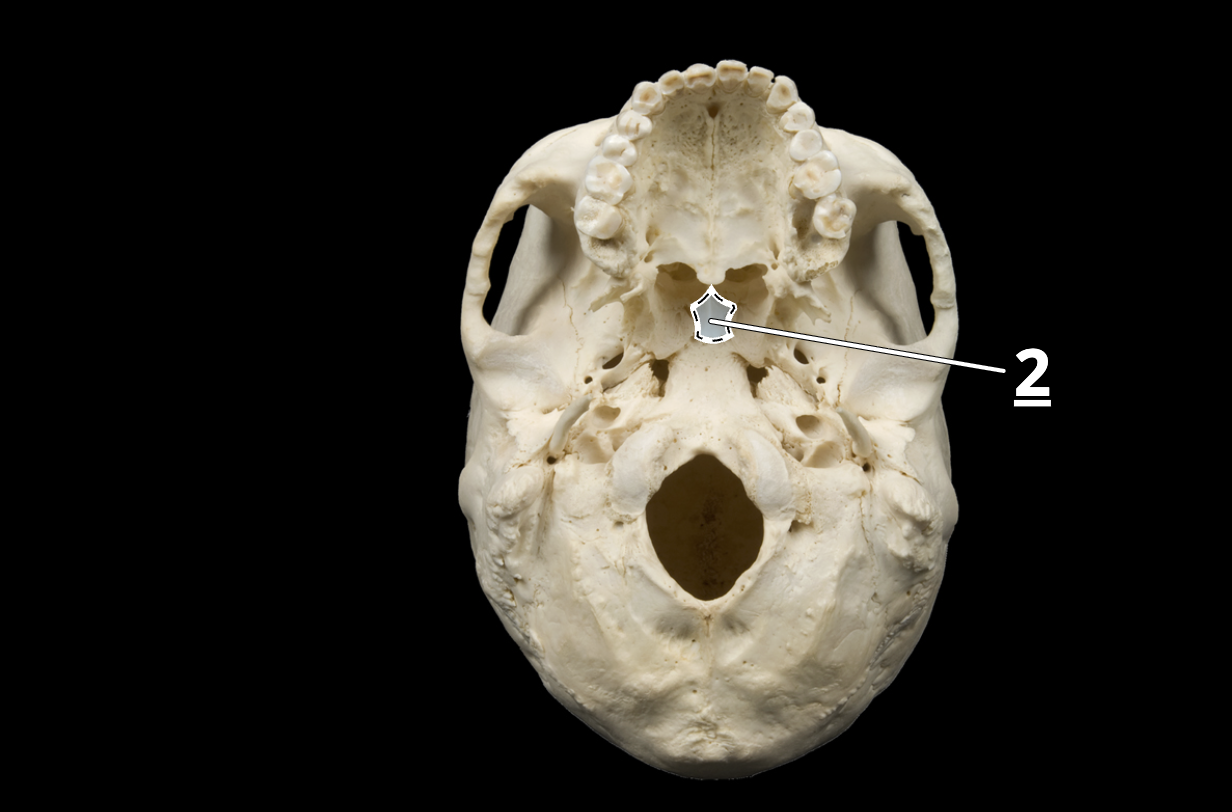
hyoid

tissue
a group of similar cells and cell products working
together to perform a specific role in an organ
Epithelial tissue
sheet of closely adhering cells, one or
more cells thick, with upper surface exposed to environment
or internal space
• Covers body surfaces and lines body cavities
• Constitutes most glands
• Avascular (does not have blood vessels)
Functions of epithelial tissue
Protection—protect deeper tissues from injury and
infection
• Secretion—produce and release mucus, sweat, enzymes,
hormones, and other substances
• Excretion—void wastes from the tissues
• Absorption—absorb chemicals, such as nutrients
• Filtration—all substances leaving the body are selectively
filtered by an epithelium
• Sensation—nerve endings in epithelia detect stimuli
General structure of epithelia:
Cells close together, small amount of extracellular material
• Avascular—no blood vessels, nourished by underlying
connective tissue
• High rate of mitosis in cells near connective tissue
• Rest on a basement membrane—layer between an
epithelium and underlying connective tissue
• Collagen, glycoproteins, other protein–carbohydrate complexes
• Anchors the epithelium to the connective tissue below it
Surfaces of epithelial cells:
Basal surface—surface facing the basement membrane
• Apical surface—surface that faces away from the basement
membrane
• Lateral surface—surface between the basal and apical surface,
the “sidewall”
Simple Squamous Epithelium
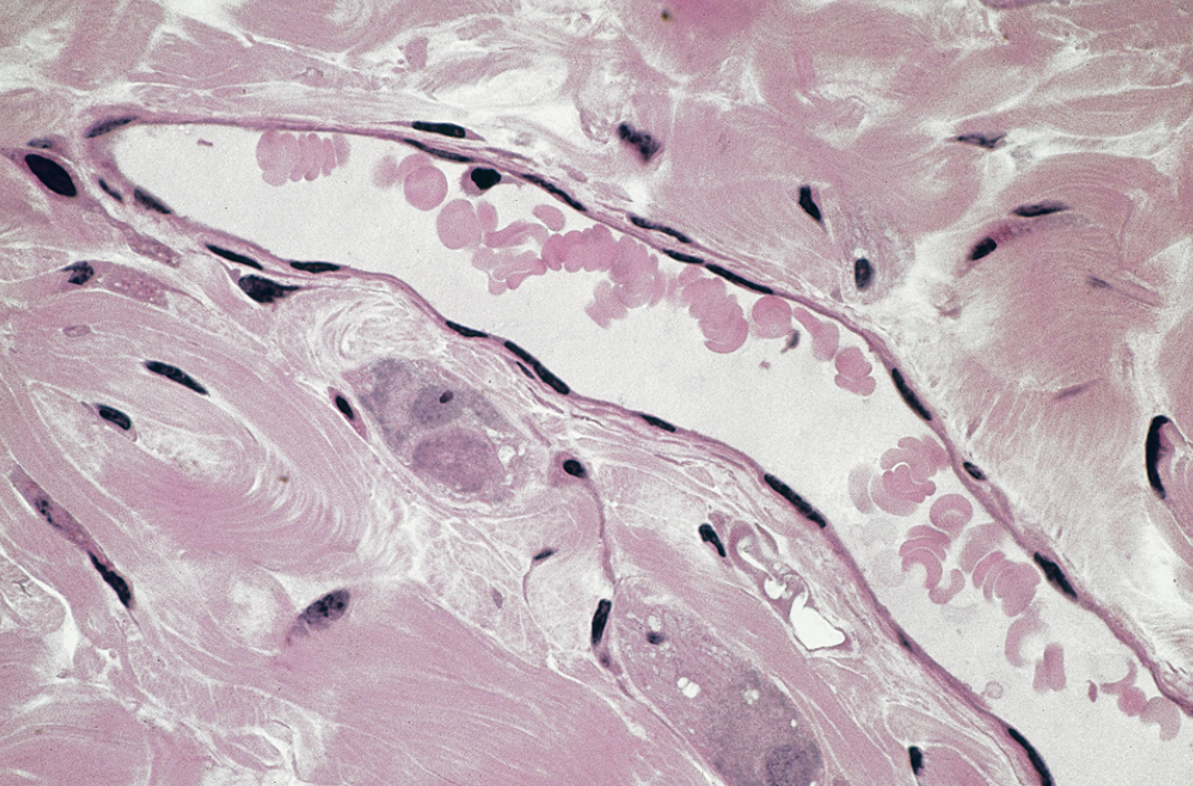
Single row of thin cells
• Permits rapid diffusion or transport of substances
• Secretes serous fluid
• Locations: alveoli, glomeruli, endothelium, and serosa
Simple Cuboidal Epithelium
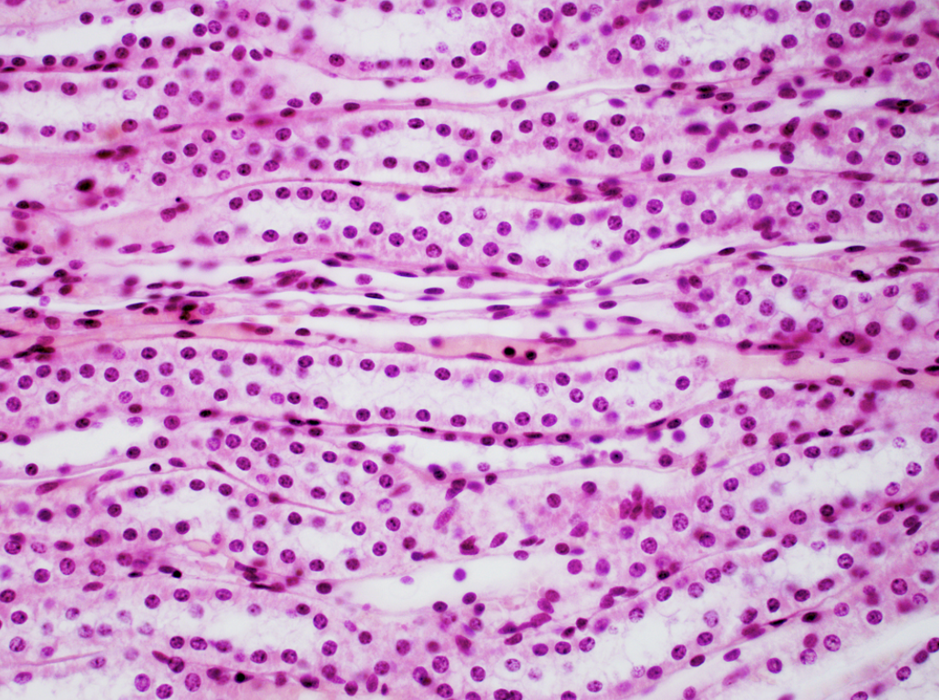
Single layer of square or round cells
• Absorption and secretion, mucus production and movement
• Locations: liver, thyroid, mammary and salivary
glands, bronchioles, and kidney tubules
Simple Columnar Epithelium

Single row of tall, narrow cells
• Oval nuclei in basal half of cell
• Brush border of microvilli, sometimes ciliated, may possess goblet cells
• Absorption and secretion; secretion of mucus
• Locations: lining of GI tract, uterus, kidney, and uterine tubes
Ciliated Pseudostratified Columnar Epithelium
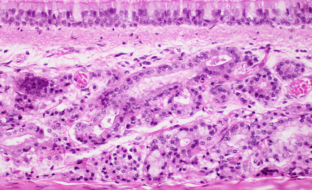

Looks multilayered, but all cells touch basement membrane
• Nuclei at several layers
• Has cilia and goblet cells
• Secretes and propels mucus
• Locations: respiratory tract and portions of male urethra
Nonkeratinized Stratified Squamous Epithelium
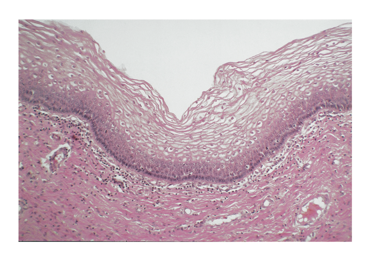
Same as keratinized epithelium without surface layer of
dead cells
• Resists abrasion and penetration of pathogens
• Locations: tongue, oral mucosa, esophagus, and vagina
Keratinized Stratified Squamous Epithelium

Multiple cell layers; cells become flat and scaly toward surface
• Resists abrasion; retards water loss through skin; resists
penetration by pathogenic organisms
• Locations: epidermis; palms and soles heavily keratinized
Stratified cuboidal epithelium

Urothelium

Connective tissue
most abundant, widely distributed, and
variable type of tissue in which cells occupy less space than
matrix
Functions of connective tissues
Binding of organs—connect one bone to another,
muscles, to bones, skin to muscle, and hold organs in
place
• Support—support the body and its organs, form internal
framework of organs
• Physical protection—protect, cushion delicate organs
• Immune protection—connective tissue cells attack
foreign invaders