1 CP180 Principle of Immunology
 Call Kai
 Learn
Learn Practice Test
 Spaced Repetition
 Match
Match Flashcards
 Knowt Play
1/103

There's no tags or description
Looks like no tags are added yet.
Name | Mastery | Learn | Test | Matching | Spaced | Call with Kai | Chat |
|---|
No analytics yet
Send a link to your students to track their progress
104 Terms
● Body’s defense against invading pathogens
● Comprised of barriers, organs, cellular elements, molecules
IMMUNE SYSTEM
IMMUNE SYSTEM Two distinct divisions:
○ Innate
○ Adaptive
MAJOR TISSUES AND ORGANS OF THE IMMUNE SYSTEM
PRIMARY LYMPHOID
SECONDARY LYMPHOID
This serves as the “training ground” or “headquarters” of the soldiers or army.
This is where the immune system components are primarily produced.
PRIMARY LYMPHOID
Major actors, This is where the immune system components are primarily produced.
○ Bone marrow
○ Thymus
● This is where the maturation process happens.
● This is where they also learn or go to “Education” for Self-tolerance. ○ Self-tolerance → They do NOT attack their own selves. They recognize the enemies between those who are not.
● Competent enough
PRIMARY LYMPHOID
This is the warzone, where the action happens.
○ Spleen
○ Lymph node
○ Mucosa-associated Lymphoid Tissue (MALT)
SECONDARY LYMPHOID
● Sites of interaction between the immune system and antigens from your pathogens.
● Site of immune response
SECONDARY LYMPHOID
● Predominant primary lymphoid tissue of the body
● Source of all cellular elements: RBC, WBC, platelet,
● Through the process of Hematopoiesis
BONE MARROW
– self-renewing pluripotent stem cells differentiate to produce all blood cell types.
Hematopoiesis
Through the process of Hematopoiesis – self-renewing pluripotent stem cells differentiate to produce all blood cell types. ○ Could diverge into
Myeloid or Lymphoid lineage
They drive responses such as growth, survival, proliferation, differentiation, maturation, and finally functional activation of each of the cell types in the immune system
Soluble mediators that control hematopoietic process
Hematopoietic growth factors / cytokines or Colony-Stimulating Factor (CSF)
● This shows the basic model of Hematopoiesis.
● It outlines the various pathways of blood cells taken from their origin as bone marrow stem cells through stages in which they are progressively selected to become
From pluripotent (marami) to
monopotent.
Currently, there are 4 human hematopoietic cytokines that have one or more combinant products that are FDA approved:
○ EPO: Erythopoietin
○ G-CSF: Granulocyte-Colony-Stimulating Factor
○ GM-CSF: Granulocyte MonocyteColony-Stimulating Factor
○ IL-11: Interleukin-11
● Bilo primary lymphoid organ located in the superior mediastinum between the aorta and the sternum.
THYMUS
Primary function of Thymus
Produce mature T-cells
Produce mature T-cells
○ Thymus-dependent lymphocytes
○ Responsible for cell-mediated immunity
○ Cytotoxic actions and immunoregulation
○ An intricate multi-step process that T cells are produced that do NOT react to self (Self-tolerance).
○ They also have all the (+) appropriate receptors for it to be beneficial to the immune system.
Allowed to leave thymus (headquarters) and take part in the immune response (deployed into the warzone)
”Thymic education”
> 99% fail leads to
Apoptosis
■ T cells that fail the thymic education test are eliminated via Apoptosis.
■ Similar to Sparta where only the strong and able-bodied are able to survive because they need to be soldiers, and if they do not reach the standard, they are eliminated
● T cells have a similar scenario to this
● A secondary lymphoid tissue (in the “warzone”)
● Immunologic filter of the blood
● Destroys defective and old erythrocytes
SPLEEN
Sequestration of cellular elements
splenomegaly
When in states of chronic or hyperinflammatory states, it could result into Splenomegaly (enlargement of the spleen)
Two important parts of the spleen:
White Pulp
Red Pulp
■ Site of interaction of APCs, T-cells, B-cells
■ Cell-mediated immune response
■ Antibody production (Humoral immune response)
White Pulp
■ RBC degradation
Red pulp
● Immunologic filters for interstitial lymphatic fluid from body’s tissues
● Site of interaction of debris with APCs and other immune cells
LYMPH NODES
filters the blood
spleen
filter the Lymph or Lymphatic Fluid..
Lymph Nodes
Sequester activated immune cells (or tumor cells) → inflamed and engorged
lymphadenopathy
That is why if the tumor cells are too active, it would sequester more, it could eventually get inflamed and engorged, resulting in Lymphadenopathy.
● Consists of well-defined networks of primary lymphoid follicles and other associated immuno-competent cells, such as adenoids, appendix, intestinal Peyer’s patches, tonsils, small solitary lymph nodes, and loosely organized clusters of lymphoid cells in the intestinal villi. ● Most extensive component of lymphoid tissue ● Distributed along mucosal linings of the body
MALT
● Mucosa-Associated Lymphoid Tissue
MALT
● Mucosa-Associated Lymphoid Tissue
Primary function
○ Filter, trap, remove pathogens that breach mucosal surfaces
○ Generate plasma cells (activated B-cells) → antibodies
MALT are lined with a very specific antibody which is generated by your activated B-cells upon encounter with these foreign pathogens and antigens.
(Secretory IgA)
● Nonspecific barriers - both physical and chemical
● First line of defense
● Composed of cellular and molecular components
● Strategically deployed and positioned
● Prevent or quickly neutralize infection
INNATE IMMUNITY
INNATE IMMUNITY
PHYSICAL DEFENSE
Skin
Intestinal Cells
Respiratory
Stomach
● Normal urine flow
● Lysozymes in tears and saliva
● Normal flora
○ Throat
○ Lower GIT, Colon
○ Genitourinary tract
○ In an immunocompetent individual, these normal flora are part of the physical defense.
○ Largest organ of the body
○ Breaks in the skin are one of the significant portals of entry of systemic infections
○ First Line of Defense
Skin
Characterized by Rapid Cell Turnover
Intestinal Cells
■ Meaning, they are intermittently sloughed off. There is intermittent sloughing of mucosal cells.
Rapid Cell Turnover
Rapid Cell Turnover
Why is that a good defense mechanism?
Because it minimizes the contact time and prevents any infected cells from propagating, resulting in an invasion.
If mabilis siyang matanggal or slough off, madadala niya yung antigens as well as other pathogens, and it doesn’t give these pathogens enough time to stay long enough to wreak more havoc in an area and cause an invasive disease.
In clinical application, some cancer patients on chemotherapy have inhibitors of the cell cycle. A faster cell cycle completion means faster turnover of cells; therefore, if the cell cycle progression is stopped, they are all halted. The turnover of the cells would also be inhibited.
Systemic chemotherapeutic agents could also affect normal cells (not just tumor cells) and when it does affect intestinal cells, that particular defense mechanism will be disrupted, and therefore more prone to breach and invasive gastrointestinal infections.
Mucus + Cilia + Reactive Cough Reflex
Respiratory
■ Provides a natural barrier to invasion via the respiratory tract.
Mucus + Cilia + Reactive Cough Reflex
coats the epithelial cells and serves in part to prevent the microorganisms from adhering to cell surfaces
Mucus
lining the epithelium of the lungs help to repel inhaled organisms
Cilia
helps in the expulsion of the foreign invading bodies.
Cough Reflex
This is why we should be more judicious in giving or taking antitussives, because we are actually disrupting the body’s natural defense mechanism which is coughing.
Cough Reflex
○ Acid pH 1-2
○ The very acidic environment of the stomach is also very important. It is a hostile environment to several pathogens, it would result in their protein denaturation in an acidic environment, resulting in their deactivation or neutralization.
○ In the same way in clinical application, especially in patients who are admitted in the ICU and hospitals where we give PPIs or H2 blockers for hyperacidity, we actually lower the defense of the stomach and they would be prone to bacterial translocation because bacteria or pathogens that would normally be neutralized by the hostile acidic pH would NOT get neutralized due to the higher pH, and they would be able to propagate and even be translocated to the different parts of the body.
Stomach
innate immunity.
CELLULAR COMPONENTS
Phagocytes:
○ Monocytes/macrophages
○ Neutrophils
○ Eosinophils
○ Mast Cells
Basophils
secrete inflammatory mediators
These inflammatory mediators would result in the inflammatory cascade, chemotaxis of the other immune cells, and to start mounting the immune response against the foreign antigen.
Mast Cells
Basophils
Recognize their target, Internalize (kakainin), Degrade invading pathogens → Phagocytosis
PHAGOCYTES
PHAGOCYTES types
○ Opsonin-dependent phagocytosis
○ Opsonin-independent phagocytosis
● Substances that coat the infectious pathogen by sticking to its conserved structures, making it more palatable or attractable.
OPSONIN-DEPENDENT PHAGOCYTOSIS
OPSONIN-DEPENDENT PHAGOCYTOSIS
● Antibodies (IgG), Complement (C3b), Lectin (CRP)
● Binds to specific receptors in the phagocytes → activate phagocytosis
OPSONIN-DEPENDENT PHAGOCYTOSIS
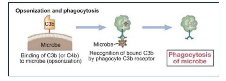
● This is an example of opsonization through complement mediated phagocytosis. ● Binding of the C3b to the microbe (natag na siya). ● These will be recognized by your phagocytes through the C3b receptors in your phagocytes resulting in recognition, internalization, and then degradation.
Opsonization and Phagocytosis
IgG
Antibodies
C3b
Complement
CRP
Lectin
● Innate leucocytes Pattern Recognition Receptors (PRRs)
● PRRs on phagocytes recognize conserved ligands or certain patterns on the surface of infectious pathogens, known as Pathogen-Associated Molecular Patterns (PAMPs)
OPSONIN-INDEPENDENT PHAGOCYTOSIS
Pattern Recognition Receptors (PRRs) are found in
Innate leucocytes
Pattern Recognition Receptors (PRRs) examples
○ Macrophage mannose receptor
○ Macrophage scavenger receptor
○ Toll-like receptor (TLR) family
OPSONIN-INDEPENDENT PHAGOCYTOSIS PROCESS
● Binding of the PAMPs from your pathogen to TRRs such as TLRs allows the phagocyte to recognize or internalize (engulf) the pathogen.
● This binding of the TRRs and PAMPs would also activate secretion of chemokines, inflammatory cytokines, and other antimicrobial peptides, as well as expression of co-stimulatory proteins and major histocompatibility complex molecules by the phagocyte that would result to a call for help.
○ Kakainin niya yung narecognize niyang pathogen, but at the same time during the process, it willsend signals, release all these mediators, and send it so that the other “soldiers” would receive some sort of a warning or call to “come here, the pathogens are here”.
Coated by the antibody yung antigen; or the bacteria was marked by C3b, the complement system.
○ The macrophage has a C3b receptor
○ The antibody has an antibody receptor
Opsonin-dependent phagocytosis
○ Lipoprotein, which is an example of a PAMP, will be recognized by the macrophages that are toll-like receptor (or TRR)
○ If it recognized the pattern even without sugar coating, it would still initiate the entire phagocytic process.
Independent phagocytosis
Steps in phagocytosis
○ Chemotaxis and adherence of microbe to phagocyte
○ Ingestion of microbe by phagocyte
○ Formation of a phagosome
○ Fusion of the phagosome with a lysosome to form a phagolysosome
○ Digestion of ingested microbe by enzymes
○ Formation of residual body containing indigestible material
○ Discharge of waste materials
○ The lysosome in step 5 contain lytic enzymes that would partially digest the microbes
○ Once completely digested and inactive, as seen in step 6 and step 7, it would release the indigestible materials. The indigestible materials are the antigens.
○ The free antigens can be recognized by your antigen-presenting cells and could be directly neutralized by your antibodies.
○ The pathogens share these antigens, and once these are recognized, they will be neutralized. This will lead to the facilitation of the overall immune response that will be mounted by the organism.
● Also known as the Polymorphonuclear cells
NEUTROPHILS
● Comprise most of the total WBC in bloodstream
● Primary human defense against invasive bacteria
○ It is breached in the physical barriers ○ It has entered the systemic circulation which means that it will be the first one in there
NEUTROPHILS
● Migrate from blood into infected or inflamed tissue in response to chemotactic factors such as IL-8 and breakdown products of C3a and C5a
NEUTROPHILS
complement systems
C3a and C5a -
Aside from opsonins, some complements function as chemotactic factors (warning signals)
Process Neutrophils
● Chemotaxis → site of inflammation → PRRs + PAMPs → adhere → phagocytosis ○ Neutrophil is a phagocyte, so it may either be opsonin-independent or opsonin-dependent ○ Opsonin-independent - there would be PRRs that would recognize PAMPs in the pathogens leading to adherence and phagocytosis
act as opsonins to neutrophil-phagocytosis ○ If these are present, they could function in the opsonin-dependent neutrophil-phagocytosis
Complement and Ab
These are the different mechanisms why which neutrophils can be able to control infections ○ It is not just phagocytosis, but it is the primary mechanism of pathogen elimination.
Mechanisms
○ Phagocytosis
○ Release of Reactive Oxygen Species and Nitric Oxide Species - highly harmful to all types of cells (even self) ■ Primarily targeted to the pathogen
○ Neutrophil Extra-cellular Traps (NET) Release
■ Neutrophils will release the nuclear contents along with its granule contents
■ It will undergo suicide or self-destruction by releasing its antigens and it own granule contents
■ This forms a “net” or a “trap” that will kill the microorganism
Neutrophil Extra-cellular Traps (NET) Release
Major role against non-phagocytable multicellular pathogens such as parasites
EOSINOPHILS
IgE
Allergy
EOSINOPHILS
Activated via high-affinity receptor for IgE → eosinophils exocytose their granules → release basic proteins or ROS → lysis of parasites
EOSINOPHILS
○ May also help with allergy control, but not so much for bacteria
○ More useful for allergies, as well as non-phagocytable multicellular pathogens such as parasites
High affinity for IgE → role in pathogenesis for allergies
EOSINOPHILS
● Express lower levels of C3 receptor and Fcy for IgG than PMNs
○ Less significant role in bacterial infections
EOSINOPHILS
. INNATE IMMUNE SYSTEM - MONOs/MPs