Biology
 Call Kai
 Learn
Learn Practice Test
 Spaced Repetition
 Match
Match Flashcards
 Knowt Play
1/170

Earn XP
Description and Tags
phosphorylate ts
Name | Mastery | Learn | Test | Matching | Spaced | Call with Kai | Chat |
|---|
No analytics yet
Send a link to your students to track their progress
171 Terms
What role do hydrogen atoms play during respiration
The substrate molecules are broken down and the hydrogen atoms become available
Hydrogen carrier molecules NAD and FAD take h turn into nadh/fadh2 and transfer to the inner mitochondrial membrane
nadh/fadh2 release the hydrogen atoms which split into protons and electrons
The protons are pumped across the inner mitochondrial membrane into the intermembrane space - forming a proton / chemiosmotic gradient
This proton gradient is used in chemiosmosis to produce ATP
After the protons have flowed back into the matrix of the mitochondria via ATP synthase they are oxidized to form water
Structure of mitochondria
Mitochondria have two phospholipid membranes, an outer and an inner membrane
The outer membrane
Smooth
Permeable to several small molecules
The inner membrane
Folded (cristae)
Less permeable
The site of the electron transport chain (used in oxidative phosphorylation)
Location of ATP synthase (used in oxidative phosphorylation)
The intermembrane space:
Has a low pH due to the high concentration of protons
The concentration gradient across the inner membrane is formed during oxidative phosphorylation and is essential for ATP synthesis
The matrix:
Is an aqueous solution within the inner membranes of the mitochondrion
Contains ribosomes, enzymes and circular mitochondrial DNA necessary for mitochondria to function
Structure and function of mitochondria
Cristae
- Large surface area holding many electron transport chain proteins & atp synthase = more atp.
4 stages of aerobic respiration.
1. Glycolysis | Phosphorylation and splitting of glucose | Cell cytoplasm |
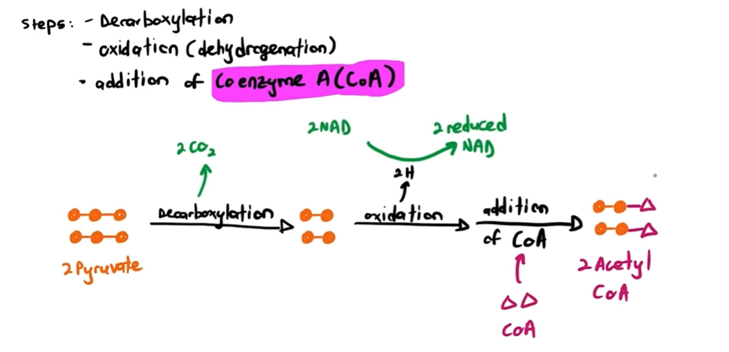
2. Link reaction | Decarboxylation and dehydrogenation of pyruvate | Mitochondrial matrix |
3. Krebs cycle | Cyclical pathway with enzyme-controlled reactions | Mitochondrial matrix |
4. Oxidative phosphorylation | Production of ATP through oxidation of hydrogen atoms | The inner membrane of mitochondria |
Aerobic respiration definition
process of breaking down a respiratory substrate in order to produce ATP using oxygen
Coenzyme
helps an enzyme carry out its function but is not used in the reaction itself
Krebs cycle
The Krebs cycle (sometimes called the citric acid cycle) series of enzyme-controlled reactions
Acetyl CoA (2C) enters the circular pathway via the link reaction
4 carbon (4C) oxaloacetate accepts the 2C acetyl fragment from acetyl CoA to form citrate (6C)
Citrate is then converted back to oxaloacetate through a series of small reactions
Series of small reactions for citrate to oxaloacetate
4c oxaloacetate reacts with acetyl CoA, oxaloacetate gets 2 carbons from it = 6C citrate, CoA released for link reaction.
2. decarboxylation and dehydrogenation, nad → nadh = 5C
3. decarboxylation and dehydrogenation, nad → nadh 4C
Atp produced = adp + Pi
2 more dehydrogenation reactions, fad THEN nad
oxaloacetate formed again
happens twice for each glucose molecule
Role of nad and fad
Reduced NAD and reduced FAD transfer the hydrogen atoms (hydrogen ions and electrons) from the different stages of respiration to the electron transport chain on the inner mitochondrial membrane
This is the site where hydrogens are removed from the coenzymes)
Electrons from reduced NAD (NADH) and reduced FAD (FADH2) are given to the electron transport chain
Hydrogen ions from (NADH) and (FADH2) are released when the electrons are lost
The e transport chain moves these (protons) across the inner mitochondrial membrane into the intermembrane space, creating a proton gradient (more protons in the intermembrane space)
The movement of protons down the proton gradient, back into the mitochondrial matrix, gives the energy required for ATP synthesis
How much nad and fad in those reactions gng
krebs = 6 nadh 2 fadh2
glycolysis = 2 nadh
Link = 2 nadh
oxidative phosphorylation
h atoms donated by nadh/fadh2, split into h+ and e
e release Energy as move through e transport chain
released Energy used to transport h+ across inner mitochondrial, from matrix to intermembrane.
conc gradient of h+ made
h+ return to matrix via facilitated diffusion through channel protein atp synthase
diffusion provides E for atp synthesis, adp+pi→atp+h20
o2 combines with h+ & e- to form water at end of transport chain
Consequences of no o2 in respiration
There is no final acceptor of electrons from the electron transport chain
The electron transport chain stops functioning
No more ATP is produced via oxidative phosphorylation
Reduced NAD and FAD aren’t oxidised by an electron carrier
No oxidised NAD and FAD are available for dehydrogenation in the Krebs cycle
The Krebs cycle stops
what type of anaerobic resp do certain organisms use
Yeast and microorganisms use ethanol fermentation
Other microorganisms and mammalian muscle cells use lactate fermentation
Ethanol fermentation
glycolysis takes place as usual (net gain 2 atp) (4 atp gained - 2atp used for glucose)
2pyruvate gained
2pyruvate decarboxylated to 2ethanal (2ch3cho)
2ethanal→2ethanol, because nad gives hydrogens from glycolysis (nad regenerated)
alcohol dehydrogenase used for ethanal to ethanol
lactate fermentation
glycolysis takes place as usual (net gain 2 atp) (4 atp gained - 2atp used for glucose)
2pyruvate gained
nad is regenerated by giving 4 hydrogens to 2pyruvate → 2lactate (pyruvate to lactate needs lactate dehydrogenase)
what happens to lactate after being formed
taken to liver where either turned into pyruvate for link reaction
or glycogen for energy storage
Chloroplast pigments
Pigment group | Name of pigment | Colour of pigment |
|---|---|---|
Chlorophylls | Chlorophyll a | Light green |
Chlorophyll b | Dark green | |
Carotenoids | β carotene | Orange |
Xanthophyll | Yellow |
Outline need for energy in organisms
anabolic reactions
Active transport
movement
bioluminescence
Examples of anabolic reactions
Dna replication
protein synthesis
active transport
movement
phosphorylation
How is atp suitable as universal currency
small
water soluble
easily transported around cell
easily hydrolysed
large quanitity of energy released
rapid turnover rate
readily available
atp equation
atp + h20 →←adp + pi
chemiosmosis
occurs in mitochondria inner membrane & thylakoid membrane
e transport chain makes h+ conc gradient
high energy e move from carrier to carrier, Energy released used to pump h+ across inner membrane to intermembrane space, matrix to intermembrane space
protons move down gradient releasing Energy
h+ move through atp synthase complex, uses released E to phosphorylate adp.
o2 accepts h+ & e- formng h20
makes most of atp, 32/34 atp per glucose.
atp structure
adenine ribose and 3 phosphates
2 ways atp synthesised
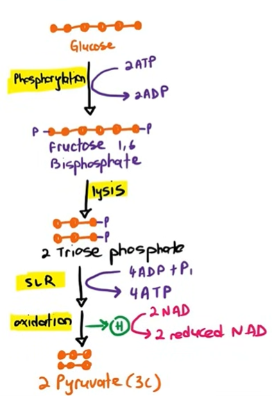
substrate level phosphorylation
uses energy from another chem reaction for adp+pi
chemiosmosis
glycolysis
link reaction
Absorption spectra
An absorption spectrum is a graph that shows the absorbance of different wavelengths of light by a particular pigment
action spectra
An action spectrum is a graph that shows the rate of photosynthesis at different wavelengths of light
Pigments and what they absorb
Pigment group | Name of pigment | Colour of pigment |
|---|---|---|
Chlorophylls | Chlorophyll a | Light green |
Chlorophyll b | Dark green | |
Carotenoids | β carotene | Orange |
Xanthophyll | Yellow |
Rf values of pigments
Carotenoids have the highest Rf values (usually close to 1)
Chlorophyll B has a much lower Rf value
Chlorophyll A has an Rf value somewhere between those of carotenoids and chlorophyll B
Purpose of photosynthesis
Produce organic molecules
sites of light dependent reaction
granum/thylakoid
sites of light independent reaction
stroma
Why do photosystems have different pigments
To increase the range of absorbed wavelengths of light
Function of photosystem
pigments absorb different wavelengths of light.
Transfer energy to reaction center, causing photoactivation of reaction center.
High energy electron released from reaction centre
PSI vs PSII
PSII has a oxygen evolving complex enzyme
Purpose of light dependent reaction
Use light energy to get ATP & Reduced nadp for light independent reaction
non cyclic phosphorylation
light hits PSI, electrons are excited out to the enzyme, produces reduced NADP.
light hits PSII, photoactivation, electrons moves through etc to PSI.
Protons pumped into thylakoid from energy from electron. Proton gradient formed.
Protons move out through atp synthase, produces atp.
electron in PSII replaced from e- from photolysis of h20 catalyzed by oxygen evolving enzyme complex. O2 waste product.
Cyclic photophosphorylation
Light absorbed by pigment in PSI, causing photoactivation of reaction center, releasing high energy e-.
Goes through etc, then returns to PSI.
This powers etc, pumps protons into thylakoid, protons leave via facilitated diffusion via atp synthase.
Produces atp.
Differences & similarities between cyclic and non-cyclic photophosphorylation
similarities:
happens in thylakoid/granum
uses PSI
produces atp
differences: (happen in non cyclic)
Uses PSII
Produces NADPH
Photolysis of h20
Calvin cycle/light independent reaction
start with rubp (5C)
Carboxylated, catalysed from rubisco
Forms 6C unstable molecule, splits apart into 3C glycerate 3-phosphate.
Hydrogenated by NADPH, uses 1 atp.
Forms TP, uses 1 atp to turn back into rubp. (5/6 TP used for this)
Uses of TP & GP
TP:
Hexose sugars
starch (energy storage)
sucrose (translocation in phloem)
cellulose (cell wall)
amino acids (when ammonium/nitrate is added)
GP:
fatty acids & glycerol
phospholipids & triglycerides
Limiting factors in photosynthesis
temp
co2 conc
strength of light
How do factors affect the systems in photosynthesis
more light
more absorption by pigment
high rate of light dependant reaction
more atp and nadph for calvin cycle
More co2
more rubp carboxylation
high rate of light independant reaction
more organic compounds produced
More temp
optimum = more enzyme substrate complexes formed (rubisco, atp synthase, nadp reductase)
higher rate of light independant reaction
Too much temp
enzymes denature (H bonds break in between)
very low rate of photosynthesis
Structure of chloroplast relate to function
Stroma for light independant reaction
Large number of grana to absorb more light
Stroma colourless so light reaches thylakoids
DNA for chloroplast protein production
starch grains to store chemical energy
Homeostasis def
maintaing an optimum internal environment
Examples of internal environments
body temp
h20 conc in blood
glucose conc in blood
Negative feedback
Body undergoes change from optimum condition (stimulus)
stimulus detected by receptor
signal sent to control centre
control centre sends signal to effector
effector produces response to correct the change
Excretion
Removal of waste products in our body
Deamination
removal of amine group (nh2) in liver
Nh3 and keto acid produced
keto acid used in respiration
nh3 = soluble = toxic
nh3 + co2 → urea = removed by kidney
Kidney function
Carries out excretion
helps wt homeostasis
Nephron
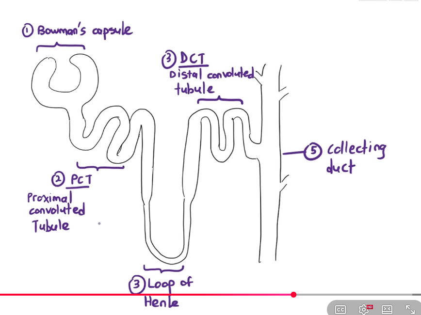
Strucutre that produces urine made out of bowmans capsule and tubules
Nephron diagram
Ultrafiltration
High pressure causing substances to separate from blood
Filters out h2o salts urea glucose amino acids
High pressure in glomerulus generated by difference in diameter of afferent and efferent arteriole. Lwk tight asl
Gaps in glomerular wall prevent RBC from leaking out.
Basement membrane filters out plasma/large proteins, mr>67000.
Podocytes = inner bowmans capsule wall allow filtrate to leak through in controlled amounts.
Selective reabsorption
Most water is reabsorbed as it moves through the nephron. Rest is turned to urine.
PCT reabsorbs all glucose and amino acids, most h2o, and some salts and urea.
Loop of henle reabsorbs some H2O and salts (tad bit urea)
Collecting duct reabsorbs some water.
Leftover stuff is urine.
PCT adaptations
1 cell thick wall of PCT
Capillary right next to it
Tight junction in between pct cells to prevent leakage of filtrates.
PCT wall has microvilli facing PCT lumen, and basal membrane facing capillaries.
Microvilli to increase surface area to contain more co-transporter proteins for more reabsorption.
Basal membrane has Na+/K+ pumps. and transport proteins for glucose/amino acid.
Many mitochondria in PCT cell to produce ATP for active transport.
Selective reabsorption in PCT
PCT cells pump out Na+ into the blood by active transport
creates a Na+ concentration gradient between the PCT cells and PCT lumen
Na+ and glucose enters the cell through co-transport (Na+ and amino acids too)
glucose and amino acids get reabsorbed from cells to blood through facilitated diffusion
water is reabsorbed through osmosis
some urea will be accidentally reabsorbed due to diffusion.
Osmoregulation
Control of water potential of the blood and tissue fluid.
Selective reabsorption in collecting duct
Posterior pituitary gland releases ADH when the amount of water in the blood is low
ADH binds to the receptor on the Collecting Duct cells
it activates phosphorylase enzyme within the Collecting Duct cells
vesicles with aquaporin will fuse to the cell surface membrane
this increases the permeability of the Collecting Duct to water
more reabsorption of water occurs
this decreases the volume of urine, making it more concentrated
How our cells process glucose
First, Glucose diffuses into the cells using GLUT proteins (Glucose Transport Proteins)
To ensure the glucose remains trapped in the cell, glucose is phosphorylated (added with phosphate) so they cannot escape. This is catalyzed by glucokinase.
Some of the glucose will then be broken down in respiration to produce ATP.
Excess glucose molecules are converted into glycogen, by an enzyme; glycogen synthase.
Action of insulin
Blood glucose concentration higher than normal.
Detected by receptors in islets of Langerhans.
B-cells secrete more insulin.
Insulin stimulates skeletal muscle cell to cause vesicles with GLUT to fuse with cell surface membrane, cell glucose permeability increased. (only for skeletal muscle cells)
Glucose diffuses into liver cells through GLUT proteins.
Insulin activates glucokinase enzyme to phosphorylate glucose to prevent its escape bc glut proteins cant transport phosphorylated glucose.
Insulin increase respiration of glucose.
Insulin activates glycogen synthase, converts excess glucose into glycogen.
Action of glucagon
Blood glucose concentration lower than normal.
Detected by islets of Langerhans in pancreas.
Alpha cells secrete more glucagon.
Only affects liver cells.
Endocrine vs nervous
Hormones vs electrical impulse
Slow vs faster
Low atp vs high atp
Sensory neurone function
transmit impulses from receptor to relay neurone
Relay neurone function and location
transmit impulses from sensory to motor neurones.
Only in CNS
motor neurone function
transmit impulses from relay neurone to effector
Dendrites
Increase surface area when receiving signals
receive incoming signals (neurotransmitters) from other neurons at the synapse and transmit these electrical impulses toward the cell body
Sensory vs motor neurone
similarities
Cell body wt nucleus
Axons (elongated structures to transmit impulses over long distances)
Dendrites (branched structure to increase surface area for receiving signals)
Terminal branches with synaptic knobs
Axons may have myelin sheath
Differences
- Cell body of motor neurone in CNS.
Function (sensory is receptor to CNS etc)
Action potential def
rapid change in the electrical charge distribution across a cell surface membrane.
resting membrane potential
Before the neurone can send an impulse, it first has to generate and maintain a resting membrane potential of -70mV.
This uses the Na+/K+ pump, that actively pumps out 3Na+ and pumps in 2K+ into the axon.
Depolarisation
Resting membrane potential created first
Voltage gated Na+ channels open
Na+ rush into axon down conc gradient.
Inside has higher voltage than outside, at +30mv voltage gated Na+ channels close.
Repolarisation
At +30mv voltage gated Na+ channel close, but K+ voltage gated channel opens.
K+ Rushes out down concentration gradient.
Goes slight beyond resting potential and voltage gated K+ channel closes to -80mv.
refractory period
period of time where axon is unresponsive to stimulus
how does action potential move along axon
Strong stimulus
1 section of axon depolarises
Local circuits cause next section of axon to depolarise.
Myelin sheath function
provides area of insulation so ions cannot pass through
Nodes of ranvier no electrical insulation, ions can pass through
Speeds up action potential
Synapse
Junction between 2 neurones
Cholinergic synapse
Impulse reaches end of presynaptic neurone.
Voltage gated Ca2+ channels open causing Ca2+ ions to rush in
Vesicles move towards cell surface membrane.
exocytosis of ACh (acetylcholine)
ACh diffuses across synaptic cleft.
ACh binds to complementary receptors on postsynaptic neurone.
Ligand gated Na+ channels open, Na+ rush in.
Postsynaptic membrane depolarises and a new action potential is generated.
ACh detaches from receptor and is broken down by acetylcholinesterase.
Into acetate and choline.
Choline reabsorbed by presynaptic neurone and regenerated back into ACh.
Role of synapse
Ensure impulses travels in one direction.
Presynaptic contains vesicles wt neurotransmitters, post synaptic contains receptors.
Allow interconnection of nerve pathways
Individual neurones can connect with multiple other neurones = more efficient response.
How do receptor generate action potential
Na+ causes Na+ channels to open
Na+ rush in
Receptor cells depolarise.
Ca2+ channels open and Ca2+ rush in.
exocytosis of neurotransmitters.
depolarisation in sensory neurone.
Myofibril
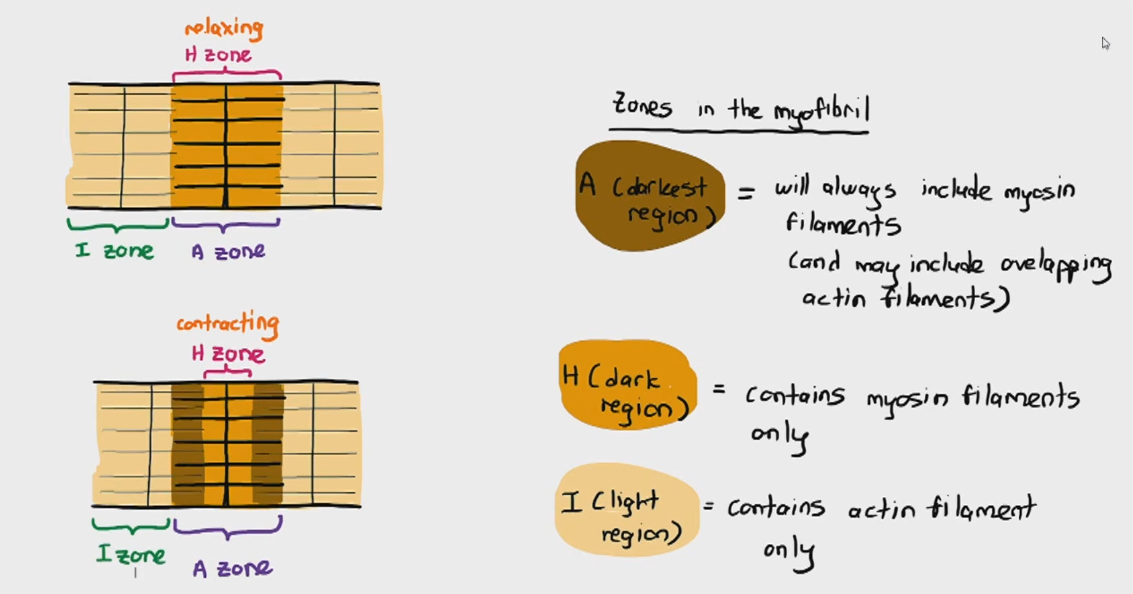
Dark and light parts
Thick and thin filaments.
dark = myosin light = actin
Myosin has M line, actin has Z line
Distance between Z lines = sarcomere.
Filament structure
Actin Filaments are THIN filaments:
made out of actin globular proteins, Troponin and Tropomyosin (prevents actin & mysin heads from attaching)
Myosin Filaments are THICK filaments:
made out of Myosin heads that face away from the M-line
myosin heads can also function as an ATP-ase enzyme; to hydrolyse ATP into ADP + Pi
Zones in myofibril
Sliding filament theory
Myosin head pulls actin filaments closer together (cross links) making the sarcomere distance shorter.
Ca2+ binds to troponin on actin filament
Causes tropomyosin shape to change, exposes actin filaments to myosin head.
Myosin head attaches to the actin filament.
Myosin head tilts back towards M-line, pulling actin filament in process.
Sarcomere distance reduces.
Myosin head hydrolyses ATP into ADP + Pi, releasing the molecules.
Myosin head detaches from actin filament.
New atp molec reattaches to myosin head and restarts whole process.
Neuromuscular junction
1. Depolarization/Action potential along motor neurone axon
2. Ca2+ channel opens and Ca2+ rushes in
3. Vesicles with ACh moves to the cell surface membrane
4. Exocytosis of ACh and diffusion of ACh along the neuromuscular junction
5. ACh binds to receptors on the sarcolemma
6. Na+ channel opens and Na+ rushes into the skeletal muscle fibre
7. Depolarization of sarcolemma, which causes depolarization of T-Tubule
8. Ca2+ transporters pump out Ca2+ into the sarcoplasm which binds to troponin on the Actin Filament
Why do venus flytraps eat insects
Grow in places where there is not enough nitrogen in soil.
Insects provide nitrogen that plants need for nucleotides or amino acids.
Venus fly trap structure
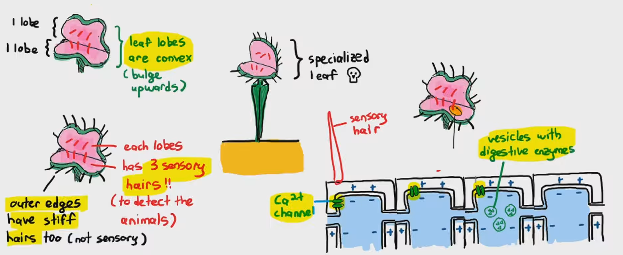
Venus flytrap whole process
Insect stimulates sensory hairs twice withing 20-35 seconds.
Ca2+ channels open and Ca2+ rush in generating action potential.
Travels along cells
Leaf lobe changes from convex to concave rapidly
Insect further stimulates sensory hairs.
More ca2+ influx → exocytosis of digestive enzymes.
Complete closure of trap.
Adaptation of flytrap
Stimulation of single hair not causing immediate closure, drop of water can do this = wasted energy.
If lots of rain = doesnt completely close
Doesnt completely close = tiny insects escape because not worth the energy compared to closing.
Gibberellins
causes stem elongation
Causes seed germination
Auxin process
Auxin binds to auxin receptor
Causes proton pump to actively pump H+ into cell wall.
Acidifies cell wall
Activates expansins which disrupt hydrogen bonds
K+ and H2O rush into cell (increases internal pressure)
cellulose microfibrils move further apart allowing cells to elongate.
Gibberellin function
XET breaks bonds between hemicellulose
Cellulose microfibrils move further apart
Allows expansion of cell wall leading to cell elongation.
Seed structure
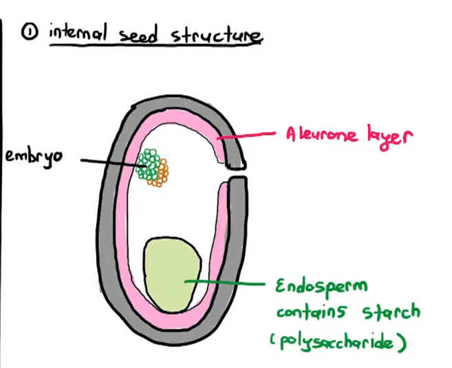
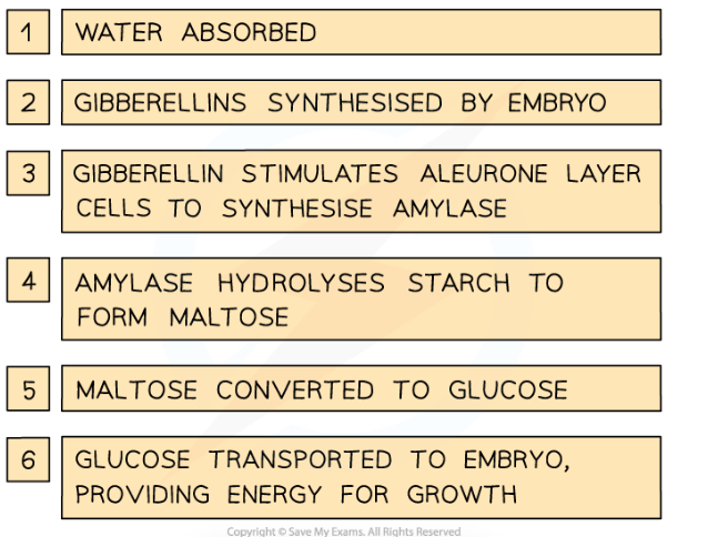
Gibberellin for seed germination
H2O uptake by seed
Embryo releases gibberellins.
Gibberellins diffuse to aleurone layer
Cells of aleurone layer synthesis amylase
Amylase hydrolyses starch → maltose → glucose
Embryo uses glucose to respire and grow
Gibberellin germination
Diploid cell def
The cell contains 2 sets of chromosomes
Haploid cell def
The cell contains 1 set of chromosomes
Homologous chromosomes
Pair of chromosomes of same length, positions of centromere, and same gene loci.
Meiosis 1
Seperation of homologous chromosomes
Meiosis 2
separation of sister chromatids
Why is meiosis reduction division
halves no. of chromosomes in nucleus
Changes a diploid cell to haploid cells.