Neuro Anatomy
 Call Kai
 Learn
Learn Practice Test
 Spaced Repetition
 Match
Match Flashcards
 Knowt Play
1/118

There's no tags or description
Looks like no tags are added yet.
Name | Mastery | Learn | Test | Matching | Spaced | Call with Kai | Chat |
|---|
No analytics yet
Send a link to your students to track their progress
119 Terms
Q: In the forebrain, which direction does rostral (cranial) point?
A: Towards the front.
Q: In the forebrain, which direction does caudal point?
A: Towards the back.
Q: In the forebrain, which direction does ventral point?
A: Towards the bottom.
Q: In the forebrain, which direction does dorsal point?
A: Towards the top.
Q: In the brainstem and spinal cord, which direction does rostral (cranial) point?
A: Towards the top.
Q: In the brainstem and spinal cord, which direction does caudal point?
A: Towards the bottom.
Q: In the brainstem and spinal cord, which direction does ventral point?
A: Towards the front.
Q: In the brainstem and spinal cord, which direction does dorsal point?
A: Towards the back.

Label this diagram

what are the 3 planes of the CNS?
coronal (frontal)
horizontal (axial)
sagittal
Q: The coronal (frontal) plane divides the CNS into which parts?
A: Rostral and caudal parts.
Q: The horizontal (axial) plane divides the CNS into which parts?
A: Dorsal and ventral parts.
Q: The sagittal plane divides the CNS into which parts?
A: Left and right parts.
Q: What is a midsagittal plane?
A: A sagittal plane that divides the CNS into equal parts.
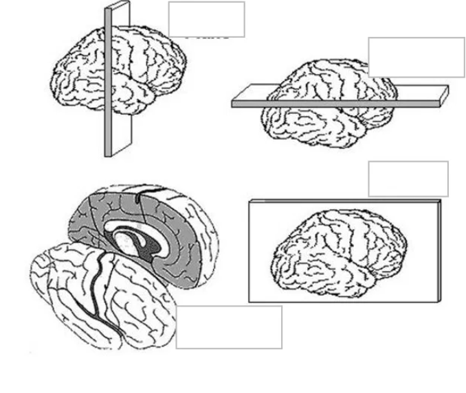
label the planes of the CNS


label the planes of the CNS
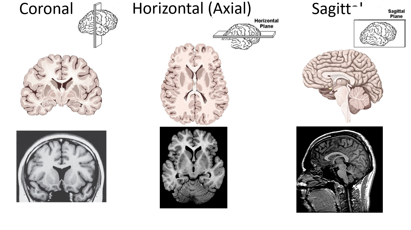

label

Q: Why is the surface of the cerebral hemispheres highly folded?
A: To increase cortical surface area.
Q: What is a gyrus?
A: A ridge or elevation of cortex.
Q: What is a sulcus?
A: A groove of cortex.
Q: What is a fissure?
A: A very prominent and deep sulcus, usually dividing lobes.
Q: What are cerebral lobes?
A: Well-defined regions of cortex named after their overlying bones
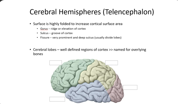
Q: Name the four cerebral lobes shown on this slide.

Q: Which hemisphere is usually the dominant hemisphere?
A: The left.
Q: The dominant hemisphere is responsible for what, regarding motor tasks?
A: Step-by-step formulation and execution of motor tasks.
Q: The non-dominant hemisphere is more important for what in most individuals?
A: Attention mechanisms.
Q: Lesions of which areas cause hemineglect syndrome?
A: The right parietal or frontal cortex.
Q: Which lobe contains motor areas?
A: Frontal.
Q: Which lobe contains somatosensory areas?
A: Parietal.
Q: Which lobe contains auditory areas?
A: Temporal.
Q: Which lobe contains visual areas?
A: Occipital.
Q: What is the limbic lobe interconnected with, and where are those structures located?
A: Other limbic structures, buried in the temporal lobe.
Q: What role does the limbic lobe play?
A: A role in emotional behavior.
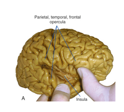
Q: Broadly, what is the insula involved in?
A: The integration of many sensorimotor and cognitive functions.
Q: Which sensory functions listed in the lecture does the insula integrate?
A: Gustation, visceral sensations, olfaction, somatosensation, and pain.
Q: Besides sensory functions, which cognitive functions is the insula involved in?
A: Attention, language, and speech.
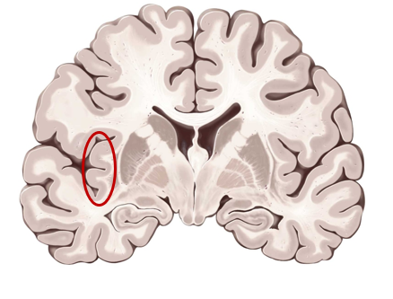
what is this structure?
insula

what is this structure?
insula
what system in the brain includes both cortical areas and non-cortical structures.
the limbic system
is the limbic lobe/system defined structurally or functionally?
it is a functionally-defined lobe
Q: What is the limbic lobe involved with?
A: Emotion and memory.

label


label


label
Q: Which lobes does the central sulcus separate?
A: The frontal and parietal lobes.
Q: Which two gyri does the central sulcus separate?
A: The pre-central gyrus (motor cortex) from the post-central gyrus (sensory cortex)
Q: What does the lateral fissure separate?
A: The temporal lobe from the frontal and parietal lobes.
Q: What does the longitudinal fissure separate?
A: The left and right hemispheres.
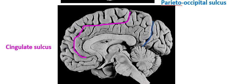
Q: What does the cingulate sulcus separate?
A: Cortical tissue of the frontal lobe from the cingulate lobe.
Q: What does the parieto-occipital sulcus separate?
A: The parietal and occipital lobes.
Q: From which view is the parieto-occipital sulcus only visible?
A: The midsagittal view.
Q: What is gray matter rich in?
A: Neuron soma and dendrites, and glial cells.
Q: What is white matter?
A: Dense areas of axons.
Q: What cells does white matter contain, and what does it have few of?
A: It contains glial cells but few neuron soma.
Q: What accounts for the white coloration of white matter?
A: Myelinated axons.
Q: How many kinds of white matter pathways/tracts are there, and what are they?
A: Three — association tracts, commissural tracts, and projection tracts.
Q: What do association tracts connect?
A: Parts of the same hemisphere (ex. anterior and posterior)
Q: What do commissural tracts connect?
A: The left and right hemispheres.
Q: What are projection tracts?
A: Afferent or efferent tracts connecting the cortex with the brainstem/spinal cord (superior-inferior)
Q: What type of tract is the corpus callosum?
A: The major commissural tract.
Q: What does the corpus callosum connect?
A: The left and right hemispheres.
Q: What type of tract is the internal capsule?
A: The major projection tract.

Q: What does the internal capsule connect?
A: The cortex with the brainstem.
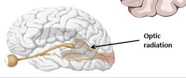
Q: What type of tract is the optic radiation?
A: A projection tract.
Q: What pathway is the optic radiation part of?
A: The visual pathway.
Q: What does the optic radiation connect?
A: The thalamus with the visual cortex (occipital lobe).
Q: What does it mean that most motor and sensory pathways decussate?
A: They cross over between the brain and the periphery.
Q: Which sensory and motor functions does the left hemisphere process?
A: Right-sided functions. (and vice versa)
Q: What is the homunculus?
A: A visual representation of spatial topography useful for mapping out deficits with brain injury

Q: What type of tissue are the basal ganglia, and where are they found?
A: Gray matter found deep within the cerebrum.
Q: What does the basal ganglia function in?
A: Control of movement.
Q: List the disorders associated with the basal ganglia in this lecture.
A: Huntington's, Parkinson's, cerebral palsy, Tourette's disorder, and tardive dyskinesia, among others.
Q: What are the four divisions of the diencephalon
A: Thalamus, hypothalamus, epithalamus, and subthalamus.
Q: What is the thalamus the gateway to?
A: The cerebral cortex and all sensory inputs to cerebrum EXCEPT OLFACTION
Q: What does the hypothalamus regulate?
A: Autonomic functions, drive-related behavior, and the pituitary gland
Q: What structure makes up the epithalamus?
A: The pineal gland.
Q: What does the pineal gland secrete?
A: Melatonin — circadian rhythms
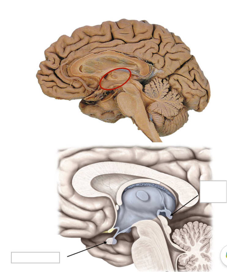
label

Q: Where does the cerebellum sit?
A: On the dorsal surface of the brainstem and the 4th ventricle.
Q: with what does the cerebellum communicate with the rest of the brain?
A: By 3 paired peduncles.
Q: What does "arbor vitae" mean and what does it refer to?
A: means “tree of life” and refers to the gray/white matter arrangement of the cerebellum seen in sagittal cut..
Q: List the functions of the brainstem given in this lecture
A: 1. Autonomic functions (breathing, heartbeat, BP, consciousness, sleep, alertness)
Q: Which cranial nerves originate in the brainstem?
A: CN III–XII.
Q: the brainstem serves as a conduit for what?
A: It serves as a conduit for many ascending and descending pathways.
Q: What are the three structures of the brainstem
A: Midbrain, pons, and medulla.
Q: What are the ventricles?
A: Elaborations of the developmental neural canal in the adult brain. (aka fluid filled spaces in the brain)
Q: The ventricles are interconnected and continuous with what two structures?
A: The central canal of the spinal cord and the subarachnoid space.
Q: What is the primary function of the ventricles?
A: Production and housing of cerebrospinal fluid (CSF).
Q: How many ventricular spaces are there, and what are they referred to as?
A: Four — the paired lateral ventricles, the third ventricle, and the fourth ventricle.
Q: what structure is located within the walls of the ventricles serving as the major site for CSF production?
A: The choroid plexus.

label the ventricles

Q: What are the functions of CSF?
A: It provides nutrients and protects the brain from injury by providing a cushion against blows to the head.
Q: What kind of substance is CSF?
A: A plasma ultrafiltrate.
Q: Where is CSF found?
A: In the central spinal canal, the ventricular system, and the subarachnoid space.
Q: What is the total adult CSF volume?
A: 150 mL.
Q: How is adult CSF volume distributed?
A: 125 mL in the subarachnoid spaces and 25 mL in the ventricles.
Q: How much CSF is produced per day?
A: 400–600 mL per day at a rate of 30 mL/hr
Q: How many times per day is CSF renewed?
A: 4–5 times per day.
Q: What is normal CSF pressure?
A: 5–15 mmHg.
Q: What are the 4 functions of CSF listed in this lecture?
A: Support, shock absorber, homeostasis, and nutrition.